1. Serviço de Reumatologia, Hospital Santa Maria, Centro Hospitalar de Lisboa Norte, EPE
Musculoskeletal ultrasound in Paediatric Rheumatology:
a retrospective analysis
ACTA REUMATOL PORT. 2014;39:309-314
AbstrAct
Objectives: Musculoskeletal Ultrasound (MSK-US) has
become increasingly important in the diagnosis and fol-low-up of children with rheumatic diseases. We des-cribe the experience of a large Portuguese centre and study the added value of MSKUS in the clinical assess -ment of paediatric rheumatic diseases.
Material and methods: Patients were observed by
as-sistant Rheumatologists, a clinical diagnosis was assig-ned and MSK-US requested. 330 MSK-US exams were performed to 222 children with rheumatic inflamma-tory diseases. The children’s ages were between 1 and 18 years (mean=11.7±4.7 years) and 67.6% were fe-male. Synovial membrane proliferation, intra-articular effusion, cartilage abnormalities, erosions and periarti-cular affections were searched in each joint. Clinical and ultrasonography data were compared.
Results: MSK-US detected synovitis in 100 of 194
exams (51.5%) of patients with that clinical informa-tion and in 36 of 136 exams (26.5%) of patients who presented other clinical findings. In those in which MSK-US did not confirm the clinical information of sy-novitis (94; 48.5%), we detected tenosysy-novitis/tendi- tenosynovitis/tendi-nopathy in 13 cases (13.8%) and synovial cyst in four (4.3%). The remaining patients had no ultrasonogra -phy changes and MSK-US helped to exclude synovitis. The sensitivity for arthritis clinical assessment was good (73.5%), with modest specificity (51.5%), an accuracy of 60.6% and precision of 51.5%. Ultrasonography synovitis was mostly found in the knee (37.5%), follow -ed by the ankle (22.8%) and hip (10.3%).
Overall, 39 exams showed ultrasonographic tenosyno-vitis/tendinopathy, 15 of which had the same clinical diagnosis. Tenosynovitis/tendinopathy was mostly found in the ankle (59.0%) and knee (23.1%) areas.
Madruga Dias J1, Costa MM1, Canhão H1, Saraiva F1, da Silva JA1
Conclusions: MSK-US is an important aid to clinical
evaluation, allowing both the detection and exclusion of joint pathology in children, contributing to a better assessment.
Keywords: Paediatric; Tenosynovitis; Ultrasound;
Rheu ma tology; Synovitis;
IntroductIon
Musculoskeletal ultrasound (MSK-US) has become in-creasingly important in the diagnosis and follow-up of both adults and children with rheumatic diseases in the last 20 years1,2.
It is a non-invasive exam which not only helps the diagnosis and assessment of the disease, but it also as-sists in treatment decisions. Additionally, it allows ul-trasound-guided procedures. Undervaluation of arthri-tis may lead to delayed diagnosis and treatment, or su-boptimal suppression of joint inflammation with anti--rheumatic therapy. The issue of subclinical arthritis is particularly relevant in Juvenile Idiopathic Arthritis (JIA), but also applies to many other inflammatory rheu-matic diseases affecting children. MSK-US seems to re-present a reliable measure of JIA disease activity3.
MSK-US has a number of advantages over other ima-ging methods, including non-invasiveness, radiation--free, relative low cost, availability, ability to scan mul-tiple joints at one time, repeatability and good patient acceptance. Another advantage of US is that it can be coupled with the clinical approach to the patient in the standard rheumatology setting. Specifically conside-ring children, the innocuous nature of MSK-US and the fact that it can be done swiftly along with clinical ob-servation makes it a useful imaging technique in cur-rent medical practice.
In more advanced stages of JIA, gadolinium-enhan-ced Magnetic Resonance Imaging (MRI) seems to be
superior when evaluating synovial proliferation, arti-cular cartilage, loculated effusions, menisci and liga-ments4. Nevertheless, considering cartilage thickness,
no significant joint size-related differences were found between MRI and MSK-US5. This gives weight to the
usage of ultrasonogra phy in children for evaluating ar-ticular changes, as MSK-US is a more accessible exam than MRI.
There are a small number of articles regarding the use of MSK-US in children, and as far as we know, no Portuguese study was published in this area of know-ledge. In our work we discuss the experience in a lar-ge centre of MSK-US in the study of rheumatic condi-tions in children, comparing clinical observation with MSK-US assessment.
MAterIAl And Methods
We performed a retrospective analysis of 330 MSK-US exams performed to 222 children with rheumatic com-plaints in our department in the last 11 years (2001--2011). The children’s ages were between 1 and 18 years (mean= 11.7±4.7 years) and 67.6% were fema-le. They were observed in the Paediatric Rheumatolo-gy outpatient clinic by consultant Rheumatologists, with training and expertise in Paediatric Rheumatolo-gy and more than a decade of practice. The great ma-jority of ultrasonographic exams were performed non--blinded in the same day as clinical observation by one of two rheumatologists with more than 15 years of ul-trasound experience, using Diasus (Dynamic Imaging) ultrasonograph equipped with 3 linear probes (5-10 MHz, 8-16 MHz and 10-22 MHz) and Logiq E9 (Ge-neral Electric Medical Systems, Milwaukee, WI), equipped with an 8–15 MHz volumetric probe (4D16L) and 2 linear probes (ML6-15 and L8-18i).
We compared the clinical information accompa-nying the MSK-US request (inflammatory arthritis, swollen joint, tender joint, tenosynovitis/tendinitis or others) with the ultrasonographic findings. Synovial membrane proliferation, intra-articular effusion, car-tilage abnormalities, erosions and soft tissue affections (tendinopathy, tenosynovitis, and bursitis) were sear-ched at each joint6. Each joint was scanned in both the
longitudinal and transversal view using grey-scale. Overall, the following joints were scanned: hand dis-tal and proximal interphalangeal, metacarpophalan-geal, wrist, elbow, shoulder, sternoclavicular, sacroco-cígeal, hip, knee, ankle, metatarsophalangeal and feet
proximal interphalangeal.
Continuous variables are summarized by mean, standard deviation and range. Categorical (dichoto-mous) variables such as synovitis or tenosynovitis are shown as absolute numbers or summarized as fre-quency (in percentage). The performance of clinical evaluation compared with MSK-US was made with de-termination of sensitivity, specificity, accuracy and pre-cision. Clinical and ultrasonographic data were eva-luated using Chi-Square test and Spearmans rank cor-relation coefficient; p values < 0.05 were considered to be statistically significant.
results
We analyzed a total of 330 exams in 222 children. In most children (70.3%) only one MSK-US exam was done per observation.
MSK-US detected synovitis in 100 of 194 exams (51.5%) of patients who had the clinical information of arthritis. In contrast, synovitis was found in 36 of 136 exams (26.5%) of patients with other clinical diagnosis: tendinitis, tenosynovitis, joint pain or other (Figure 1). Using Chi-Square, there is a statistically significant dif-ference between the total number of ultrasound-con-firmed synovitis and the total number of clinically diag-nosed arthritis (p<0.0001). Nevertheless, if we compa-re the number of clinical arthritis with the number of ul-trasonographic synovitis for each individual joint, there is a significant correlation (r=0,88; p=0,002).
In 48.5% of patients with arthritis at observation, MSK-US did not support the clinical findings. In the-se patients with clinical, but not ultrasonographic sy-novitis (n=94), we detected tenosysy-novitis/tendinopa thy in 13 cases (13.8%) and the presence of synovial cyst in 4 (4.3%). The 77 remaining patients had no ultra-sonographic changes whatsoever.
Ultrasonographic synovitis was mostly found in the knee (37.5%), followed by the ankle (22.8%) and hip (10.3%). (Table I) If we consider the relative frequen-cy of synovitis, the elbow (72.7%), shoulder (55.6%), knee (50.5%) and foot joints (50.0%) were the anato-mical areas where MSK-US mostly confirmed the cli-nical findings.
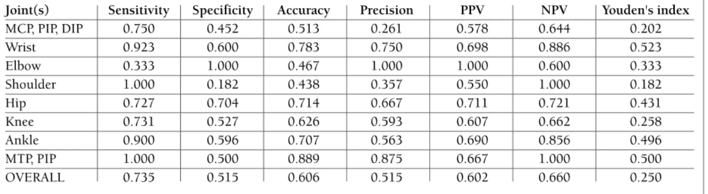
Compared to MSK-US, the overall sensitivity for arthritis clinical assessment was 73.5%, with a 51.5% specificity. There was a 60.6% accuracy and 51.5% pre-cision (Table II).
specificity, accuracy and precision for clinical arthritis compared to MSK-US. The shoulder, wrist, ankle and feet joints have the highest sensitivity when using cli-nical evaluation (Table II). Hip, knee and hand joints also present sensitivity over 70%. As for specificity the elbow and hip present the best values. The most accu -rate clinical assessment for synovitis was in the wrist and feet joints, although both hip and ankle clinical evaluations proved to have an accuracy above 70%. Precision was highest in assessment of the elbow, wrist and feet joints.
Overall, 39 exams showed ultrasonographic teno-synovitis/tendinopathy, 15 (38.5%) of which had the same clinical diagnosis associated. In the remaining 24 exams, 14 (35.9%) had clinical arthritis, 7 (17.9%) had joint pain and 3 (7.7%) had clinical information of bur-sitis. Of 26 patients with clinical tenosynovitis/tendi-nitis only 15 (57.7%) had ultrasonographic tenosyno-vitis/tendinopathy. There is a statistically significative difference between MSK-US and clinical evalua tion at detecting tenosynovitis/tendinopathy (p< 0.0001).
Tenosynovitis/tendinopathy was mostly found in the ankle (59.0%) and knee (23.1%) areas (Table III). If we consider the relative frequency of tenosynovi-tis/tendinopathy, the ankle (28.4%) is still the most af-fected area.
MSK-US also identified erosions in 7 patients (2 of
them had no ultrasonographic synovitis). Considering synovial cyst, there were 9 ultrasonographic findings, one third in patients with the same clinical diagnosis, four in patients with the clinical (but not ultrasono-graphic) diagnosis of arthritis and 2 in patients with popliteal pain.
dIscussIon
The first publications using MSK-US in children with osteo-articular pathologies date back over 20 years1,2.
MSK-US has seen a growing implementation in daily practice in the last decade in the field of Paediatric Rheumatology. Our work confirms the need of a wide implementation of MSK-US in Paediatric Rheumato-logy. In fact, it is an added value in the diagnosis and monitoring of children with rheumatic inflammatory conditions, confirming or denying synovitis and iden-tifying other pathologies.
Our results are in agreement with a previous study7
which also concluded that MSK-US is more accurate at detecting synovitis in children than clinical exami-nation, allowing either confirmation or denial, or re-classification of diagnosis. Several other authors have reached the same conclusions when studying peri -pheral joints8, ankles9, knees10-12and hips11,13,14.
Ove-other020406080100120 140 160 180 200synovial cystbursitistenosynovitis/tendinitisjoint contractureswollen jointtender jointarthritis100 (51,5%)Clinical dianosis19427 (33,3%)8112103 (10,7%)1 71 31 1328Ultrasonographic synovitis
other 0 20 40 60 80 100 120 140 160 180 200 synovial cyst bursitis tenosynovitis/tendinitis joint contracture swollen joint tender joint arthritis 100 (51,5%) Clinical dianosis 194 27 (33,3%) 81 1 2 10 3 (10,7%) 1 7 1 3 1 13 28 Ultrasonographic synovitis
rall, for multiple joint assessments, MSK-US also pro-ved to be an indispensable complement to clinical exa-mination, allowing enhanced evaluation7,8,15-17.
Other interesting finding of our work is that MSK--US could exclude synovitis in nearly half (48.5%) of the children with the clinical diagnosis of arthritis. It also excluded synovitis in two thirds of children with tender joints and in most children with apparent swol-len joints. This emphasises the utility of MSK-US in identifying children without joint pathology, sparing unnecessary treatment and possible iatrogenesis.
We used an ultrasonograph without Power Doppler
for most exams, because no other device was available at the time. Although Power Doppler is important in accessing information about joint active inflammation in children18-20a recently published study15compared
both Power Doppler and grey-scale MSK-US, showing that there were more findings in children’s joints using grey-scale than Power Doppler. Other authors3have
reported that MSK-US parameters, not using Power Doppler, represent a reliable index of JIA disease acti-vity, especially considering knee synovial thickness and knee effusion. Knee synovial thickness and effusion were always scanned in our patients.
tAble I. ultrAsonoGrAphIc synovItIs by AnAtoMIcAl locAtIon
Joints with US synovitis Total joints scanned Relative frequency Absolute frequency
Knee 51 101 50.5 % 37.5 % Ankle 31 81 38.3 % 22.8 % Hip 14 49 28.6 % 10.3 % Wrist 11 22 50.0 % 8.1 % Elbow 8 11 72.7 % 5.9 % MTP, PIP 8 16 50.0 % 5.9 % MCP, PIP, DIP 7 39 17.9 % 5.1 % Shoulder 5 9 55.6 % 3.7 % Sternoclavicular 1 1 100.0 % 0.7 % Sacrococígeal 0 1 0.0 % 0.0 % TOTAL 136 330 100 %
(US – ultrasonographic, MCP – metacarpophalangeal, MTP – metatarsophalangeal, PIP – proximal interphalangeal, DIP – distal interphalangeal)
Relative frequency is the percentage of positive findings per anatomical area. Absolute frequency is the percentage of positive findings in an anatomical area compared with the total number of joints with US synovitis (136)
tAble II. sensItIvIty, specIFIcIty, AccurAcy And precIsIon oF clInIcAl evAluAtIon oF ArthrItIs coMpAred to MsK-us, by joInt
Joint(s) Sensitivity Specificity Accuracy Precision PPV NPV Youden's index
MCP, PIP, DIP 0.750 0.452 0.513 0.261 0.578 0.644 0.202 Wrist 0.923 0.600 0.783 0.750 0.698 0.886 0.523 Elbow 0.333 1.000 0.467 1.000 1.000 0.600 0.333 Shoulder 1.000 0.182 0.438 0.357 0.550 1.000 0.182 Hip 0.727 0.704 0.714 0.667 0.711 0.721 0.431 Knee 0.731 0.527 0.626 0.593 0.607 0.662 0.258 Ankle 0.900 0.596 0.707 0.563 0.690 0.856 0.496 MTP, PIP 1.000 0.500 0.889 0.875 0.667 1.000 0.500 OVERALL 0.735 0.515 0.606 0.515 0.602 0.660 0.250
(MCF – metacarpophalangeal, MTF – metatarsophalangeal, PIP – proximal interphalangeal, DIP – distal interphalangeal, PPV – Positive Predictive Value, NPV – Negative Predictive Value)
The most frequent locations for MSK-US synovitis in our study were the knees (37.5%) and the ankles (22.8%), while other authors studying only JIA chil-dren found the knees and wrists16and others establi
-shed the feet as more prevalent8. However, we
analy-sed also the relative frequency of synovitis and we found that the elbow (72.7%), shoulder (55.6%), knee (50.5%) and foot joints (50.0%) were the anatomical areas where MSK-US mostly confirmed the clinical fin-dings. The small number of scanned shoulder and elbow joints makes these results to be confirmed by fur -ther studies. The hip (28.6%) and hand joints (17.9%) were the anatomical areas where MSK-US less corro-borated clinical evaluation.
Our work also evaluated clinical assessment of te-nosynovitis/tendinitis. There seems to be a remarkable discordance between clinical and MSK-US evaluation of tenosynovitis. In fact, most (61.5%) of the ultraso-nographic diagnosis of tenosynovitis were found in pa-tients with no clinical signs or symptoms of this affec-tion. This may be due to subclinical tenosynovitis and illustrates the limitations of physical examination in children. The most common location for tenosynovi-tis was in the ankles (59.0%). Other studies concur with our results about tenosynovitis/tendinopathy and in fact they have very similar figures to ours9,17.
If we consider MSK-US as the gold standard, we ve-rified that clinical assessment in general had a good sensitivity but a modest specificity for synovitis detection. Despite having a correlation with ultrasonogra -phy findings and being fundamental in assessing chil-dren with rheumatic inflammatory conditions, clinical evaluation does not seem to be specific enough for sy-novitis detection, and lacks precision and accuracy.
MSK-US, being a bedside, painless and radiation-free procedure has an added value and can help in decision making on the spot.
One limitation is that our work is a retrospective analysis, which requires cautious interpretation. An important aspect to note is that MSK-US was perfor-med promptly in children observed in the Rheumato-logy clinic. The importance of this timing to exclude possible time-related differences between clinical ob-servation and MSK-US examination has been underli-ned by other authors3,7.
Globally, our results support the suggestion that MSK-US should be a screening procedure7for children
with suspected joint pathology.
conclusIon
MSK-US is an important aid to clinical evaluation, allowing both the detection and exclusion of joint pa -thology in children, contributing to a better assessment and quality of care.
correspondence to
João Alexandre Costa Madruga Dias Serviço de Reumatologia
Hospital de Santa Maria, Centro Hospitalar Lisboa Norte Avenida Professor Egas Moniz
1649-035 Lisboa, Portugal reFerences
1. Goldenstein C, McCauley R, Troy M, Schaller JG, Szer IS. Ul-trasonography in the evaluation of wrist swelling in children. J Rheumatol 1989; 16:1079-1087.
2. Kallio P, Ryöppy S, Jäppinen S, Siponmaa AK, Jääskeläinen J, Kunnamo I. Ultrasonography in hip disease in children. Acta
tAble III. MsK-us tenosynovItIs/tendInItIs by AnAtoMIcAl AreA
Joints with US
tenosynovitis/tendinitis Total joints scanned Relative frequency Absolute frequency
Ankle 23 81 28.4 % 59.0 % Knee 9 101 8.9 % 23.1 % MCP, PIP, DIP 4 39 10.3 % 10.2 % Wrist 2 22 9.1 % 5.1 % Shoulder 1 9 11.1 % 2.6 % TOTAL 39 252 100.0 %
(US – ultrasonographic, MCP – metacarpophalangeal, PIP – proximal interphalangeal, DIP – distal interphalangeal)
Relative frequency is the percentage of positive findings per anatomical area. Absolute frequency is the percentage of positive findings in an anatomical area compared with the total number of joints with US tenosynovitis/tendinitis (39)
Orthop Scand 1985; 56 :367-371.
3. Algergawy S, Haliem T, Al-Shaer O. Clinical, laboratory, and ul-trasound assessment of the knee in juvenile rheumatoid arth-ritis. Clin Med Insights Arthritis Musculoskelet Disord 2011; 25: 21-27.
4. El-Miedany YM, Housny IH, Mansour HM, Mourad HG, Me-hanna AM, Megeed MA. Ultrasound versus MRI in the evalua-tion of juvenile idiopathic arthritis of the knee. Joint Bone Spi-ne 2001; 68: 222-230.
5. Spannow AH, Pfeiffer-Jensen M, Andersen NT, Herlin T, Sten-bøg E. Ultrasonographic measurements of joint cartilage thick-ness in healthy children: age- and sex-related standard refe-rence values. J Rheumatol 2010; 37: 2595-2601.
6. Wakefield RJ, Balint PV, Szkudlarek M, et al; OMERACT 7 Spe-cial Interest Group. Musculoskeletal ultrasound including de-finitions for ultrasonographic pathology. J Rheumatol 2005;32:2485-2487.
7. Filippou G, Cantarini L, Bertoldi I, Picerno V, Frediani B, Ga-leazzi M. Ultrasonography vs. clinical examination in children with suspected arthritis. Does it make sense to use poliarticu-lar ultrasonographic screening? Clin Exp Rheumatol 2011; 29: 345-350.
8. Breton S, Jousse-Joulin S, Cangemi C, et al. Comparison of Cli-nical and Ultrasonographic Evaluations for Peripheral Synovi-tis in Juvenile Idiopathic ArthriSynovi-tis. Semin ArthriSynovi-tis Rheum 2011; 4.
9. Pascoli L, Wright S, McAllister C, Rooney M. Prospective eva-luation of clinical and ultrasound findings in ankle disease in juvenile idiopathic arthritis: importance of ankle ultrasound. J Rheumatol 2010; 37: 2409-2014.
10. Kakati P, Sodhi KS, Sandhu MS, Singh S, Katariya S, Khandel-wal N. Clinical and ultrasound assessment of the knee in chil-dren with juvenile rheumatoid arthritis. Indian J Pediatr 2007; 74: 831-836.
11. Frosch M, Foell D, Ganser G, Roth J. Arthrosonography of hip and knee joints in the follow up of juvenile rheumatoid arthri-tis. Ann Rheum Dis 2003; 62: 242-244.
12. Sureda D, Quiroga S, Arnal C, Boronat M, Andreu J, Casas L. Juvenile rheumatoid arthritis of the knee: evaluation with US. Radiology 1994; 190: 403-406.
13. Friedman S, Gruber MA. Ultrasonography of the hip in the eva-luation of children with seronegative juvenile rheumatoid arth-ritis. J Rheumatol 2002; 29: 629-632.
14. Sureda D, Quiroga S, Arnal C, Boronat M, Andreu J, Casas L. Ultrasonography in the early diagnosis of hip joint involvement in juvenile rheumatoid arthritis. J Rheumatol 1997; 24: 1820--1825.
15. Rebollo-Polo M, Koujok K, Weisser C, Jurencak R, Bruns A, Roth J. Ultrasound findings on patients with juvenile idiopa -thic arthritis in clinical remission. Arthritis Care Res (Hobo-ken) 2011 ; 63: 1013-1019.
16. Haslam KE, McCann LJ, Wyatt S, Wakefield RJ. The detection of subclinical synovitis by ultrasound in oligoarticular juveni-le idiopathic arthritis: a pilot study. Rheumatology (Oxford) 2010; 49: 123-127.
17. Magni-Manzoni S, Epis O, Ravelli A, et al. Comparison of cli-nical versus ultrasound-determined synovitis in juvenile idio-pathic arthritis. Arthritis Rheum 2009; 61: 1497-1504. 18. Spârchez M, Fodor D, Miu N. The role of Power Doppler
ul-trasonography in comparison with biological markers in the evaluation of disease activity in Juvenile Idiopathic Arthritis. Med Ultrason 2010; 12: 97-103.
19. Shanmugavel C, Sodhi KS, Sandhu MS, et al. Role of power Doppler sonography in evaluation of therapeutic response of the knee in juvenile rheumatoid arthritis. Rheumatol Int 2008; 28: 573-578.
20. Shahin AA, el-Mofty SA, el-Sheikh EA, Hafez HA, Ragab OM. Power Doppler sonography in the evaluation and follow-up of knee involvement in patients with juvenile idiopathic arthritis. Z Rheumatol 2001; 60:148-155.