A R T I G O O R I G I N A L
I S O K I N E T I C S T R E N G T H M E A S U R E M E N T S I N E A R L Y K N E E O S T E O A R T H R I T I S
Demirhan Dıraço
ˇg
lu,*Akın Bas,
kent,*˙ll
ker Yaˇg
cı,** Levent Özçakar,***,****Resa Aydın*in patients with knee OA compared to healthy sub-jects (p<0.05). Patients with stage I OA had greater muscle strength than those of stage II (p<0.05). Conclusions: Whether being a cause or a con-sequence of knee OA, muscle strength loss which cannot be detected during clinical examination ap-pears to be present during isokinetic measure-ments.
Keywords: Osteoarthritis; Knee; Muscle strength; Isokinetic testing.
Introduction
Osteoarthritis (OA) is the most commonly seen jo-int disease throughout the world.1The knee joint is often involved and locomotor dysfunction and di-sability in these patients ensue due to muscle weak-ness of the lower extremity.2
Stability of the knee joint is achieved through two ways, first of which is the active neuromuscu-lar control provided by muscle strength and propri-oceptive sense. The second is the passive resistan-ce formed by surrounding ligaments and joint cap-sule. Any problem arising from these factors may disturb the stability of the joint, thereby rendering it susceptible to degenerative processes.3 Pre-viously, isokinetic muscle strength of the knee mus-cles has been shown to decrease significantly in el-derly patients with chronic knee OA.4-9However, it is still controversial whether muscular weakness is also present in early stages of OA.
Objectives
The aim of this study was to compare the manual and isokinetic knee muscle strength of patients with early knee OA with those of subjects without clinical or radiological evidence of knee OA. *Istanbul University Istanbul Medical Faculty, Department of
Physical Medicine and Rehabilitation, Istanbul,Turkey **Marmara University Medical School, Department of Physical Medicine and Rehabilitation, Istanbul,Turkey
***Gülhane Military Medical Academy Haydarpas,a Training Hospital, Department of Physical Medicine and Rehabilitation, Istanbul, Turkey
****Hacettepe University Medical School, Department of Physical Medicine and Rehabilitation,Ankara,Turkey
Abstract
Objectives: One of the most important reasons for locomotor dysfunction and disability in patients with knee osteoarthritis (OA) is muscle weakness in the lower extremity. The aim of this study was to compare the isokinetic knee muscle strength of patients with early knee OA with those of healthy people.
Patients and Methods: Fifty-one patients with bi-lateral knee osteoarthritis who were radiologically graded as stage I or II and forty-three healthy sub-jects were enrolled. Western Ontario and McMas-ter Universities Osteoarthritis Index and 100 mm VAS were used to assess patients with knee OA. Ma-nual muscle strength testing for quadriceps mus-cle and circumference measurements 10 cm abo-ve the midpatellar line were performed. Bilateral isokinetic (concentric/concentric) knee flexion and extension with the protocol of 60 degrees/sec (four repetitions), 180 degrees/sec (four repetitions) and 240 degrees/sec (20 repetitions) were performed. Results: Regarding manual muscle testing of knee OA group, quadriceps muscle strength in six knees were 4/5 and in 96 knees were 5/5; whereas in the control group only two knees had 4/5 and the rest 84 knees had 5/5 muscle strengths (p=0.22). Thigh circumference measurements were statistically si-milar in this regard (all p values > 0.05). In all velo-cities knee flexor and extensor isokinetic muscle strength values were found to be significantly lower
D E M I R H A N DıR A Ç OG L U E C O Lˇ .
Patients and Methods
Fifty-one female subjects who were diagnosed with bilateral knee osteoarthritis according to American College of Rheumatology (ACR) criteria10and whose X-rays were graded as stage I or II according to Kell-gren & Lawrence Scale11were enrolled. Forty-three female subjects without clinical or radiological evi-dence of knee OA who volunteered to participate were also recruited (from hospital staff and their re-latives) as a control group. Patients were excluded if they had any of the following: active synovitis, arthroscopic/surgical intervention or intraarticular injection within the last six months, any pathology (e.g. lumbar nerve root compression, polyneuro-pathy, myopathy) that would cause muscle we-akness and inability to perform isokinetic testing.
Western Ontario and McMaster Universities Oste-oarthritis Index (WOMAC) (5-point Likert 3.0) ques-tionnaire which had been validated in our language12 was performed and pain levels were measured with 100 mm VAS in patients with knee OA. Manual mus-cle strength testing for quadriceps musmus-cle (Lovett scale; 0-5)13and circumference measurements 10 cm above the midpatellar line were performed by the same physician at baseline. Informed consent was taken before subjects agreed to participate.
Isokinetic testing
In both groups, isokinetic quadriceps/hamstring strengths were measured by using the Biodex Sys-tem 3-Pro (Biodex Medical Systems, Inc, New York, USA) dynamometer with the knee attachment.
Orientation of the dynamometer was kept at 0°, tilt at 0°, seat orientation at 0°.The patients were sea-ted and secured to the apparatus with chest and thigh straps. The attachments of the dynamometer were adjusted so that the centre of motion of the le-ver arm was aligned as accurately as possible with the slightly changing flexion-extension axis of the joint. The resistance pad was placed on the distal tibia. The range of motion of the knee joint was kept at 0-90°. Bilateral isokinetic (concentric/concen-tric) knee flexion and extension with the protocol of 60 degrees/sec (four repetitions), 180 degrees/ /sec (four repetitions) and 240 degrees/sec (20 re-petitions) were performed. Enough resting was pro-vided between the sessions and vocal encourage-ment was standardized.
Statistical Analysis
SPSS 13.0 software was utilized for statistical
as-sessment. Student’s t test was used for comparison of the mean values regarding isokinetic test results and Mann Whitney U test was used to compare isokinetic test results between different radiologi-cal stages. Chi-square test was used for compari-son of manual muscle test results. Pearcompari-son coeffi-cients were used for correlation analysis. Statisti-cal significance was set at p<0.05.
Results
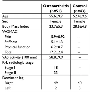
Demographic and clinical features of the subjects are summarized in Table I. The two groups were found to be statistically indifferent with regard to age and BMI values (p=0.11 and p=0.32, respecti-vely).
Thigh circumference measurements were 47.41±4.57 (right) and 47.44±4.47 (left) in the OA group, and 49.20±4.84 (right) and 48.86±4.85 (left) in the control group. The groups were found to be statistically similar in this regard (all p values > 0.05). Regarding manual muscle testing of knee OA group, quadriceps muscle strength in six knees were 4/5 and in 96 knees were 5/5; whereas in the control group only two knees had 4/5 and the rest 84 knees had 5/5 muscle strengths (p=0.22).
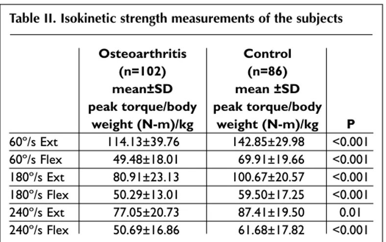
Table II lists isokinetic muscle strength values of
Table I. Demographic and clinical features of the subjects
Osteoarthritis Control
(n=51) (n=43)
Age 55.6±9.7 52.4±9.6
Sex Female Female
Body Mass Index 23.7±5.3 28.6±4.8 WOMAC Pain 5.9±0.92 – Stiffness 5.1±1.3 – Physical function 6.2±0.7 – Total 17.2±2.4 – VAS activity (100 mm) 58.8±9.9 – K-L radiologic stage Stage I 18 – Stage II 33 – Dominant leg Right 49 40 Left 2 3
I S O K I N E T I C S T R E N G T H M E A S U R E M E N T S I N E A R LY K N E E O S T E O A R T H R I T I S
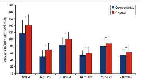
both groups. In all velocities knee flexor and exten-sor isokinetic muscle strength values were found to be significantly lower in patients with knee OA compared to healthy subjects. When the right and left knee strengths were compared separately, the two groups were again found to be statistically dif-ferent (Figures 1 and 2).
Isokinetic strength measurements regarding stage I and II (knees) are given in Table III. Patients with stage I had greater knee muscle strength when compared with stage II. There were negative cor-relations between isokinetic strength values and WOMAC-pain and VAS scores but the correlations were only significant for measurements at 60°/s velocity, both during extension (p values being 0.04 and 0.03 respectively) and flexion (p values being
0.02 and 0.01 respectively).
Discussion
In this study, manual and isokinetic knee muscle strength testing of female subjects with early OA were compared with other females without any eviden-ce of knee OA. Although manual mus-cle testing were similar between the groups, patients with OA were found to have significantly lower flexor and ex-tensor isokinetic knee strength. Further, stage I patients had greater muscle strength when compared with stage II patients.
Regarding OA, a close association was establi-shed between dynamic isokinetic muscle strength measurements and the progression level of disea-se and clinical signs.14It is known that isokinetic muscle strength measurements in patients with knee OA is a validated and reliable method in a re-peatable manner.15,16Many deficits rarely detecta-ble through manual muscle measurement can be revealed using isokinetic measurement.
There might be many reasons for muscle weak-ness in knee OA. Young et al17reported that “arthro-genic muscle inhibition” of quadriceps muscles in patients with knee OA might lead to weakness. This term refers to inhibition of motor neurons due to afferent signals from affected joint or periarticular tissues, i.e. inhibition of muscular contraction. This reflex inhibition may also be detected following joint sur-gery18 or with joint effu-sions.19It has been recently shown that in early OA mus-cle weakness was related with changes in motor unit physiology.20
Impaired neuromuscular control, decreased muscle strength and muscle atrophy are related to less muscle use due to pain and dysfunction consequent to the degenera-tive process. On the other hand, Slemenda et al5found significantly lower isokinetic muscle strength in 462
pati-Table II. Isokinetic strength measurements of the subjects
Osteoarthritis Control
(n=102) (n=86)
mean±SD mean ±SD
peak torque/body peak torque/body weight (N-m)/kg weight (N-m)/kg P 60º/s Ext 114.13±39.76 142.85±29.98 <0.001 60º/s Flex 49.48±18.01 69.91±19.66 <0.001 180º/s Ext 80.91±23.13 100.67±20.57 <0.001 180º/s Flex 50.29±13.01 59.50±17.25 <0.001 240º/s Ext 77.05±20.73 87.41±19.50 0.01 240º/s Flex 50.69±16.86 61.68±17.82 <0.001 Ext: Extension, Flex: Flexion
Figure 1. Comparative isokinetic muscle strength measurements of the right extremity (OA vs healthy).
D E M I R H A N DıR A Ç OG L U E C O Lˇ .
the joint due to deterioration of shock absorption during walking.9There is a complex relationship between disabi-lity and cartilage degenera-tion, and sensorimotor dys-function originating from muscles around the joint.
The increase of load on the joint accelerates the pro-gression of knee OA, espe-cially in medial compart-ment.21van der Esch et al have shown that isokinetic muscular weakness had more influences on the limi-tation of functional ability in knee OA patients with poo-rer proprioception.6Their isokinetic testing protocol comprised only 60º/sec measurements; in our study, we have measured at three velocities and found that both in low and high speeds the muscle strength loss was significant. Brandt et al detec-ted that people with knee pain but wi-thout radiological signs of OA, had more weakness in quadriceps and ham-string muscles when compared to sub-jects with pain and radiological signs.8 In clinical practice, severity of the dise-ase is frequently assessed according to the radiological signs. Nonetheless, mainly in knee OA, the correlation between radiological scores, clinical findings and pain is poor.22In our study, although being in the early stage, we have observed muscle weakness in the OA group. However, since the muscle strengths of Stage 1 pa-tients were greater than Stage 2 papa-tients; we may not definitely propose that muscle weakness is a primary factor that has an adverse effect in the di-sease process.
On the other hand, concerning the negative cor-relations between pain scores and muscle strength, it would rather be wise to relate these results to some technical features of isokinetic testing. Yet, the correlations were statistically significant only at the velocity of 60°/s and values regarding flexion measurements were more significant than those of extension. During isokinetic testing, a patient may exert less force when it is painful. Considering the
Table III. Isokinetic strength measurements of the subjects
Stage I Stage II
(n=36) (n=66)
mean±SD mean ±SD
peak torque peak torque
(N-m) (N-m) P 60º/s Ext 103.0±25.7 70.3±18.4 <0.001 60º/s Flex 44.8±12.8 30.6±8.6 <0.001 180º/s Ext 70.1±15.0 51.7±9.0 <0.001 180º/s Flex 39.4±10.0 35.0±8.9 0.02 240º/s Ext 64.3±12.5 50.5±9.1 <0.001 240º/s Flex 42.9±11.0 33.0±9.0 <0.001 Ext: Extension, Flex: Flexion
Figure 2. Comparative isokinetic muscle strength measurements of the left extremity (OA vs healthy).
Ext: Extension; Flex: Flexion; (*: p<0,001; †: p<0,05)
ents without knee pain or muscular atrophy though having radiological signs of knee OA. Ac-cordingly, they suggested that quadriceps we-akness was the primary risk factor for the progres-sion of joint damage, disability and pain. Similarly, our study also showed that while people with ear-ly stage knee OA had isokinetic muscle strength loss, they had no significant change in their mus-cle mass.
Most important factors impairing the function of periarticular muscles in degenerative processes are advanced age, increased fatigue, delayed mus-cle reaction time, previous minor trauma, abnor-mal articular sensory input, impaired neuromus-cular protective reflexes, and abnormal loads on
I S O K I N E T I C S T R E N G T H M E A S U R E M E N T S I N E A R LY K N E E O S T E O A R T H R I T I S
fact that knee flexion is generally more painful than extension and these patients need to apply greater forces during isokinetic measurements at low ve-locities, our findings seem to be reasonable.
One possible drawback of our study would be related with the difficulties of isokinetic testing. While providing accurate and objective data con-cerning muscle weakness, isokinetic devices may not be available at every clinic. The required time and manpower further challenge the routine use of this method. Regarding some confounding fac-tors for isokinetic measurements, the absence of pain and stiffness measurements (WOMAC and pain VAS) in control subjects could be another li-mitation of our study.
To summarize, in the light of our results, we may conclude that muscle strength loss which cannot be detected during clinical examination appears to be present in female subjects with early stage knee OA; moreover, isokinetic measurements seem to uncover such muscle weakness. It should be the-refore emphasized that knee strengthening exerci-ses should be given to patients as soon as they are diagnosed to have knee OA. Further studies con-cerning the effect of exercise therapy on strength measurements are warranted.
Correspondence to Demirhan Dıraçoˇglu I.U. Istanbul Tip Fakultesi
Fizikel Tip ve Rehabilitasyon Anabilim Dali 34093, Istanbul/Turkey
Fax: +90 212 4142000
E-mail: demirhan1@yahoo.com References
1. Cooper C. Osteoartritis and Related disorders, epidemiology. In: Klippel JH, Dieppe PA, eds. Rheumatology Second Ed, Philadephia: Mos-by;1998:8-2.1.
2. Hurley MV. The role of muscle weakness in the pathogenesis of osteoarthritis. Rheum Dis Clin North Am 1999;25:283-298.
3. Schipplein OD, Andriacchi TP. Interaction betwe-en active and passive knee stabilizers during level walking. J Orthop Res 1991;9:113-119.
4. Messier SP, Glasser JL, Ettinger WH Jr et al. De-clines in strength and balance in older adults with chronic knee pain: a 30-month longitudinal, observational study. Arthritis Rheum 2002;47:141--148.
5. Slemenda C, Brandt KD, Heilman DK et al. Qua-driceps weakness and osteoarthritis of the knee.
Ann Intern Med 1997;127:97-104.
6. van der Esch M, Steultjens M, Harlaar J et al. Joint proprioception, muscle strength, and functional ability in patients with osteoarthritis of the knee. Arthritis Rheum 2007;57:787-793.
7. Gür H, Cakin N. Muscle mass, isokinetic torque, and functional capacity in women with osteo-arthritis of the knee. Arch Phys Med Rehabil 2003;84:1534-1541.
8. Brandt KD, Heilman DK, Slemenda C et al. A com-parison of lower extremity muscle strength, obe-sity, and depression scores in elderly subjects with knee pain with and without radiographic evidence of knee osteoarthritis. J Rheumatol 2000;27:1937--1946.
9.Hurley MV. Muscle dysfunction and effective reha-bilitation of knee osteoarthritis: what we know and what we need to find out. Arthritis Rheum 2003;49:444-452.
10. Altman R, Asch E, Bloch D, Bole G et al. Develop-ment of criteria for the classification and reporting of osteoarthritis. Classification of osteoarthritis of the knee, Diagnostic and Therapeutic Criteria Committee of the American Rheumatism Associa-tion. Arthritis Rheum 1986;29:1039-1049.
11. Kellgren JH, Lawrence JS. Radiological assessment of osteoartritis. Ann Rheum Dis 1957;16:494-502. 12. Tüzün EH, Eker L, Aytar A et al. Acceptability,
reli-ability, validity and responsiveness of the Turkish version of WOMAC osteoarthritis index. Osteo-arthritis Cartilage 2005;13:28-33.
13. Lovett RW, Martin EG. Certain aspects of infantile paralysis with a description of a method of muscle testing. JAMA 1916;66:729-733.
14. Hurwitz D, Ryals A, Case J et al. The knee adduc-tion moment during gait in subjects with knee osteoarthritis is more closely correlated with static alignment than radiographic disease severity, toe out angle and pain. J Orthop Res 2002;20: 101–108.
15. Carpenter MR, Carpenter RL, Peel J et al. The relia-bility of isokinetic and isometric leg strength mea-sures among individuals with symptoms of mild osteoarthritis. J Sports Med Phys Fitness 2006;46: 585-589.
16. McCarthy CJ, Callaghan MJ, Oldham JA. The relia-bility of isometric strength and fatigue measures in patients with knee osteoarthritis. Man Ther 2008;13:159-164.
17. Young A. Current issues in arthrogenous inhibi-tion. Ann Rheum Dis 1993;52:829-834.
18. Stokes M, Young A. The contribution of reflex inhi-bition to arthrogenous muscle weakness. Clin Sci 1984;67:7-14.
19. Spencer JD, Hayes KC, Alexander IJ. Knee joint ef-fusion and quadriceps reflex inhibition in man. Arch Phys Med Rehabil 1984;65:171-177.
20. Ling SM, Conwit RA, Talbot L et al. Electromyo-graphic patterns suggest changes in motor unit physiology associated with early osteoarthritis of the knee. Osteoarthritis Cartilage 2007;15:1134--1140.
21. Miyazaki T, Wada M, Kawahara H et al. Dynamic load at baseline can predict radiographic disease progression in medial compartment knee osteo-arthritis. Ann Rheum Dis 2002;61:617–622. 22. Claessens AA, Schouten JS, van den Ouweland FA
et al. Do clinical findings associate with radio-graphic osteoarthritis of the knee? Ann Rheum Dis 1990;49:771-774.
D E M I R H A N DıR A Ç OG L U E C O Lˇ .