Universidade de Lisboa
Faculdade de Farmácia
Advances in the development of a stable transfection system for
Babesia ovis
Catarina Maria da Silva Rosa
Supervisor: Sandra Isabel da Conceição Antunes, PhD, Instituto de Higiene e Medicina Tropical, Universidade Nova de Lisboa
Co-supervisor: Professor Madalena Maria Vilela Pimentel, Faculdade de Farmácia, Universidade de Lisboa
Dissertation
Master degree in Biopharmaceutical Sciences
I The author would like to acknowledge the upmost support and guidance from Doctor Ana Domingos during the present study. My sincere gratitude for allowing me to have the opportunity to develop such a challenging project.
The results discussed in this dissertation were or will be presented in the following scientific meetings:
Catarina Rosa, Ana Domingos, Masahito Asada, Hassan Hakimi, Madalena Pimentel and Sandra Antunes. “Development of Methods for Babesia ovis Genetic Manipulation”. Trends in Biodiversity and Evolution- Host-Parasite interactions. 5th – 7th December 2018, CIBIO-InBIO (Research Center in Biodiversity and Genetic Resources), Vila do Conde. [Accepted for oral communication]
Catarina Rosa, Ana Domingos, Masahito Asada, Madalena Pimentel and Sandra Antunes. “Development of Methods for Babesia ovis Genetic Manipulation”. International Congress on Tropical Veterinary Medicine and 2nd Joint AITVM-STVM Meeting. 23rd - 28th September 2018. Buenos Aires, Argentina. [Poster presentation]
Catarina Rosa, Ana Domingos, Masahito Asada, Madalena Pimentel and Sandra Antunes. “Development of Methods for Babesia ovis Genetic Manipulation”. 10th iMed.ULisboa Postgraduate Students Meeting and 3rd i3DU. 24th – 25th July 2018, Faculty of Pharmacy, Universidade de Lisboa [Poster presentation]
This work was developed at the Institute of Hygiene and Tropical Medicine, Universidade Nova de Lisboa and at the Research Institute for Medicines (iMed.ULisboa), Faculty of Pharmacy, Universidade de Lisboa.
Project was partially supported by National Funds through Fundação para a Ciência e a Tecnologia (FCT) (PTDC/CVT-EPI/4339/2012 and PTDC/CVT-WEL/1807/2014)
II
Abstract
Babesia species, etiological agents of babesiosis, a recognized emerging vector-borne zoonose, are a significant animal and human health concern with a worldwide socio-economic impact. Genetic manipulation methods are pivotal to improve knowledge regarding the biology of these poorly studied parasites towards better disease control strategies. For Babesia ovis, responsible for ovine babesiosis, a tick-borne disease of small ruminants, these tools are not yet available.
Transfection technologies have a wide range of applications, being particularly useful in the study of the parasite-host cell interactions. The establishment of parasites expressing a fluorescent marker has allowed imaging of tick vector colonization, an approach that can also be applied to clarify B. ovis and related Babesia spp. life cycle events in the tick vector tissues. Thus, the main goal of this study was to set up basis for the development of a stable transfection system, driving expression of a fluorescent marker for B. ovis.
The present work has four objectives, (1) screening of regulatory regions in B. ovis genome and/or identification of heterologous promoters from other Babesia species, (2) development of transient transfection systems with the previously selected regions and evaluation of transfection conditions, promoter activity, transfection efficiency, (3) identification of a proper selection system for upcoming stable transfection of B. ovis and (4) the development of the plasmid construct for B. ovis stable transfection.
The study was based on the existence of interchangeable cross-species functional promoters between Babesia species. Therefore, first steps consisted in the development of transient transfection constructs with elongation factor 1-alpha (ef-1α) promoter regions from B. bovis and B. ovata to drive expression of the luciferase reporter gene. A promoterless plasmid was also developed to use as negative control in the luminescence assays.
Herein, we describe for the first time B. ovis transient transfection using heterologous promoters, the ef-1α-B intergenic regions from B. bovis and B. ovata. Their ability to drive expression of a reporter luciferase in B. ovis supports their cross-species functionality. The ef-1α-B promoter region from B. ovata resulted in the expression of high luciferase levels, thus an appropriate promoter for stable gene expression. Transfection efficiency was evaluated through qPCR to preclude the hypothesis that higher luminescence values were related with a higher transfection efficiency.
Based on available sequences from B. bovis, B. bigemina and B. divergens, PCR experiments were performed aiming the search for regulatory elements in B. ovis genome.
III Analysis of the actin 5’ flanking region revealed homology with B. bovis actin promoter but curiously the absence of a transcriptional start site consensus motif common between B. bovis and Theileria species. Regarding ef-1α and rhoptry associated protein -1 (rap-1) locus, their organization in B. ovis remains unclear but it was possible to retrieve a rap-1 intergenic region with transcription termination signals, essential for the development of future autologous transfection systems. Also, a potential region for stable construct integration has been sequenced, the ef-1α gene.
Evaluation of B. ovis sensitivity to WR99210 and blasticidin-S, antibiotics commonly used to select apicomplexan transfectants, has been performed and blasticidin-S was identified as a selectable marker for future stably transfected B. ovis.
Currently, efforts are being conducted in the construction of a stable transfection plasmid with the ef-1α-B promoter region from B. ovata driving expression of a fluorescent reporter gene and an antibiotic resistance gene. The establishment of a B. ovis lineage expressing a fluorescent reporter gene will be applied to study B. ovis life cycle, enlightening interactions with the host cells.
Keywords: Babesia ovis, transient transfection, elongation factor-1alpha, plasmid
iv
Resumo
Os parasitas do género Babesia são agentes etiológicos de babesiose, uma zoonose emergente transmitida por carraças, reconhecida com uma ameaça importante para a saúde animal e humana e com um impacto socioeconómico a nível mundial. Métodos de manipulação genética são essenciais para o conhecimento da biologia destes pouco estudados organismos, de forma a melhorar as estratégias para o seu controlo. Em Babesia ovis, agente responsável pela babesiose ovina, tais metodologias não foram, até à data, desenvolvidas.
A transfecção é uma metodologia que têm inúmeras aplicações, sendo particularmente útil no estudo de interações parasita-célula hospedeira. O estabelecimento de uma linhagem de parasitas a expressar um marcador fluorescente permitiu monitorizar a colonização do vector pelo agente patogénico. Esta estratégia pode ser também aplicada para clarificar eventos do ciclo de vida de B. ovis e de outras espécies do género Babesia, na carraça vector. Desta forma, o principal objetivo do presente estudo foi o de criar os pilares necessários ao desenvolvimento de um sistema para transfecção estável capaz de expressar um marcador fluorescente em B. ovis.
O presente trabalho apresenta quatro objetivos, (1) o screening de regiões reguladoras no genoma de B. ovis e/ou a identificação de promotores heterólogos noutras espécies do género Babesia, (2) o desenvolvimento de sistemas para transfecção transiente com as regiões reguladoras previamente selecionadas bem como a avaliação de parâmetros da transfecção, atividade das regiões promotoras e eficiência da transfecção, (3) identificação de um sistema de seleção apropriado para isolar B. ovis transfectada de forma estável e (4) o desenvolvimento de um plasmídeo para transfecção estável de B. ovis.
Este estudo baseou-se na existência de promotores funcionais e intercambiáveis entre espécies do género Babesia. Sendo assim, o trabalho iniciou com o desenvolvimento de plasmídeos para transfecção transiente contendo uma das regiões promotoras do factor de elongação-1alfa (ef-1α) de B. bovis e B. ovata permitindo a expressão do gene repórter que codifica para a luciferase.
O presente trabalho descreve, pela primeira vez, a transfecção transiente de B. ovis, sendo que ambas as regiões promotoras utilizadas permitiram a expressão da luciferase em B. ovis corroborando a sua funcionalidade entre espécies. Em particular a região promotora de B. ovata induziu a expressão de níveis elevados de luciferase, revelando ser uma região promotora adequada para incorporar num sistema que permita uma transfecção estável. A eficiência de
V transfecção foi avaliada através de análise por qPCR de forma a validar que as diferenças observadas nos valores de luminescência não estavam relacionadas com diferenças na eficiência do processo de transfecção.
Tendo por base sequências de B. bovis, B. bigemina e B. divergens, foram realizados PCR com objetivo de amplificar regiões reguladoras no genoma de B. ovis. Análise da região 5’ flanqueante da actina revelou homologia com a região promotora da actina em B. bovis, embora, curiosamente, a sequência consenso em B. bovis e espécies do género Theileria para iniciação da transcrição estivesse ausente. Relativamente ao locus do ef-1α e da rhoptry associated protein -1 (rap-1), a sua organização ainda não está esclarecida tendo sido possível obter a região intergénica de rap-1. Esta região apresenta sinais de terminação podendo ser utilizada para um futuro sistema para transfecção constituído por regiões reguladoras autólogas. Foi ainda sequenciada uma possível região para integração de um construto estável no genoma de B. ovis, o gene ef-1α.
A blasticidina-S foi selecionada como agente selector de B.ovis transfectados após ensaios de suscetibilidade aos antibióticos WR99210 e blasticidina-S, comummente utilizados para selecionar espécies transfectada do filo apicomplexa.
O desenvolvimento de um plasmídeo para transfecção estável está em curso o que permitirá estabelecer uma linhagem de B. ovis a expressar a proteína fluorescente vermelha (RFP). Os resultados do presente trabalho decerto irão contribuir para melhor compreender o parasita bem como interações com células hospedeiras.
Palavras-chave: Babesia ovis, transfecção transiente, factor de elongação-1alpha, construção
VI
Agradecimentos
Esta dissertação de mestrado foi marcada por inúmeros desafios e são muitas pessoas às quais devo o meu mais sincero agradecimento.
O meu primeiro agradecimento é dirigido à minha orientadora, Doutora Sandra Antunes. Obrigada por ter acreditado em mim, pela confiança nas minhas capacidades e pela oportunidade de levar a cabo este projeto desafiante. Agradeço toda a orientação ao longo deste ano, a disponibilidade, o conhecimento transmitido e o entusiasmo sempre demonstrado ao longo do processo! Foi sem dúvida um pilar fundamental para que esta dissertação se tornasse uma realidade!
Os meus mais sinceros agradecimentos à Professora Madalena Pimentel por me ter dado a conhecer o mundo fascinante da construção de plasmídeos! Obrigada por todo o conhecimento transmitido, o apoio e a orientação dados ao longo deste ano. Agradeço a confiança depositada em mim e todo o investimento neste projeto! Foi sem dúvida indispensável à concretização deste trabalho!
I am very grateful to professor Masahito Asada and Hassan Hakimi for sending me the plasmid constructs that were the basis for part of the work developed and for advice concerning plasmid construction and the sensitivity assays.
Agradeço a todo o grupo do IHMT que me acompanhou ao longo deste trabalho. Agradeço à Joana Couto pelo tempo despendido neste projeto, pelas palavras de incentivo, pela alegria contagiante e pela frase “Paciência para a ciência” que muitas vezes me acompanhou! Agradeço à Joana Ferrolho pelo apoio, pelas conversas e por ter sido uma das primeiras pessoas a fazer despertar em mim o gosto pela investigação! Agradeço à Samira d’Almeida por todas as conversas e pelas inúmeras palavras de encorajamento!
Agradeço de forma muito especial à Filipa Dias, a minha companheira de laboratório de todas as horas! Obrigada por estares lá sempre, por me apoiares continuamente e por viveres comigo esta jornada que espero, seja apenas o início de uma longa caminhada! Obrigada pela amizade Filipa!
Ao Francisco Olivença, um agradecimento especial, por todos os ensinamentos, pelo tempo despendido para o fazer, pelas conversas de dias longos no laboratório! Obrigada pelo teu apoio ao longo deste projeto e obrigada pela tua amizade!
VII Aos meus colegas do mestrado o meu sincero agradecimento por terem partilhado estes dois anos comigo e pela entreajuda demonstrada! Quero agradecer de forma particular ao André Gomes, Catarina Fialho, Filipe Carralves, Márcia Costa, Nuno Paiva e Victor Martin, por terem feito parte do meu percurso de uma forma muito presente e por todo o apoio mostrado! Foi para mim um privilégio poder conhecer um grupo de pessoas tão maravilhoso, tão cheio de sonhos e de uma insaciável curiosidade científica!
Aos meus colegas de outros laboratórios o meu profundo agradecimento por toda a entreajuda e o companheirismo! Em particular, quero agradecer ao Pedro Dionísio pelo tempo despendido neste projeto e pelas cantorias que enchiam o laboratório e sempre alegraram os meus dias!
Agradeço aos meus colegas e amigos das farmácias onde trabalhei ao longo destes dois anos. As palavras de incentivo, as conversas sobre o “meu” protozoário e a compreensão demonstrada por imprevistos decorrentes do trabalho de investigação foram cruciais para conseguir atingir os objetivos a que me propus.
Agradeço à Joana e à Ana, amigas de todas as horas, pelo apoio incondicional ao longo deste ano. As vossas palavras de incentivo constante e as vossas chamadas de atenção para descansar de vez em quando foram essenciais para levar este último desafio avante!
Por último, mas não menos importante, quero agradecer à minha família e em particular aos meus pais e ao meu irmão. Obrigada pelo vosso apoio e por mostrarem pelo exemplo que devemos lutar pelos nossos sonhos! Serei eternamente grata por tudo o que fizeram por mim!
VIII
Table of Contents
Abbreviations ... XIV
1. Introduction ... 1
1.1. Babesia and Babesiosis ... 2
1.1.1. Babesia life cycle ... 4
1.1.2. Babesia evolution and comparative genomics: applications ... 5
1.2. Babesia ovis ... 6
1.3. Genetic manipulation Methods for Babesia spp... 8
1.3.1. DNA transfer system: nucleofection ... 10
1.3.2. Regulatory sequences ... 11
1.3.3. Selectable markers ... 14
1.3.4. Genome integration targets ... 15
2. Objectives ... 17
3. Materials and Methods ... 18
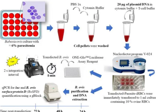
3.1. In vitro Babesia ovis cultures ... 18
3.2. Evaluation of Babesia ovis sensitivity to WR9910 and blasticidin-S ... 18
3.3. Bacterial strains, plasmids and growth conditions ... 19
3.4. Preparation and transformation of chemically and electrocompetent cells ... 20
3.5. Babesia ovis DNA extraction ... 21
3.6. PCR, enzymatic restriction and purification ... 21
3.7. Amplification, cloning and sequencing ... 22
3.8. Screening for suitable regulatory regions in Babesia ovis genome ... 22
3.9. Transient transfection plasmid constructs ... 25
3.10. Plasmid construct validation and preparation for transfection ... 27
3.11. Procedures for transfection ... 28
3.12. Chemiluminescence quantification to evaluate promoter activity and optimization of transfection process ... 28
3.13. Transfection efficiency by quantitative real-time PCR ... 29
3.14. Development of a stable transfection plasmid construct ... 30
3.15. Statistical Analysis ... 31
4. Results and Discussion ... 32
4.1. Screening and identification of suitable regulatory regions in B. ovis genome ... 32
IX
4.1.2. Exploring elongation factor 1-alpha locus ... 34
4.1.3. Exploring the rhoptry associated protein 1 locus ... 42
4.2. Transient plasmids construction ... 43
4.2.1. Development of plasmid with Babesia bovis elongation factor-1alpha intergenic region-B driving expression of a reporter luciferase ... 43
4.2.2. Development of plasmid with Babesia ovata elongation factor-1alpha intergenic region-B driving expression of a reporter luciferase ... 47
4.2.3. Development of a promoterless plasmid ... 48
4.3. Preliminary transfection of Babesia ovis by nucleofection ... 49
4.3.1. Transfection of Babesia ovis by nucleofection: a comparison between two nucleofection buffers ... 49
4.3.2. Optimal plasmid DNA amount ... 51
4.4. Babesia bovis and Babesia ovata elongation factor-1alpha intergenic region-B have heterologous promoter activity in Babesia ovis ... 53
4.5. Growth inhibition of B. ovis by blasticidin-S and the effect of WR99210 in in vitro cultures... 56
4.6. Construction and preliminary validation of a recombinant plasmid expressing an RFP-BSD fusion protein ... 58
5. Conclusions and Future Perspectives ... 60
6. Bibliography ... 61 7. Appendix ... 76 Appendix I ... 76 Appendix II ... 78 Appendix III ... 79 Appendix IV ... 81 Appendix V... 82 Appendix VI ... 83 Appendix VII ... 84 Appendix VIII... 87 Appendix IX ... 90 Appendix X... 92
X
Index of Figures
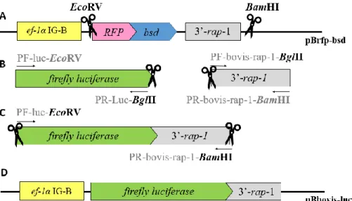
Figure 1. Babesia spp. generic life cycle. ... 4 Figure 2. Babesia ovis forms inside lamb erythrocytes in in vitro cultures. ... 7 Figure 3. Schematic diagrams of two different transfections. ... 8 Figure 4.Timeline of selected landmark publications on the development of transient and stable
transfection systems in Babesia spp. ... 10
Figure 6. Schematic representation of a stable transfection construct integrating elongation factor-1alpha open reading frame-B through a double crossover recombination event. ... 16 Figure 7. Expected organization of Babesia ovis elongation factor-1alpha locus based on Babesia bovis sequence. ... 23 Figure 8. Scheme of the PCR-mediated genome walking method for Babesia ovis elongation factor-1alpha locus. ... 24 Figure 9. Schematic diagram representing the development of the transient transfection
plasmid Bbovis-luc.. ... 25
Figure 10. Schematic diagram representing the development of the transient transfection
plasmid Bovata-luc. . ... 26
Figure 11. Schematic diagram representing the development of the promoterless plasmid,
pBS-luc. ... 26
Figure 12. Schematic representation of the methodologies used to evaluate activities of elongation factor-1alpha intergenic region-B candidate promoters from Babesia bovis and Babesia ovata in Babesia ovis. ... 30 Figure 13. Amplification of part of Babesia ovis actin gene. ... 32 Figure 14. Expected amplicon sizes for PCR with primers ORF-B-RV1 and
PR-Ef-ORF-B-RV3. ... 35
Figure 15. Amplification and isolation of a Babesia ovis 1500 base pairs (bp) size fragment.
... 35
Figure 16. Attempts for Babesia ovis elongation factor 1-alpha intergenic region amplification
... 36
Figure 17. Amplification of part of Babesia ovis elongation factor-1alpha gene. ... 37 Figure 18. Expected amplicon sizes for PCR with primers PR-Ef-IG1/ PR-Ef-ovis-specific and
PR-Ef-IG3/ PR-Ef-ovis-specific. ... 38
Figure 19. Attempts for amplification of Babesia ovis elongation factor-1α 5’ flanking region.
XI
Figure 20. Attempts for amplification of Babesia ovis elongation factor-1alpha 5’ flanking
region with annealing temperatures between 36ºC and 40ºC. ... 39
Figure 21. Alignment of Elongation factor 1-alpha protein sequences from Babesia species 42 Figure 22. Amplification of Babesia ovis rhoptry associated protein-1 gene and 3' end. ... 43
Figure 23. Digestion of plasmid Brfp-bsd and amplification of firefly luciferase (luc) gene and rhoptry associated protein-1 terminator region (3’rap-1). ... 44
Figure 24. Amplification of luciferase-rhoptry associated protein 3’ cassette. ... 44
Figure 25. Escherichia coli JM109 colonies after transformation with different ligation reactions to originate pBbovis-luc. ... 45
Figure 26. Colony screening PCR in transformed Escherichia coli JM109 with expected pBbovis-luc targeting luc-3’rap-1 cassette amplification. ... 46
Figure 27. Development and validation of plasmid with Babesia bovis elongation factor-1alpha intergenic region-B driving expression of firefly luciferase gene (pBbovis-luc). ... 47
Figure 28. Schematics representing the development and validation of plasmid with Babesia ovata elongation factor-1alpha Intergenic region-B driving expression of firefly luciferase (luc) gene (pBovata-luc).. ... 48
Figure 29. Development and validation of the promoterless plasmid (pBS-luc). ... 48
Figure 30. Transfection of Babesia ovis by nucleofection. ... 51
Figure 31. Transfection of Babesia ovis using varying amounts of plasmid DNA... 52
Figure 32. Fluorescence microscopy image of Babesia ovis expressing green fluorescent protein (GFP) after transfection with pmaxGFPTM vector. ... 53
Figure 33. Schematic diagram of the plasmids used for transient transfection and evaluation of Babesia bovis and Babesia ovata elongation factor-1alpha intergenic region-B heterologous promoter activity in Babesia ovis. ... 55
Figure 34. Time course of luciferase activity and parasitemia measured at 24h, 48h and 72h post-transfection with pBovata-luc. ... 55
Figure 35. In vitro growth curve and growth inhibition of Babesia ovis Israeli strain with WR99210 ... 58
Figure 36. In vitro growth curve and growth inhibition of Babesia ovis Israeli strain with blasticidin-S ... 58
XII
Figure 37. Schematics of the development of a plasmid with Babesia ovata elongation factor-1alpha Intergenic region-B region driving expression of red fluorescent-blasticidin-S deaminase fusion protein.. ... 59
Figure 38. Fluorescent microscopy image of Babesia ovis transiently expressing the red
XIII
Index of Tables
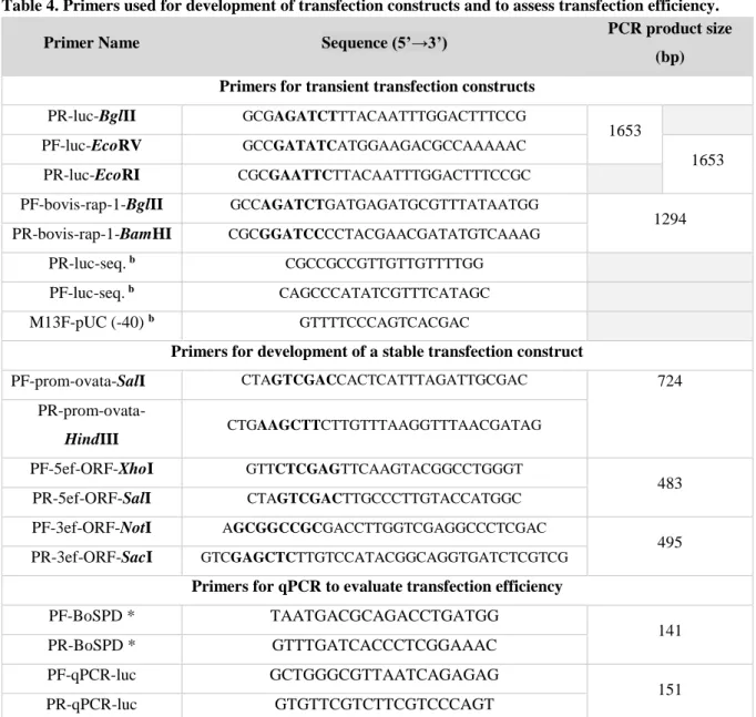
Table 1. Distribution of several Babesia spp., their main tick vectors and hosts. ... 3 Table 2. Strains and plasmids used in the present study. ... 19 Table 3. List of primers for amplification and sequencing of Babesia ovis elongation factor-1alpha and rhoptry associated protein-1 locus ... 24 Table 4. Primers used for development of transfection constructs and to assess transfection
efficiency. ... 27
Table 5. Babesiidae annotated sequences producing significant alignments with Babesia ovis actin gene sequence. ... 33 Table 6. Babesiidae annotated protein sequences producing significant alignments with Babesia ovis actin translated sequence. ... 33 Table 7. Babesiidae annotated sequences producing significant alignments with the cloned Babesia ovis 1500 base pairs sequence. ... 36 Table 8. Babesiidae sequences producing significant alignments with Babesia ovis elongation factor-1alpha gene sequence. ... 38 Table 9. Babesiidae sequences producing significant alignments with the Babesia ovis obtained
sequences from the genome walking PCR method. ... 40
Table 10. Babesiidae sequences producing significant alignments with the Babesia ovis
obtained sequences from the genome walking PCR method and their genomic location. ... 40
Table 11. Babesiidae Elongation factor 1-alpha protein sequences producing significant
XIV
Abbreviations
5’ act 5’ flanking region of actin gene
BLAST Basic Local Alignment Search Tool
BoSPD Babesia ovis surface protein D gene
Bp Base pair
Bsd blasticidin deaminase coding gene
CMV Cytomegalovirus
DMSO Dimethyl sulfoxide
hdhfr human dihydrofolate reductase coding gene ef-1α elongation factor 1-alpha gene
gDNA Genomic DNA
GFP Green Fluorescent Protein
IC50 50 % inhibitory concentration
IG Intergenic
iRBC Infected red blood cells
IRES Internal ribosome entry sites
LB Luria-Bertani
Luc firefly luciferase coding gene
TES N-Tris (hydroxymethyl)methyl-2-aminoethanesulfonic acid
ORF Open reading frame
PAS Polyadenylation signal
PCR Polymerase chain reaction
Pol II RNA Polymerase II
PPE Percentage of parasitized erythrocytes
qPCR Quantitative real-time PCR
rap-1 rhoptry associated protein-1 gene
RFP Red Fluorescent Protein
RLU Relative luciferase unit
rRNA Ribosomal RNA
SD Standard deviation
s.l. Sensu lato
XV
spp. Species
s.s. Sensu stricto
SV40 Simian virus 40
TSS Transcription Start Site
UTR Untranslated region
VESA Variant erythrocyte surface antigens
1
1. Introduction
Development of genetic manipulation methods for Babesia ovis can provide an essential breakthrough for the study of this parasite basic biology, including its life cycle dynamics, parasite-vector and parasite-host interactions, ultimately enabling rational design of more effective drugs and vaccines towards disease control 1,2.
Currently, scarce information regarding intra-tick Babesia species (spp.) dynamics exists, even though targeting key developmental stages in tick tissues presents as an attractive way towards the development of transmission-blocking vaccines. Such approach demands a deep knowledge of conserved and divergent developmental features between Babesia spp. 3.
Transfection technology (incorporation and expression of foreign DNA or RNA) has a wide range of applications, particularly useful for the study of parasite-host cell interactions. Recently, it was possible to image pathogen colonization in its tick vector by transforming the bacteria Borrelia sp. to stably express a fluorescent marker, thus the same strategy could be applied to understand B. ovis and related Babesia spp. life cycle events in the tick vector tissues 4. Even though there is very limited knowledge regarding B. ovis-Rhipicephalus bursa interactome, some candidate protective antigens have been identified recently in the tick vector salivary glands, as having an important role in biological functions, such as tick development, feeding and pathogen transmission 5. Transfection methods stand out also in the vaccinomics research currently ongoing for the B. ovis-R. bursa interface, by enabling targeted disruption of genes of interest leading to the identification of key/essential parasite gene products for successful transmission 2,5–8. Moreover, the same gene manipulation strategy can be applied to the study of virulence factors, particularly those that interact with effectors produced by the host immune system. The phenotypic characterization of mutants lacking or with partially functional virulence factors can be a powerful method to characterize these factors, which can be potential targets for vaccine development 2,9. Other possible endpoint of this tool is the design of novel-life transfection-based vaccines, in which the host is vaccinated with attenuated-live vector vaccine containing a stably transfected tick antigen, eliciting the host protective immune response against both the parasite and the tick 10,11.
Babesia spp. have many characteristic features that make this parasite especially suitable for the development of genetic manipulation methods and posterior identification of drug targets 12. A small genome size in comparison to other apicomplexan parasites simplifies comparative genomic studies and, the relatively reduced number of genes concomitantly with being haploid in the asexual cycle also facilitates the discovery of drug targets 13–15. Moreover,
2 it appears that the integration of exogenous DNA in Babesia spp. genome occurs exclusively by a homologous recombination mechanism which supports the development of gene knockout experiments for functional studies 8. Currents challenges for the development of such tools are related with scarce of appropriately annotated genomes and, particularly for B. ovis, the unavailability of its genome sequence in public databases 13,15–18.
The “One Health” concept recognizes that Human, animal and environment Health are tightly intertwined, demanding synergistic interdisciplinary collaborations to, among other purposes, pave the way for the expansion of scientific knowledge. Research projects that aim the control of tick and tick-borne diseases have been positioned in the framework provided by this initiative, prioritizing the use of “omnics” and systems biology approaches concomitantly with the development of genetic manipulation methods 2,19.
1.1. Babesia and Babesiosis
The genus Babesia (phylum Apicomplexa, order Piroplasmida) comprises protozoan hemoparasites responsible for babesiosis, a recognized emerging vector-borne zoonose. More than one-hundred Babesia spp. have been identified so far, remarkably able to establish infections in a wide variety of domestic and wild animals, with some species having the ability to infect humans. These organisms are considered the second most prevalent blood-borne parasites of mammals after trypanosomes and, giving the worldwide distribution of their tick vectors, babesiosis is recognized as the most common blood disease of free living animals 20– 25.
Babesia spp. were named after Victor Babes, the bacteriologist that provided their first description in 1888, after observation of these microorganisms inside erythrocytes of cattle presenting hemoglobinuria and later in sheep blood. In 1893, Smith and Kilbourne identified this intraerythrocytic parasite as the etiological cause of Texas Cattle Fever and also the role of ticks as vectors, being the firsts to demonstrate that an arthropod was the carrier of a disease agent 26–28. Indeed, transmission usually occurs through a Babesia-infected tick bite but it can also happen via blood transfusion or congenital acquisition during pregnancy 27.
Babesiosis is acknowledged for having a great impact at economic and social levels, particularly in developing countries. In addition, the intensification of human and animal movements combined with environmental changes and the geographical expansion of several tick species is contributing to the rise of this tick-borne disease as a global threat 29–31.
From the veterinary point of view, bovine babesiosis, caused by Babesia bovis and Babesia bigemina, has drawn more attention due to the economic burden imposed, linked to
3 mortalities, abortions, decreased milk and meat production, control measure costs, losses in potential production and cattle trade restrictions 21,32,33. Despite existence of tick vector eradication programs disease outbreaks occur often and it is considered that most of the 1.2 billion cattle worldwide is exposed to babesiosis 34–36. Moreover, ovine babesiosis caused mainly by B. ovis has a major economic impact and companion animals, such as dogs, are also severely affected with infections caused by Babesia vogeli and Babesia gibsoni 21,37–40.
From the medical point of view, human babesiosis, caused by an increasing diverse range of species, mainly attributed to Babesia microti and Babesia divergens, is still not recognized as a neglected disease 27,41–45. Even though humans are only considered accidental
hosts for Babesia spp., there is a global concern regarding this emerging zoonosis since human babesiosis incidence is increasing with several clinical cases being reported recently from many countries worldwide, as reviewed by Yabsley and Shock., 2013 and Ord and Lobo, 2015 27,43. Moreover, babesial infections are often overlooked due to the relative unspecific clinical signs and lack of medical awareness for this disease, with underreporting of human cases being a major issue 45,46.
Babesiosis clinical signs arise when the rate of erythrocyte (infected) loss surpasses the rate of their replacement, giving rise to anemia and its associated health issues. Therefore, babesial infections progress with quite different degrees of severity depending of hosts’ age, immunological status, co-infections with other pathogens, and/or genetic factors. Acute babesia infections manifestations include, besides anemia, fever, hemoglobinuria, jaundice, malaise, lethargy and anorexia while the chronic status is usually asymptomatic. Nonetheless, these surviving hosts frequently become carriers and maintain ticks infection cycle 21. Babesia spp. are responsible for diseases of veterinary and medical importance through transmission by their different tick vectors (Table1).
Table 1. Distribution of several Babesia spp., their main tick vectors and hosts.
Babesia spp. Main vectors Main hosts Distribution Ref
B. bovis Rhipicephalus microplus / Rhipicephalus annulatus
Cattle (Bos taurus)
Africa, America, Asia, Australia, Europe
21 B. bigemina
B. ovata Haemaphysalis longicornis Asia 47
B. divergens * Ixodes ricinus Europe and Asia 48,49
B. ovis Rhipicephalus bursa Sheep (Ovis aries) and
goat (Capra aegagrus) Africa, Asia, Europe 50
B. gibsoni
Rhipicephalus sanguineus Dog (Canis lupus) Africa, America, Asia, Australia, Europe
39,51,52
B. vogelli
B. microti * Ixodes scapularis White-footed mouse (Peromyscus leucopus)
America, Europe and Asia
53–55
4
1.1.1. Babesia life cycle
Babesia life cycle is maintained in a complex system of tick vectors and vertebrate reservoirs (Figure 1). A generic Babesia life cycle is currently accepted, even though there is the major controversy regarding variants across the genus, as reviewed by Jalovecka et al., 2018 3. In the vertebrate hosts these parasites reproduce asexually within the erythrocytes (merogony) and are referred as piroplasms or piroplasmids due to their pear-shaped appearance, sharing the term with Theileria, a phylogenetically related protozoan. The initial Babesia sexual stages, referred as gametocytes, appear with successful transmission to its vector, reaching the tick midgut together with the blood meal, differentiating into gametes. After fertilization these give rise to a zygote that undergoes a meiotic division to originate kinetes (gamogony) and a final cycle of asexual multiplication originates sporozoites (sporogony), the host-invasive stage, guarantying a successful infection of the host. In the tick vector occurs transstadial transmission, between the tick stages of development and, for most Babesia spp. transovarial transmission is present, a process mediated by parasite invasion of the ovarian cells and transmission via larval progeny to tick larvae. Babesia sensu stricto (s.s.) is the informal classification for the organisms that exploit this transovarial transmission strategy while Babesia sensu lato (s.l.) refers to species that are morphologically similar to Babesia but have different life cycle features or cannot be designated either Babesia or Theileria with certainty. Nonetheless, this transovarial transmission is recognized as a unique feature among all the apicomplexan parasites 3,14,21,25,43,56–59.
Figure 1. Babesia spp. generic life cycle. A susceptible tick, upon its blood meal, ingests these parasites, gametocytes, from an infected host. In the tick midgut, they undergo sexual development into mature gametocytes
5 and gametes, whose fusion results in zygotes, able to invade midgut cells. These zygotes go through meiosis and differentiate into motile prolonged kinetes, ookinetes, (gamogony) that escape midgut cells and spread to different tissues throughout the haemolymph, including the salivary glands. Here, kinetes undergo a cycle of asexual multiplication originating sporozoites (sporogony) that will infect a naïve vertebrate host during feeding. The infection is maintained throughout tick developmental stages (transstadial transmission). For Babesia sensu stricto, kinetes invade tick ovaries and eggs resulting in infected larvae (transovarial transmission). In the vertebrate host, upon erythrocytes invasion, parasites reproduce asexually (merogony). 14,21,25. Adapted from: Schnittger et al., 2012 21.
1.1.2. Babesia evolution and comparative genomics: applications
Molecular phylogeny data contributes to more robust Babesia spp. taxonomic classifications, establishment of co-evolution patterns with its tick vectors and vertebrate hosts, enlightening evolutionary lineages 21,60. Phylogenetic studies using the 18S ribosomal RNA (rRNA) gene separates Babesia spp into six broad clades with different degrees of support with the clear distinction between two major lineages: Babesia s.s. and Babesia s.l. 21,61. Based on this 18S rRNA sequence, B. ovis is placed in Babesia sensu stricto closely related to B. bovis, assembling with B. orientalis in a group of moderate support, separated from the branch comprising B. bigemina, Babesia ovata, Babesia major, Babesia crassa and Babesia motassi. Nonetheless, all of these species are placed in the same phylogenetic clade, clade VI. In a more recent study using C1A cysteine proteinases sequences this hypothesis was further supported. Also, it has been considered that, within Piroplasmida, the most rapidly evolving lineages are those in the clade with B. bovis and B. ovis 21,62–64.
As an alternative to analyzing 18S rRNA sequences alone, mitochondrial genome sequences and structures have proven to be useful for elucidation of evolutionary relationships and recently with was possible to identify five distinct Piroplasmida lineages with Babesia spp. being assigned to three different groups, referred as the Babesia microti group, the Western Babesia group, and Babesia s.s..59.
Babesia bovis genome was the first to be reported with a total size of 8.6 mega base pairs (Mbp) 13. Since then other Babesia species genomes have been sequenced, B. divergens (10.8 Mbp), B. bigemina (13.8 Mbp), B. microti (6.5 Mbp), Babesia sp Xinjiang (8.4 Mbp), Babesia orientalis and B. ovata (14.4 Mbp) 15,17,18,65,66. B. ovis genome has also been sequenced however it is not publicly available 62 . For B. bovis it was found that genome organization and structure was remarkably similar to Theileria, presenting microregional similarity to Plasmodium falciparum despite the similarities with the last concerning the infection clinical outcomes.
Exploring evolutionary lineages alongside with genome sequencing provides a potential avenue to the development of a pan-babesiacidal or even a more wide-range therapeutic
6 strategy. Genomic comparative analyses between T. parva and B. bovis has allowed to identify potential antigens in B. bovis that were already under study as vaccine candidates for theileriose
13,67. Particularly, in B. ovis, a simple gene comparative analysis using C1A family cysteine
protease sequences from B. bovis, resulted in the identification of papain-like cysteine protease, ovipain-2, an attractive potential vaccine target 68.
Using the same approach, comparing B. ovata to close related apicomplexan, such as B. bigemina, B. bovis, B. microti, P. falciparum and Toxoplasma gondii, allowed to conclude that the limited diversity of the ves genes, which encode variant erythrocyte surface antigens (VESAs) related with the parasite ability to evade or suppress host immune responses, could explain the lower virulence of this species 18. Likewise, this multi-gene family ves in Babesia sp. Xinjiang, a species known for having a limited virulence/pathogenicity in sheep was found to be substantially distinct both in number and sequence from those of B. bovis, B. bigemina and B. divergens 17.
In sum, genomic comparison facilitates the identification of genes that are undergoing positive selection and gene family expansions or contractions providing insights into the evolution of gene families and their possible roles in virulence and pathogenicity, which can be improved by using model apicomplexan parasites for functional studies 17. The ability to genetically manipulate Babesia spp. genome, namely B. ovis, a sheep and goat-infecting pathogen phylogenetically close to B. bovis and recognized for having a high virulence for its hosts, could be a platform for understanding the role of key molecules in host invasion and intra-tick development.
1.2 Babesia ovis
Babesia ovis is the main etiological agent of ovine babesiosis, a tick-borne disease of small ruminants prevalent in Eastern Asia, Southern Europe (Mediterranean basin), the Middle East and Northern Africa, a geographical distribution overlapping the one of its main vector, the tick R. bursa 38,69. Ovine babesiosis is an acute disease whose onset is characterized by high fever, a scenario that progresses to other clinical symptoms such as hemolytic anemia, hemoglobinuria, icterus and, in more severe cases, pancytopenia occurs. The untreated cases usually lead to death and even upon treatment the animal may die as a result of a heavy infection or suffer a disease relapse after the therapy period 70. Recently, an outbreak has occurred in Spain, demonstrating the deleterious effect of B. ovis in naïve sheep transferred from a tick-free region to a R. bursa-infested region with endemic piroplasmosis 71.
7 Ovine babesiosis is acknowledged for having an important economic impact related with animal mortality, productivity losses (decrease of milk, meat and wool production) and costs of the livestock treatment. Particularly for underdeveloped countries, social impact is quite significant in the livehood of resource-poor farmers and in food security72,73. The major disease control method consists in the use of a babesiacide, imidocarb dipropionate, to manage the clinical signs. Even though its efficacy has been long-established, safety issues have emerged related with milk contamination and potential carcinogenicity of the drug 74–76. The control of the tick vector infestations is another approach to manage this tick-borne disease however, emergence of acaricide resistance, including in R. bursa species, exposes the vulnerability of this strategy 77. The need for safer and more effective strategies is clear and for areas with a B. ovis unstable endemic status, the requirement of an immunization programme has been established 77,78. Some efforts were conducted in the context of the development of a live attenuated vaccine but the inability to eliminate B. ovis virulence after several blood passages invalidates, to date, this therapeutic approach 79. Alternatively, the research paradigm can be focused in the development of subunit vaccines that target the reduction of tick infestations and block pathogen transmission 80–82. Insight on the molecular interactions in the tick-pathogen interplay and the consequent identification of suitable antigenic targets are presently key to implement this strategy 83. Research focusing Babesia spp., as well as other apicomplexan parasites, encompasses significant constraints such as maintenance of in vitro parasite cultures, unavailability of complete and well annotated genomes and practical tools for their genetic manipulation 2. Regarding the first, in vitro B. ovis Israeli strain cultures have been successfully established (Figure 2), however genome sequencing availability and methods for genetic manipulation remain a necessity 84.
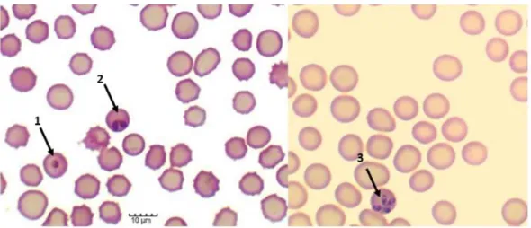
Figure 2. Babesia ovis forms inside lamb erythrocytes in in vitro cultures. 1. Single round trophozoite. 2. Merozoite characterized by piriform shapes forming a pair. 3. Merozoite characterized by piriform shapes
8 forming a tetrad (“Maltese cross” pattern). Intraerythrocytic parasites were observed under a 400x original magnification of a Motic BA210 LED trinocular compound microscope (original from the author).
1.3 Genetic manipulation Methods for Babesia spp.
Transfection is the process of introducing exogenous nucleic acid molecules (DNA or RNA) into eukaryotic cells. The transferred nucleic acid might reside in the cell either stably or transiently depending of the nature and destination of the genetic material (Figure 3). Briefly, transient transfection techniques are designed to introduce exogenous DNA and short-term expression of the foreign genetic material. Thus, the introduced nucleic acid does not integrate into the genome of the target cells, and the transfected DNA will not be replicated. On the other hand, in stable transfection, the introduced genetic material, that usually has a marker gene for selection (transgene) integrates the host genome and maintains the transgene expression along host cells replication 85.
Figure 3. Schematic diagrams of two different transfections. (A) Stable transfection. Foreign DNA (red wave) is delivered to the nucleus by passage through the cell and nuclear membranes. Foreign DNA is integrated into the host genome (black wave) and expressed sustainably. (B). Transient transfection. Foreign DNA is delivered into the nucleus but is not integrated into the genome. Triangles are expressed proteins from transfected nucleic acids. Black arrows indicate delivery of foreign nucleic acids. Adapted from: Kim et al., 2010 85.
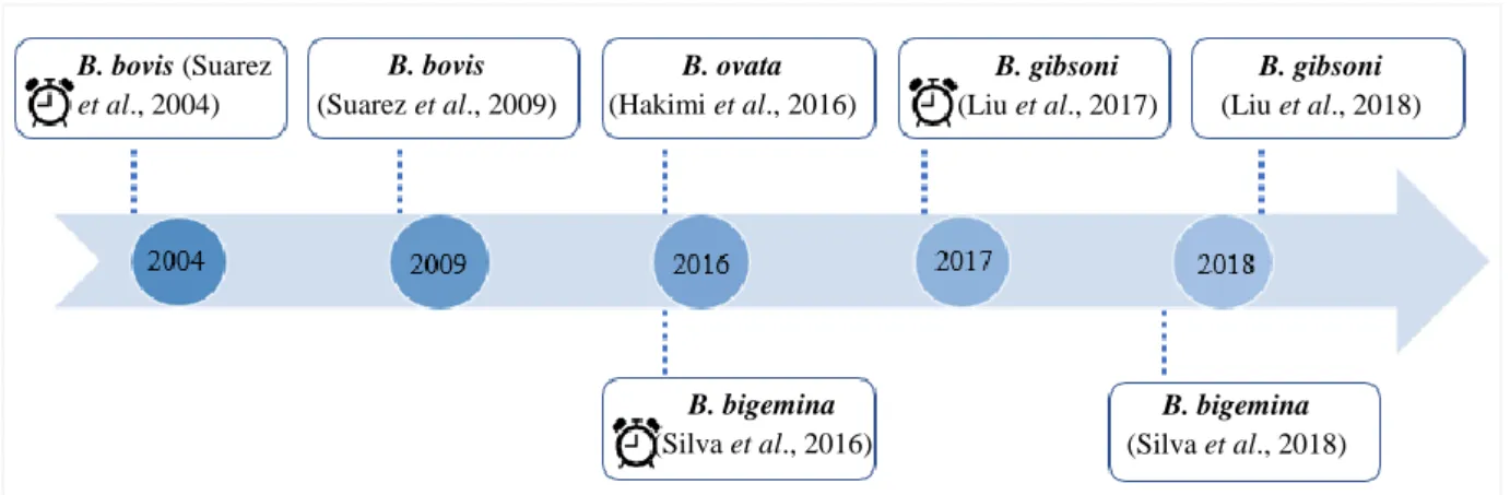
Transfection systems have been developed for several apicomplexan parasites with the initial report of the successful application of transfection technology described in T. gondii 86. Since then, transient and stable transfection systems were also developed for some Plasmodium spp. and for Neospora caninum a transient transfection was promptly developed based on the work done with T. gondii 87–89. For Babesia spp. such manipulation emerged in the last decade after availability of B. bovis genome sequence with reports of transient transfections system for this specie 136,90–92. A stable transfection system for B. bovis was developed and it is currently acknowledged that for at least T. gondii and B. bovis a stable transfection baseline
9 protocol was defined 2,93. More recently, transient transfection systems followed by stable transfection were performed in B. ovata after the unravelling of its genome 18,94. Similarly, stable transfection systems have been developed for B. bigemina and B. gibsoni 95–98 (Figure
4).
Figure 4. Timeline of selected landmark publications on the development of transient and stable transfection systems in Babesia spp. indicates transient transfection system.
Transient transfection systems establishes the appropriate parameters for introduction of the exogenous DNA (definition of an electroporation/nucleofection protocol that carries the minor impact for the parasite) and, more importantly, permit to identify and test function and efficacy of promoter and termination signals (regulatory elements) mediating gene expression and regulation 6,99. In fact, these systems were essential to find suitable promoters in Babesia spp. and to reveal the existence of promoters with interchangeable cross-species function 6,94,95,97,100. The establishment of a stable transfection system demands, besides the aforementioned criteria, to identify a suitable genetic marker to select the transgenic parasites (a selectable marker) and the preparation of a cassette containing the defined selectable marker gene along with suitable targets for DNA integration, whose disruption does not compromise the parasite viability 6,2 . Therefore, to establish a stable transfection system for B. ovis that enables the expression of a fluorescent reporter protein, the methodology followed should be similar to the ones used previously for transgenic- Babesia spp., following the steps described in figure 5. B. bovis (Suarez et al., 2004) B. bovis (Suarez et al., 2009) B. ovata (Hakimi et al., 2016) B. bigemina (Silva et al., 2016) B. bigemina (Silva et al., 2018) B. gibsoni (Liu et al., 2017) B. gibsoni (Liu et al., 2018)
10 Figure 5. Flowchart describing the steps required for the development of a stable transfection system for Babesia ovis. Adapted from: Suarez et al., 2010 6.
1.3.1. DNA transfer system: nucleofection
There is a huge methodological diversity concerning transfection, with the several strategies being broadly classified as biologically, chemically, and physically mediated methods 85. Herein, focus will be on a physical method, electroporation, since it is regarded as the most robust and reproducible method to transfect Apicomplexan parasites 101.
Electroporation is the most commonly used physical method of DNA delivery and its exact mechanism is unknown. However, it is suggested that a short electrical pulse disturbs cell membranes, creating holes through which nucleic acids can pass. It is accepted that it occurs through a multistep mechanism that relies not only on the plasma membrane permeabilization but also in the electrophoretically driven interaction between the DNA molecule and the destabilized membrane, both mediating its passage across the membrane and, once inside the cell, its migration towards the nucleus 85,102.
Babesia merozoites are obligatory intraerythrocytic organisms thus, a successful transfection approach must be able to transfer the exogenous DNA through the erythrocyte, merozoite and nuclear membrane layers. This results in a severe impairment in DNA delivery as seen in the low transfection efficiencies for this parasite and related apicomplexan 92,103,104. For B. bovis the process of developing a transient transfection system involved the determination of suitable electroporation parameters and later a comparison between that process and nucleofection, a novel electroporation based method 92,99. Nucleofection consists in a transfection method that combines a specific nucleofector solution and specific electrical parameters to successfully deliver DNA into the nucleus. It is though that during nucleofection
11 a square wave pulse is produced which improves pore-formation and enables the incorporation of exogenous DNA in the nucleus without cell division 101,105.
Nucleofection has resulted in higher transfection efficiencies and enhanced parasite viability in Plasmodium 106,107. For B. bovis it was shown that nucleofection was more efficient for the delivery of smaller plasmid DNA amounts and the inverse occurred with electroporation in which efficiency would increase concomitantly with the amount of DNA to deliver (to a maximum plateau of 200 μg), whereas delivery of 20 μg of DNA corresponded in both methods to similar relative efficiencies. Regarding the detrimental effects of any of the methods for the parasite it was shown that nucleofection provided a superior parasite viability 6,99,104.
Electroporation and nucleofection have been used for transfection of several Babesia spp. but an optimization of the electroporation parameters for the first and the assessment of the best nucleofector solution for the latter needs to be performed. For B. bovis it was suggested that nucleofector program v-024 in combination with “Plasmodium” buffer was optimal and for B. ovata the same program in conjunction with Amaxa nucleofector human T-cell solution was used to stably transfect the parasite in a Nucleofector™ 2b Device, an association also used to stably transfect B. bovis 7,92,94,108. On the other hand, B. gibsoni was stably transfected using an optimized protocol consisting of Lonza buffer SF associated program FA113 of Amaxa 4D Nucleofector™ device 98,100. For B. bovis, optimized electroporation settings (1.2 kV/ 25 µF/200 Ω) have been used successive times to transfect this parasite and the same protocol was used to stably transfect B. bigemina 6,10,93,96.
1.3.2. Regulatory sequences
1.3.2.1. Promoter regions and transcription start sites
The identification of transcription start sites (TSSs) is crucial for in-silico gene discovery, to understand transcription regulation mechanisms and to aid in the definition of the core promoter109–111. The latter, allied to the identification of cis-regulatory elements, binding sites for proteins involved in transcription initiation and regulation, it is essential to define the size of the isolated promoter for cloning. This information is key for the development of more efficient systems for transient and stable transfection 112,113. Nonetheless, it must be taken in account that gene expression can also be regulated at post-transcriptional and translational levels, but little is known about this modulation in apicomplexan parasites. In P. falciparum the correlation between mRNA and protein expression is found moderately positive even though
12 there is some delay in some genes expression indicating the existence of such regulatory mechanisms 114,115.
It is known that for every eukaryotic gene there is a core promoter region in the 5′ untranslated region (UTR) containing ate least one TSS signal to which binds the RNA Polymerase 2 (Pol-II). Particularly, the 5′ UTRs between TSSs and the first in-frame initiation codons are recognized for modulating RNA stability through action of internal ribosome entry sites (IRESs) or riboswitches. This region, for B. bovis, has a median length of 152 bp, similar in size with the one from T. gondii (130 bp) 113,116–118. This gene-proximal region is crucial for a nominal gene expression and additional upstream sequences enhance the promoter activity 119.
In B. bovis a TSS initiator-like motif TYAYWWW has been identified, probably a binding site for the general transcription factors TAF1 an TAF2, and also other location-specific consensus motifs, cis elements, have been found, probably crucial for binding to other transcriptional factors 113.
The apicomplexan parasites transcriptional machinery is recognized for differing from other eukaryotes, in which the absence of canonical eukaryotic transcription factors is particularly noticeable 113,120–124. Additionally, it is acknowledged that apicomplexan transcription machinery is not able to recognize viral promoters, such as the cytomegalovirus (CMV) and simian virus 40 (SV40) that are widely used to produce heterologous transfection systems 125,126.
Herein, the most used promoter regions in Babesia spp. to drive expression of exogenous genes: the 5’ flanking region of the actin gene (5’ act) and the elongation factor-1 alpha intergenic region (ef-factor-1α IG) will be focused. The 5’ act has been used to drive expression of the selective marker cassette in stably transfected B. bovis and B. ovata 7,94. More recently it was shown that 5’ act from B. gibsoni presents higher activity in B. bovis than its homologous promoter and this promoter region was interchangeably functional between B. bovis and B. gibsoni127. The ef-1α IG contains a recognized strong constitutive promoter signal 91,128. In B. bovis and B. bigemina the ef-1α locus consists in two identical ef-1α genes arranged in a head to head orientation separated by a 1.4 kb IG, structurally similar to the locus found in Plasmodium genome. Apparently, the IG contains two independent promoters (ef-1α IG-A and ef-1α IG-B) regulating bidirectional transcription of the two ef-1α open reading frames (ORFs), a bidirectional activity first described in P. berghei 91,95,129,130 Therefore, a rational approach for the identification of B. ovis ef-1α promoters consists in searching for this locus in its genome. The most active promoter region within the ef-1α IG region consists in a 710 bp region located
13 upstream the ef-1a gene B, the ef-1α IG-B 6,91. This promoter region has been used to drive stable expression of a green and a red fluorescent-blasticidin deaminase-S (GFP-BSD and RFP-BSD) fusion proteins in B. bovis merozoites 93 and GFP expression in B. ovata and B. gibsoni 94,98. Remarkably, the bidirectional transcription activity was used to express simultaneously a GFP-BSD and a tick antigen, being this the first experience of expression of a foreign antigen in a transfected B. bovis delivery platform, thus a live-vector vaccine 10,128,108.
Recently, it was found that promoters previously identified in B. bovis ef-1α IG region are also functional in B. bigemina and ten interchangeable cross-species functional promoters, including ef-1α IG-B, were identified between B. gibsoni and B. bovis 100. Interestingly, the ef-1α IG heterologous promoter sequence showed to be more efficient driving expression of the reporter luciferase than the homologous promoter in B. bigemina, and similar results were found in the latter study with B. gibsoni 5’ act being more active than the homologous promoter in B. bovis 95,100. Finally, a study in B. bovis suggests that the use of heterologous regulatory regions such as cross-species functional promoters increases the specificity of the genome integration event 8. Since the B. ovis genome is currently unavailable, these findings together with the availability of other genome sequences from Babesia spp., particularly the one of its phylogenetically close B. bovis, provides a solid working basis for the present study 16,65,77, 95,100.
1.3.2.1. Transcription termination regions
Transcription termination in eukaryotes consists in the release of the Pol II together with the nascent RNA from the DNA template, however the underlying molecular mechanisms are still under study 120,131,132. Transcription termination is assumed to occur the same way as in the yeast system model, with the Pol II continuing transcription after the polyadenylation signal (PAS). Then, a succession of allosteric protein-protein interaction end in the transcript cleavage 11-30 bp downstream the PAS 120,132. Around the transcription termination site, it has been observed, for the apicomplexan T. parva, the presence of over-represented motifs, some of them with the potential to produce a stem-loop structure (hairpin), possibly having a structural role in transcription termination 120,133.
The intergenic regions in the rhoptry associated protein-1 (rap-1) locus were used for the first transient transfection system in B. bovis promoting exogenous gene expression 90. However, it was demonstrated that rap-1 promoter signals were relatively weak and potentially unsuitable for the development of a stable transfection system that would require a high level expression of the exogenous gene (able to confer resistance to a selectable marker) 90,91.
14 Therefore, the following transfection constructs developed for Babesia spp. used the rap-1 intergenic regions for its termination signals 6,94,95,97. Studies of rap-1 locus organization have been conducted for several Babesia spp., revealing that the rap-1 family consists in multiple gene copies arranged in tandem and separated by IG regions 134. In B. ovis, the rap-1a locus is partially described, pointing towards the existence of 5 closely linked genes (Bo60.1 – Bo60.5) with an apparent high sequence conservation between genes Bo60.1-60.4 and lower similarity to 60.5 135.
Currently, three rap-1 genes in Babesia spp. designated rap-1a, rap-1b and rap-1c are described. However, it appears that this feature is confined to a phylogenetic group comprising B. motassi-like and B. bigemina 136,137. In B. ovis all the copies have been assigned as rap-1a with two different gene types within this designation (rap-1a60-1-4 and the type rap-1a60-5). In a recent study it was verified that RAP-1a sequences from B. sp. Xinjiang formed a clade with B. ovis, B. bovis and B. orientalis clustering with all the babesial RAP-1a type sequences 136. Recent work with B. gibsoni transfection indicated that B. bovis 3’ rap-1a has cross-species function even though the interchangeability was not tested 100. Exploring B. ovis rap-1 locus can give a better insight in its structure, important not only to use the IG region for transcription termination but also because RAP-1 proteins have been proposed as a potential candidate for the development of recombinant vaccines against babesiosis due to their implication in erythrocyte invasion and high immunogenicity 134,136. Assessing the number of rap-1 copies in B. ovis genome would also provide more information to study this region regarding its importance as a virulence factor 138.
1.3.3. Selectable markers
The development of a stable transfection system requires the identification of an appropriate genetic marker in order to select the transgenic parasites, i.e., a selectable marker 2,6. The firsts reports of stable transfection in apicomplexan parasites, namely Plasmodium, used the WR99210/ human dihydrofolate reductase (hdhfr) selection system 139. The enzyme dihydrofolate reductase (dhfr) has been a target for several anti-malarial drugs since its inhibition depletes the parasite reduced folate pool, blocking de novo thymidylate biosynthesis by thymidylate synthase, and eventually preventing DNA synthesis 140. The 4,6-diamino-1,2-dihydro-1,3,5-triazine WR99210 is not available as anti-malarial drug due to its high toxicity, however it was used to successfully select transgenic P. berghei parasites harbouring the hDHFR gene, since this gene conferred resistance to the antifolate drug 141.
15 The first attempts to stably transfect B. bovis used the same strategy, however naturally resistant parasites to WR99210 emerged 6. In fact, B. bovis contains a copy of the dhfr enzyme gene, required for synthesis of precursors of purines and thymidylate. Moreover, it was found out that the presence of polymorphic versions of this gene, such as the single amino-acid change present in B. bovis dhfr enzyme, renders the parasite resistant to antifolate drugs, namely WR99210 13,140. Interestingly, subsequent studies were able to select transgenic-B. bovis parasites and more recently, this strategy was used to select B. ovata and transgenic-B. gibsoni 7,94,98,108.
The blasticidin/blasticidin-deaminase (bsd) selection system has been described for effective selection of transgenic-Plasmodium parasites and it was the first system used to select stably transfected B. bovis parasites 93,142. Blasticidin S is a nucleoside antibiotic able to inhibit protein synthesis in both prokaryotes and eukaryotes 143. For parasites infecting an anucleate host erythrocyte the drug concentration range is not restrained by toxic effects in the host cell 142,144. Also, there are no bsd homologous genes in B. bovis genome and experimental evidence demonstrated that B. bovis was highly susceptible to blasticidin, supporting it as a good selection system 13,93. In B. bigemina blasticidin-S was also used as a suitable selective inhibitory drug 96.
The previous reports support the evaluation of B. ovis sensitivity to WR99210 and blasticidin-S to assess the suitability of these selection systems.
1.3.4. Genome integration targets
The integration of exogenous DNA into a specific genome region depends on homologous recombination mechanisms, a process that consists in the exchange of genetic information between two identical DNA sequences 145. The existence of different patterns of processing transfected genes has been proposed for Toxoplasma, Plasmodium and Babesia, as it heavily depends of their available DNA repair mechanisms 8,146. Gene targeting in Toxoplasma parasites is innately ineffective due to the ability of randomly inserting the foreign DNA in different sites from the specifically targeted due the existence of a non-homologous end joining mechanism, a problem solved by knocking out KU80, an important component of the pathway 147. In contrast, for Plasmodium, the process of exogenous DNA integration depends exclusively on homologous recombination mechanisms, as reviewed by Limenitakis and Soldati-Favre (2011) 101. Even so, for Plasmodium, gene targeting is still quite inefficient due to the unique richness of A + T of its genome that impairs efficient exogenous DNA integration 104.
16
Babesia and Plasmodium both present homologous recombination mechanisms and the random insertion of exogenous genes appears to be rare, however Plasmodium parasites can use both single and double cross- over insertion recombinant mechanisms whereas for Babesia it is demonstrated in several transfection studies that gene insertion occurs exclusively through a double crossover mechanism 7,8,93,94,96,98,101,108,148 (Figure 6). Thus, Babesia spp.- specific transfection appears to be an efficient genetic manipulation technique 8.
The first report of a stable transfection system for B. bovis targeted one of the ef-1α gene copies and, several posterior transfection studies confirmed the successful and specific integration in this site 7,93,94,96,98,108. Furthermore, the disruption of one of the two ef-1α gene copies by a double-crossed homologous recombination mechanism in several Babesia spp. did not affect parasite viability and the growth of the transfected isolates is comparable to the wild-type parasite 93,94,96,98. An identical ef-1α locus arrangement in B. ovis genome is expected supporting the feasibility of using this locus for exogenous gene integration.
Figure 6. Schematic representation of a stable transfection construct integrating elongation factor-1alpha open reading frame-B through a double crossover recombination event. A. Stable transfection construct with elongation factor 1α (ef-1α) promoter driving expression of a cassette comprising a reporter gene and a selectable marker gene with 3’ rhoptry associated protein-1 (3’ rap-1) as terminator B. Representation of the ef-1α locus in a transfected parasite line. represent enzyme restriction sites for plasmid linearization.
17
2. Objectives
The main goal of this project was the development of a stable transfection system driving expression of a fluorescent marker for B. ovis.
In order to achieve this goal, the following specific aims were established:
1. Identification of suitable regulatory elements in B. ovis genome and/or identification of their
heterologous in other Babesia spp;
2. Construction of transient transfection plasmids with the previously selected promoter
regions, driving expression of a reporter luciferase;
3. Evaluation of transfection process parameters, transfection efficiency and promoter activity; 4. Identification of a suitable selection system for B. ovis transfectants;
5. Construction and validation of a transient transfection system for B. ovis with the previously
selected promoter driving expression of a fusion protein expressing the fluorescent and selectable maker genes;
This study represents a step forward in the development of methods for B. ovis genetic manipulation, an undoubtedly necessary tool to study this parasite basic biology, including its life cycle, the parasite interactions with host cells and virulence factors.