The 4 2 -kD a co at pro te in o f Ande an
po tato m o ttle virus acts as a
transcriptio nal activato r in ye ast
1Laboratório de Genética Molecular Vegetal, Departamento de Genética,
Instituto de Biologia, Universidade Federal do Rio de Janeiro, Rio de Janeiro, RJ, Brasil
2Departamento de Bioquímica, Instituto de Q uímica,
Universidade Federal do Rio de Janeiro, Rio de Janeiro, RJ, Brasil M.S. Vidal1
and R. Margis1,2
Abstract
Interactions of viral proteins play an important role in the virus life cycle, especially in capsid assembly. Andean potato mottle comovirus (APMoV) is a plant RNA virus with a virion formed by two coat proteins (CP42 and CP22). Both APMoV coat protein open reading frames were cloned into pGBT9 and pGAD10, two-hybrid system vectors. HF7c yeast cells transformed with the p9CP42 construct grew on yeast dropout selection media lacking tryptophan and histidine. Clones also exhibited ß-galactosidase activity in both qualitative and quantitative assays. These results suggest that CP42 protein contains an amino acid motif able to activate transcription of His3 and lacZ reporter genes in Saccharomyces cerevisiae. Several deletions of the CP42 gene were cloned into the pGBT9 vector to locate the region involved in this activation. CP42 constructions lacking 12 residues from the C-terminal region and another one with 267 residues deleted from the N-terminus are still able to activate transcription of reporter genes. However, transcription activation was not observed with con-struction p9CP42DC57, which does not contain the last 57 amino acid residues. These results demonstrate that a transcription activation domain is present at the C-terminus of CP42 between residues 267 and 374.
Co rre spo nde nce
R. Margis
Laboratório de Genética Molecular Vegetal
Departamento de Genética Instituto de Biologia, CCS, UFRJ 21944-970 Rio de Janeiro, RJ Brasil
Fax: + 55-21-2590-0111 E-mail: margisr@ ufrj.br
Received August 7, 2001 Accepted January 22, 2002
Ke y wo rds
·Andean potato mottle virus ·Virus assembly
·Coat protein ·Transactivation
·Yeast two-hybrid system
Intro ductio n
Andean potato mottle virus (APMoV) is a plant virus endemic in South America (1) and a member of the Comovirus genus and Comoviridae family, of which cowpea mo-saic virus (CPMV) is the type member (2). APMoV infects economically important Solanaceae plants, such as potato, tomato, eggplant and pepper. The symptoms
pro-teins, i.e., the large and the small subunits. Virus particles contain 60 copies each of large and small coat proteins arranged with pseudo T = 3 (P = 3) symmetry, the large and small subunits being situated around the three- and five-fold symmetry axes, respec-tively (5). APMoV genomic RNAs are 3'-end polyadenylated and linked to a virus-encoded peptide at their 5'-end (Figure 1). These RNAs direct the synthesis and subse-quent processing of large precursor polypro-teins. APMoV RNA-1 encodes a single poly-protein processed in cis by a poly-proteinase do-main that gives origin to proteins related to viral replication such as helicase, virus-en-coded peptide, protease and the RNA-de-pendent RNA polymerase. RNA-2, as a re-sult of additional initiation at a second in-frame AUG codon, directs the synthesis of two distinct polyproteins. Both are processed in trans by the RNA-1-encoded proteinase to produce a pair of N-terminal proteins, that allow viral infection to move throughout the plant, and the proteins of the virion (4). The two coat proteins, CP42 and CP22, have molecular masses of approximately 42 and 22 kDa, respectively (6).
Viruses need to protect their genome from nuclease degradation to establish a success-ful infection. This is achieved through en-capsidation by the viral coat proteins. The assembly of viruses involves protein fold-ing, protein assembly, and nucleic acid rec-ognition. This process occurs via discrete intermediate units sometimes aided by a tem-porary scaffold. In simple spherical viruses, the relationship between assembly units and three-dimensional structure is best docu-mented for picornaviruses in which the three larger coat proteins have the same fold and are similar to the single chain protein that is present in the capsid of many plant RNA viruses (7). A similar assembly is observed in small, spherical RNA plant viruses, in particular for Southern bean mosaic virus, tomato bushy stunt virus, and turnip crinkle virus (8). A preliminary crystallographic
anal-ysis at 3.5-Å resolution has shown that the CPMV structure is very similar to that of animal picornaviruses (9). For bean pod mottle virus, another member of the Comoviridae family, a well-ordered RNA interaction with each asymmetric unit of the coat particle was detected and enabled the construction of a model for RNA packing in the bean pod mottle virus middle component (10).
Viral coat proteins play an important role in the interaction of virus particles with their vector (11). Several studies have been con-ducted in an effort to investigate heteroen-capsidation due to risks for the generation of novel viral pathogens with altered transmis-sion properties (12). The major functions of the coat protein are assumed to be the protec-tion of viral RNAs within the infected cell and to allow transmission of the viral ge-nome from plant to plant, either mechani-cally or by vectors such as insects, nema-todes, or fungi. Studies achieved by exchang-ing the coat protein gene of the whitefly-transmitted African cassava mosaic virus (ACMV) with that of the leafhopper-trans-mitted beet curly top virus (BCTV) pro-duced a leafhopper-transmissible ACMV-BCTV:coat protein chimera, emphasizing the importance of geminivirus coat protein to dictate which insect vector may transmit the virus (13). Coat protein may be required for cell-to-cell movement of the viral RNA through modified plasmodesmata and also for systemic movement of the virus from leaf to leaf through the vascular system of the plant (14).
func-tional role in viral replication (15,16). In the present study, we examined the interactions between the two APMoV coat proteins (CP42 and CP22) using a two-hy-brid system. The results of transcriptional activation, obtained with the CP42 construct in the yeast system, will permit further ex-periments in order to determine a functional map of CP42 transactivation domains.
Mate rial and Me tho ds
DNA manipulation and plasmid constructions
Restriction endonucleases and DNA-modi-fying enzymes were purchased from New En-gland Biolabs Inc. (Beverly, MA, USA), Gibco-BRL (Gaithersburg, MD, USA) and Amer-sham-Pharmacia Biotech (Uppsala, Sweden). Chemicals for yeast culture media and assays were from Difco Laboratories (Detroit, MI, USA), Reagen (Rio de Janeiro, RJ, Brazil) or Sigma (St. Louis, MO, USA). Recombinant DNA manipulations were carried out using standard protocols (17). The APMoV CP42-and CP22-coding regions were cloned into the plasmids pGBT9 and pGAD10 (18) after spe-cific amplification by the polymerase chain reaction (PCR). Oligonucleotide primers were designed to incorporate BamHI and PstI re-striction enzyme sites at the 5'- and 3'-ends of the respective genes. Reactions were performed with 100-µl preparations containing 50 ng of template DNA - pM15, which contains the full APMoV RNA-2 open reading frame (4), 125 nM of each primer pair (as described in Table 1), 0.25 mM of each dATP, dCTP, dGTP and dTTP, 6 mM MgCl2, 50 mM KCl, 10 mM Tris-HCl, pH 8.3, and 2.5 units of Taq DNA poly-merase. Different combinations of forward primers: CP22F (ttG GAT CCG CTT TTG
TAG TCC ATG TAT AAA TGT TTG G), CP42F (ttG GAT CCT GGA CCT TGC GCA
GTT TG), N29 (ttG GAT CCT AGG TAG
ATT TTC AAT ACC CAT GAC), N160 (ttG
GAT CCT AAC AAC TGA CTG TGC TGT
CGC), N267 (ttG GAT CCT AAT GGA TCA
GGA CGT GTC TTA TAG), and N330 (ttG
GAT CCT AGT AAA GAT TGC ATA GCT
TCT ACC) and reverse primers: CP22R (ttg gat cCT GCA GCT ACT TTA GTA TTG
GAA GCA TAC CAG), CP42R (aaG GAT CCT GCA GCT ACT GAG GCC ACG CCA
CA), C57 (aag gat cCT GCA GAA CAA
CAT ACA AAT AAG GAC AAC), C120 (aag gat cCT GCA GTG CTA TAA AAG
CCA AAA GCG), C217 (aag gat cCT GCA GGG GTG TTG CAC TCC ATG G), and
C358 (aag gat cCT GCA GAC CTG CTG
CAA TTC TGC TAT TC) were used in PCR amplifications (Table 1). Primer nucleotide sequences are from 5' to 3', restriction sites used to clone the PCR fragments are given in bold and other non-viral sequences are given in lower case. CP42 PCR products were di-gested with BamHI, purified from agarose gels and than cloned into BamHI-digested pGBT9 or BamHI/BglII-digested pGAD10. The PCR product corresponding to the CP22 gene was cloned into the BamHI/PstI-digested pGBT9 and pGAD10 two-hybrid system vectors. Amino and carboxyl terminal CP42 deletion mutants were PCR generated using pM15 as template. PCR products corresponding to the six CP42 N-terminal deletions (DN) were cloned into the pGBT9 BamHI site. The six C-terminal deletions (DC) were generated by
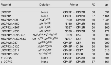
Table 1. List of the different APM oV CP22 and CP42 constructs, range of amino acid deletions, primer pairs used and annealing temperature and length of the correspond-ing PCR-amplified product.
Plasmid Deletion Primer oC bp
p9CP22 None CP22F - CP22R 68 591 p9CP42 None CP42F - CP42R 67 1161 p9CP42DN29 DM1A28 N29 - CP42R 50 1034
p9CP42DN160 DM1P160 N160 - CP42R 50 681
p9CP42DN267 DM1A266 N267 - CP42R 50 360
p9CP42DN330 DM1V229 N330 - CP42R 50 171
p9CP42DN29DC57 DM1A28DV330Q386 N29 - C57 50 903
p9CP42DN267DC57 DM1A266 DV330Q386 N267 - C57 50 189
p9CP42DC57 DV330Q386 CP42F - C57 50 990
p9CP42DC120 DM267Q386 CP42F - C120 50 801
p9CP42DC217 DT161Q386 CP42F - C217 50 510
p9CP42DC358 DR30Q386 CP42F - C358 50 87
cloning the PCR-digested amplification prod-ucts into the pGBT9 BamHI/PstI site. Con-struct p9CP42DC12 was produced by full amplification of the CP42 domain with prim-ers CP42F and CP42R followed by BamHI and PstI digestion and ligation into the corsponding sites of pGBT9. PstI digestion re-moves the sequence coding for the 12 resi-dues present at the C-terminus of CP42. In all these constructions the APMoV CP42 deletion mutants were fused to the C-termi-nus of the GAL4 DNA-binding domain pres-ent in pGBT9.
Ye ast transfo rm atio n
The yeast strain HF7c [MATa, ura3-52,
his3-200, ade2-101, lys2-801, trp1-901, leu23,112, gal4-542, gal80-538, LYS2:: GAL1UAS-GAL1TATA-HIS3 URA3::GAL4 (17-mers)3-CYC1TATA-lacZ], which contains two reporter genes (His3 and lacZ), was used in all two-hybrid system analyses. Trans-formation of yeast with the pGBT9 and pGAD10 constructs was performed as previ-ously described (19). Transformed cells were plated onto yeast dropout selection media lacking tryptophan and leucine (SD-W-L) for testing the transformation efficiency, or lacking tryptophan, leucine and histidine (SD-W-L-H) for testing and detecting any pro-tein-protein interactions. The positive colo-nies were obtained from selective plates af-ter 2 days of growth and then submitted to more stringent growth conditions on selec-tive plates containing increasing concentra-tions of 3-AT (3-amino-1,2,4-triazole). To test the transactivation characteristics of APMoV pGBT9-CP42 constructs, SD with-out tryptophan was used as the non-selective culture medium and SD lacking tryptophan and histidine was used as the selective me-dium.
ß-Galacto sidase assays
Two different methodologies were used to
evaluate lacZ gene expression in HF7c yeast cells. The first consists of freezing colony filter in liquid nitrogen followed by incubation with X-Gal in Z buffer (60 mM Na2HPO4, 40 mM NaH2PO4, 10 mM KCl, 1 mM MgSO4, pH 7.0, 50 mM ß-mercaptoethanol, and 0.3 mg/ml X-Gal) for blue/white screening. The second approach was the liquid culture ß-galactosidase assay, also performed in Z buf-fer (20). The ß-galactosidase activity present in a solution of freeze-and-thaw disrupted cells was measured using o-nitrophenyl-ß-D-galactopyranoside (ONPG) as substrate. One ß-galactosidase unit is defined as the amount of enzyme necessary to hydrolyze 1 µmol of ONPG to o-nitrophenol and D-galactose per minute (21).
Co m pute r-assiste d analysis o f pro te in
se co ndary structure
The APMoV CP42 GenBank accession number is L16239. The analyses of the sec-ondary and tertiary structure were carried out using the Swiss-Model ProMod package (22) which provides predictions based on the search for homology with sequences of proteins of known structure and integrates the What-If and the What-Check softwares (23).
Re sults and D iscussio n
Analysis o f inte ractio n be twe e n the APMo V
co at pro te ins
and p10CP22 constructs. Each of these plas-mids has the respective wild-type APMoV coat protein fused to the C-terminus of the GAL4 DNA-binding domain in the pGBT9 vector, or to the GAL4 transcription activa-tion domain in pGAD10 (18).
The testing of each construct for self-transactivation of selective or reporter gene
Figure 1. Schematic representa-tion of APM oV RNA-2 w ith poly-protein-P2 functional domains (M P, CP42 and CP22). A detail predicted of CP42 secondary structure is reproduced above the amino-terminal (DN) or
car-boxy-terminal (DC) deletion
mu-tants. Boxes correspond to the relative position of each CP42 mutant draw n in a proportional scale to the w ild-type CP42. The tw o ß-sheet structures flanking the loop w ith putative relevance to coat protein transactivation are in gray. VPg, virus-encoded peptide.
Table 2. Analysis of APM oV CP42 and CP22 yeast transformants and co-transformants grow ing in different sets of selective media.
Plasmid SD mediuma ß-Galactosidase assayb
-W -W-H -L -L-H -W-L -W-L-H
pGAD10 96; 152 0; 0 w
p10CP42 200; 100 0; 0 w
p10CP22 208; 250 0; 0 w
pGBT9 88; 256 0; 0 w
p9CP42 100; 300 80; 104 B
p9CP22 50; 296 0; 0 w
p9CP22/p10CP42 26; 20 0; 0 w
aUpper and low er values of the number of colonies obtained in three independent assays.
bß-Galactosidase activity in qualitative filter assays. “ w ” corresponds to normal yeast colony color and “ B” corresponds to blue colonies w ith ß-galactosidase activity.
transcription is one of the first steps that must be checked in order to use the two-hybrid system as a methodology to study the GAL4-based protein-protein interaction. The coat protein constructs p9CP42, p10CP42, p9CP22 and p10CP22 and the control plas-mids pGBT9 and pGAD10 were used to transform HF7c yeast cells that were plated
RNA-2 VPg
ß1 ß2 ß3 ß4 ß5 ß6 ß7 ß8 ß9 ß11 ß13 ß15 ß16 ß18 ß19 ß20 ß21 ß17
ß14 ß10 ß12
a3
CP42 CP42DC12
CP42DC57
CP42DC120
CP42DN29
CP42DN160
CP42DN267
CP42DN330
CP42DN29DC57
CP42DN267DC57
CP42DC358
CP42DC217
An
CP42 CP22
M P
386
a2 a1
onto a series of selective media (Table 2). Among these four constructs, plasmid p9CP42 produced a high number of growing colonies when transformed cells were plated onto the selective medium depleted of histidine.
This result indicates that CP42, when fused to the GAL4-binding domain, was able to activate the transcription of the His3 and lacZ reporter genes. The same was not ob-served when the CP42 protein was fused to the GAL4-activation domain (Table 2). Con-sequently, it was not possible to test the interaction properties of the two APMoV coat proteins using p9CP42 and p10CP22 in double transformation experiments. Thus, the analysis of heterodimer formation by the expression of the CP42 and CP22 core pro-teins in yeast transformed cells was limited to the p9CP22-p10CP42 combination.
In fact, no colony was obtained when the double transformed cells were plated onto the SD-W-L-H media (Table 2), but the effi-ciency of double transformation was assured by the presence of growing colonies on the SD-W-L selective medium. This result was quite surprising because there are sev-eral reports demonstrating that comovirus coat proteins should interact with one an-other.
Three independent components can be distinguished when members of the Picorna-viridae virus family are purified by centrifu-gation on density gradients, i.e., top, middle, and bottom. The three components have iden-tical protein composition and contain 60 copies of each coat protein. The middle com-ponent contains a single molecule of RNA-2 and the bottom component contains a single molecule of RNA-1, but the top component is devoid of RNA (10). The proportion of the top component in wild-type virus prepara-tions is relatively small, suggesting that virus assembly may proceed by mechanisms in which the top component may function as a provirion (25). Another experimental ap-proach revealed that transient expression of the two CPMV coat proteins in protoplasts
resulted in the formation of virus-like par-ticles (26). These results showed that the coat proteins were able to stabilize each other, this possibly being the first step in the CPMV assembly process. The same results were obtained when the single capsid pro-tein of arabis mosaic virus was expressed in transgenic plants or in insect cells (27).
The absence of interactions between the two coat proteins has led to the assumption that a constraint should exist in this experi-mental system. A set of factors may be nega-tively affecting the interactions of the two APMoV coat protein subunits: the depend-ence of a third host or viral factor, the differ-ences between the yeast and plant intracellu-lar environment, and especially the second-ary and tertisecond-ary structures derived from the fusion of the coat proteins at the GAL4 C-terminal domains.
Merits et al. (28) also reported that inter-actions between potato A virus P1, P3 and other proteins did not give identical results when three methods were used, two of them based on in vitro binding assays and the third being the yeast two-hybrid system. The low expression efficiency or quick turnover of the proteins, the incorrect post-translational modification and unfavorable folding of pro-teins in the yeast nucleus, or the masking of the DNA-binding or transcriptional activa-tion domain by the fusion proteins are as-sumptions that cannot be overlooked (28). Finally, detection of protein-protein interac-tions in the two-hybrid system depends on which protein is fused to which part of the GAL4 transcription factor. Interaction of plant potyvirus RNA polymerase with the viral coat protein was ten times higher when the polymerase was fused to the DNA-bind-ing domain and the coat protein was fused to the activator domain (29).
Transactivatio n pro pe rtie s o f the APMo V
m ajo r co at pro te in
construction were plated onto non-selective media (SD-W) and selective media contain-ing increascontain-ing concentrations of 3-AT (SD-W-H + 3-AT) in order to confirm the trans-activation properties of the APMoV CP42. Yeast cells transformed with the p9CP42 were able to grow on selective media supple-mented with 3-AT concentrations as high as 100 mM (Figure 2). These yeast clones were used in ß-galactosidase assays and exhibited ß-galactosidase activities (Table 2).
Strong evidence supports the involve-ment of viral coat proteins in replication. In the case of alfalfa mosaic virus, the coat protein is necessary for viral RNA synthesis and initiation of the infection cycle (15). Indeed, the coat protein was found to be tightly associated with the viral RNA poly-merase, which was purified by immunoaf-finity chromatography with antibodies raised against the coat protein (30). Mutations of basic N-terminal residues of alfalfa mosaic virus coat protein abolished viral replication and its RNA-binding activity (31). The con-firmation of reporter gene transactivation by the p9CP42 construct has redirected the origi-nal work. Efforts were made to map the functional domains of the APMoV 42-kDa coat protein, since the two-hybrid methodol-ogy permits to map functional regions of several other viral proteins (32,33).
Lo calizatio n o f the transcriptio n activatio n
se que nce in the APMo V CP42
In order to identify the CP42 region able to transactivate the reporter genes, several in-frame CP42 deletions were cloned into the pGBT9 expression vector. Figure 1 shows a schematic representation of the ten CP42 deletion mutants used in this study. Yeast cells were transformed with pGBT9-based constructs and transformants were plated onto selective (SD-W-H) media. Only two CP42 derivatives were able to transactivate the transcription of the lacZ and His3 genes in yeast cells (Table 3): a mutant with a
12-Figure 2. Transactivation of the His3 reporter gene by GAL4 DNA-binding domain-CP42 fragment fusion proteins. Transformed HF7c yeast cells w ere grow n on selective medium lacking tryptophan (SD-W), lacking tryptophan and histidine (SD-W-H), and lacking tryp-tophan and histidine and supplemented w ith 75 mM 3-amino-1,2,4-triazole (3-AT) (SD-W-H + 75 mM 3-AT). Samples: A) HF7c; B) pGBT9; C) p9CP42; D) p9CP42DC12; E) p9CP42DN267
and F) p9CP22.
Table 3. Analysis of the transactivation properties of APM oV CP42 and CP42 deletions.
SD mediuma
Plasmid ß-Galactosidase assayc -W -W-H +3-ATb
HF7c - - - 0.33 ± 0.03
pGBT9 1768; 900 0; 0 0.70 ± 0.01 p9CP42 1998; 1155 105; 120 ++ 2.63 ± 0.20 p9CP42DN29 1600; 865 0; 0 nd
p9CP42DN160 1200; 970 0; 0 nd
p9CP42DN267 952; 918 292; 150 ++ 1.98 ± 0.02
p9CP42DN330 895; 768 0; 0 nd
p9CP42DC12 2000; 997 100; 103 ++ 1.91 ± 0.09
p9CP42DC57 1568; 1200 0; 0 nd
p9CP42DC120 1200; 1168 0; 0 nd
p9CP42DC217 1700; 1003 0; 0 nd
p9CP42DC358 998; 670 0; 0 nd
p9CP42DN29DC57 904; 899 0; 0 nd
p9CP42DN267DC57 1064; 978 0; 0 nd
aUpper and low er values of the number of colonies obtained in three independent assays.
bResults obtained by plating transformed yeast onto selective media (SD-W-H) con-taining 3-AT.
cUnits of ß-galactosidase activity. Values correspond to the means ± SD of three independent cultures. nd, not determined.
SD-W SD-W-H SD-W-H 75 mM 3-AT amino acid deletion in the C-terminus
re-insect cells and that this protein contains a transcriptional activation domain of the acidic type. They also reported that the minimal activation domain comprised the AL2 pro-tein 15 C-terminal amino acids.
According to the present results, the oc-currence of an activation domain present at the CP42 C-terminal region was postulated. By analyzing the predicted tertiary structure of the wild-type CP42 and those of several deletions of the CP42 core protein (Figure 4), we identified the presence of a loop that may be the interactive region of the protein with the yeast RNA polymerase II. This re-gion is located between amino acid residues 332 and 342 and contains a loop region stabilized by two ß-sheets (Figures 1 and 4). The APMoV CP42 loop contains a glycine and an acidic residue and both are conserved
A B C D E
F G H I
K L
J
Figure 4. Predicted three-dimensional structure of w ild-type and deletion mutants of APM oV CP42. M odel A corresponds to the w ild-type CP42. M odels B to E represent CP42DN29, CP42DN160, CP42DN267 and CP42DN330 amino-terminal deletions; F to J correspond to CP42DC12, CP42DC57,
CP42DC120, CP42DC217 and CP42DC358 carboxy-terminal deletions, and K and L are the predicted models for the CP42DN29DC57 and
CP42DN267DC57 double deletion mutants, respectively. The hypothetical CP42 transactivation loop domain containing one cysteine and tw o acid
amino acid residues is present and identified in models A, B, C, D, E and F.
gions of the maize streak virus RepA and Rep proteins are involved in transcriptional activation and regulation of viral DNA repli-cation by using the two-hybrid system. Hartitz et al. (34) reported the phosphorylation of the geminivirus AL2 protein expressed in
Figure 3. Assay of APM oV CP42 direct transactivation in yeast. CP42 constructs w ere intro-duced into HF7c yeast cells as a fusion w ith the GAL4 DNA-bind-ing domain. Transactivation prop-erties w ere assayed by the GAL4-mediated activation of the ß-Gal activity present in trans-formed cells. The displacement of yeast transformants on the ni-trocellulose filter is: A) pGBT9; B) p9CP42; C) p9CP42DC12 and
D) p9CP42DN267.
A
B
C
Re fe re nce s
1. Fribourg CE, Jones RAC & Koening R (1977). Andean potato mottle, a new member of the cow pea mosaic virus group. Phytopathology, 67: 969-974. 2. Adams M J, Antoniw JF, Barker H, Jones
AT, M urant AF & Robinson D (1998). De-scriptions of Plant Viruses on CD-ROM. Association of Applied Biologists, Welles-bourne, Warw ick, UK.
3. Krengiel R, Vicente ACP, Weyne M , Shindo N, Brioso PST, Félix DB, Villaroel R, de Oliveira DE & Timmerman B (1993). M olecular cloning and sequence analysis of a segment from Andean potato mottle virus B RNA encoding the putative RNA polymerase. Journal of General Virology, 74: 315-318.
4. Shindo N, Vicente ACP, Krengiel R & de Oliveira DE (1993). Nucleotide sequence analysis of an Andean potato mottle virus middle component RNA cDNA clone: Comparisons of the encoded proteins w ith those of other comoviruses. Intervi-rology, 36: 169-180.
5. Lomonossoff GP & Johnson JE (1991). The synthesis and structure of comovirus capsids. Progress in Biophysics and M o-lecular Biology, 55: 107-137.
6. Brioso PST, Pimentel JP, Louro RP, Kitajima EW & de Oliveira DE (1993). Andean potato mottlevirus - Caracteriza-ção de uma estirpe infectando natural-mente berinjela (Solanum melanogena).
Fitopatologia, 18: 526-532.
7. Achary R, Fry E, Stuart D, Fox G, Row lands D & Brow n F (1989). The
three-dimen-sional structure of foot-and-mouth disease virus at 2.9 A resolution. Nature, 337: 709-716. 8. Rossmann M G & Johnson JE (1989). Icosahedrical RNA virus structure. Annual Review of Biochemistry, 58: 533-573. 9. Stauffacher CV, Usha R, Harrington M ,
Schmidt T, Hosur M & Johnson JE (1987). The structure of cow pea mosaic virus at 3.5 A resolution. In: M oras D, Drenth J, Strandberg B, Suck D & Wilson K (Edi-tors), Crystallography in M olecular Biol-ogy. Plenum Publishing Corporation, New York, NY, USA, 293-308.
10. Chen Z, Stauffacher CV, Schmidt T, Fisher A & Johnson JE (1990). In: Brinton M A & Heinz FX (Editors), New Aspects of Posi-tive-Strand RNA Viruses. American Soci-ety for M icrobiology, Washington, DC, USA, 218-226.
11. Andrejeva J, Puurand Ü, M erits A, Raben-stein F, Järvekülg L & Valkonen JPT (1999). Potyvirus helper component-pro-teinase and coat protein (CP) have coordi-nated functions in virus-host interactions and the same CP motif affects virus trans-mission and accumulation. Journal of Gen-eral Virology, 80: 1133-1139.
12. Candelier-Harvey P & Hull R (1993). Cu-cumber mosaic virus genome is encap-sidated in alfalfa mosaic virus coat protein expressed in transgenic plants. Trans-genic Research, 2: 277-285.
13. Briddon RW, Pinner M S, Stanley J & M arkham PG (1990). Geminivirus coat protein gene replacement alters insect specificity. Virology, 177: 85-94.
14. Rojas M R, Zerbini FM , Allison RF, Gilbertson RL & Lucas WJ (1997). Capsid protein and helper component-proteinase function as potyvirus cell-to-cell move-ment proteins. Virology, 237: 283-295. 15. de Graaff M , M an in’t Veld M R & Jaspars
EM J (1995). In vitro evidence that the coat protein of alfalfa mosaic virus plays a direct role in regulation of plus and minus RNA synthesis: Implications for the life cycle of alfalfa mosaic virus. Virology, 208: 583-589. 16. Bol JF (1999). Alfalfa mosaic virus and ilarviruses: Involvement of coat protein in multiple steps of the replication cycle.
Journal of General Virology, 80: 1089-1102. 17. Sambrook J, Fritsch EF & M aniatis T (1989). M olecular Cloning: A Laboratory M anual. 2nd edn. Cold Spring Harbor Laboratories, Cold Spring Harbor, NY, USA.
18. Bartel PL, Chien C-T, Sternglanz R & Fields S (1993). Using the tw o-hybrid system to detect protein-protein interactions. In: Hartley DA (Editor), Cellular Interactions in Development: A Practical Approach. Ox-ford University Press, OxOx-ford, UK, 153-179. 19. Gietz D, St Jean A, Woods RA & Schiestl RH (1992). Improved method for high effi-ciency transformation of intact yeast cells.
Nucleic Acids Research, 20: 1425. 20. Reynolds A & Lundblad V (1997).
Saccha-rom yces cerevisiae. In: Ausubel FM , Brent R, Kingston RE, M oore DD, Seid-man JG, Smith JA & Struhi K (Editors),
Current Protocols in M olecular Biology, Chap. 13. Wiley, New York, NY, USA. in the coat protein sequence of six other
comoviruses (alignment not shown). Similar ß-sheet-turn-ß-sheet motifs have been found in other yeast and plant virus transactivators of transcription (32,35). The deletions that abolished the formation of this loop, like CP42DC57, CP42DC120, CP42DC217, CP 42DC358, CP42DN29DC57 and CP42D N267DC57, were not able to activate the transcription of the reporter genes (Table 3). When the loop was formed the transcription of the genes analyzed in the two-hybrid sys-tem should be expected, but the deletion of 29 amino acids in the N-terminal region of the CP42 protein (CP42DN29) did not acti-vate the reporter genes. According to the
predicted structure of this protein, the N-terminal ß-sheet was deleted but the remain-ing N-terminal unstructured region could spatially block the access to the loop (Figure 4). This may occur with the CP42DN160 mutant, that has 160 deleted amino acids in the N-terminal region of the coat protein.
21. M iller JH (1972). Experiments in M olecu-lar Genetics. Cold Spring Harbor Labora-tories, Cold Spring Harbor, NY, USA. 22. Schw ede T, Diemand A, Guex N & Peitsch
M C (2000). Protein structure computing in the genomic era. Research in M icrobi-ology, 151: 107-112.
23. Hooft RWW, Vriend G, Sander C & Abola EE (1996). Errors in protein structures.
Nature, 381: 272.
24. Fields S & Song O (1989). A novel system to detect protein-protein interactions. Na-ture, 340: 245-246.
25. King AM Q, Lomonossoff GP & Ryan M D (1991). Picornaviruses and their relatives in the plant kingdom. Seminars in Virol-ogy, 2: 11-17.
26. Wellink J, Verver J, van Lent J & van Kammen A (1996). Capsid proteins of cow pea mosaic virus transiently ex-pressed in protoplasts form virus-like par-ticles. Virology, 224: 352-355.
27. Bertioli DJ, Harris RD, Edw ards M L,
Coo-per JI & Haw es WS (1991). Transgenic plants and insect cells expressing the coat protein of arabis mosaic virus produce empty virus-like particles. Journal of Gen-eral Virology, 72: 1801-1809.
28. M erits A, Guo D, Järvekülg I & Saarma M (1999). Biochemical and genetic evidence for interactions betw een potato A potyvi-rus-encoded proteins P1 and P3 and pro-teins of the putative replication complex.
Virology, 263: 15-22.
29. Hong Y, Levay K, M urphy JF, Klein PG, Shaw JG & Hunt AG (1995). A potyvirus polymerase interacts w ith the viral coat protein and VPg in yeast cells. Virology, 214: 159-166.
30. Quadt R, Rosdorff HJM , Hunt TW & Jaspars EM J (1991). Analysis of protein composition of alfalfa mosaic virus RNA-dependent RNA polymerase. Virology, 182: 309-315.
31. Yusibov V & Loesch-Fries LS (1998). Func-tional significance of three basic
N-termi-nal amino acids of alfalfa mosaic virus coat protein. Virology, 242: 1-5. 32. Horváth GV, Pettkó-Szandtner A, Nikovics
K, Bilgin M , Boulton M , Davies JW, Gutiérrez C & Dudits D (1998). Prediction of functional regions of the maize streak virus replication-associated proteins by protein-protein interaction analysis. Plant M olecular Biology, 38: 699-712. 33. Tenllado F & Bol JF (2000). Genetic
dis-section of the multiple functions of alfalfa mosaic virus coat protein in viral RNA rep-lication, encapsidation, and movement.
Virology, 268: 29-40.
34. Hartitz M D, Sunter G & Bisaro DM (1999). The tomato golden mosaic virus transacti-vator (TrAP) is a single stranded DNA and zinc-binding phosphoprotein w ith an acidic activation domain. Virology, 263: 1-14. 35. Leuther KK, Salmeron JM & Johnson SA