lessons from
Drosophila
oogenesis
Barbara M.I. Vreede
Successfully defended on November 30th, 2012 at the Instituto Gulbenkian de Ciˆencia in Oeiras, Portugal, before a jury presided over by:
Dr. Carlos Jos´e Crispim Rom˜ao
And consisting of:
Prof. Dr. Gertrud M. Sch¨upbach Dr. M. Abderrahman Khila
Dr. Jos´e B. Pereira Leal Dr. Sara Newbery R. de Magalh˜aes
de 2008 e Julho de 2012 no laborat´orio do Dr. ´Elio Sucena, Instituto Gulbenkian de Ciˆencia em Oeiras, Portugal, no ˆambito do Programa Gulbenkian de Doutora-mento (edi¸c˜ao 2007-2008). Parte do cap´ıtulo 2 est´a aceite para publica¸c˜ao no BMC EvoDevo como “Co-option of a coordinate system defined by the EGF and Dpp pathways in the evolution of a morphological novelty”, B.M.I. Vreede, J.A. Lynch, S. Roth, e ´E. Sucena. O modelo computacional apresentado no cap´ıtulo 4 est´a integrado num manuscrito em prepara¸c˜ao para submiss˜ao com autoria de A. Faur´e, B.M.I. Vreede, ´E. Sucena, e C. Chaouiya.
This dissertation is the result of my own research, carried out between January 2008 and July 2012 in the laboratory of Dr. ´Elio Sucena, Instituto Gulbenkian de Ciˆencia in Oeiras, Portugal, in the Gulbenkian Doctoral Programme (edition 2007-2008). A part of chapter 2 has been accepted for publication in BMC EvoDevo as “Co-option of a coordinate system defined by the EGF and Dpp pathways in the evolution of a morphological novelty”, B.M.I. Vreede, J.A. Lynch, S. Roth, and ´E. Sucena. The computational model presented in chapter 4 is part of a manuscript in preparation, authored by A. Faur´e, B.M.I. Vreede, ´E. Sucena, and C. Chaouiya.
Apoio financeiro / Financial support
Apoio financeiro da FCT e do FSE no ˆambito do Quadro Comunit´ario de Apoio, bolsa de doutoramento #SFRH/BD/33216/2007, e projecto #FCT-PTDC/BIA-BCM/74583/06.
Financial support for this thesis was provided by FCT and FSE through the Quadro Comunit´ario de Apoio, doctoral fellowship #SFRH/BD/33216/2007 and project grant #FCT-PTDC/BIA-BCM/74583/06.
This thesis was written in LATEX. All images were made with Gimp and Inkscape.
Printed by jm artes gr´aficas, Carnaxide, Portugal
Barbara M.I. Vreede
Dissertation presented to obtain the Ph.D degree in Evolutionary Biology
Instituto de Tecnologia Química e Biológica | Universidade Nova de Lisboa
Research work coordinated by:
Oeiras,
November 2012
lessons from
Drosophila
oogenesis
This thesis would not exist without the following people: first, my father, whose idea it was that I should study biology. Without the encouragement of Bas Zwaan and Patr´ıcia Beldade, I may not have continued my scientific career beyond grad-uation. It was Wallace Arthur’s writing more than anything else that led me to evo-devo. Patr´ıcia Beldade pointed me to the IGC, and Henrique Teot´onio ac-cepted me to the doctoral programme. Finally, ´Elio Sucena provided the fantastic idea ofDrosophilaoogenesis as the perfect model for a question I had long wanted to answer, and invited me to join his lab.
But many, many more were instrumental in the course of this work, and I would like to take this opportunity to thank them all. While I will try my best not to forget any, I will undoubtedly do so. If this is the case for you, please accept my apologies on top of my gratitude.
It has been an honour to work at the IGC these few years. Ant´onio Coutinho made it into the absolute best institute I know, and the many scientists and non-scientists working there have all done their part to uphold his high standards, and add their own character to create this extraordinary place. The borders of labs at IGC are vague, which allowed me to be an adopted member of the Computational Genomics journal clubs. This lab, as well as the ‘evo-devo community’ between the Beldade, Mirth, and Sucena labs, and their past and present members, have been important for my own evolution and development as a scientist. I thank you all.
In particular I need to thank my thesis committee, Patr´ıcia Beldade and Jos´e Leal, and my supervisor ´Elio Sucena, for their unwavering dedication to the cause, and for their advice—both the scientific and the utterly practical.
Throughout the entire process of my PhD there have been several people who graciously shared unpublished data and expertise, to an unknown girl on the other side of an email address. Many things in this thesis are a direct result of these interactions, and I feel immensely grateful to those who took the time to help me. In particular I’d like to thank Andrew Jessup, who helped me keep the Cerati-tis alive, Al Handler, who gave us access to the Ceratitis genome, and Giorgos Pyrowolakis, and Leonard Dobens, who shared unpublished data.
This thesis results in part from several important collaborations: with Adrien Faur´e and Claudine Chaouiya we worked on modelling eggshell patterning, and with V´ıtor Barbosa and Triin Laos, we worked on the mutant troya. I am very grateful for the discussions that followed in these collaborations, about oogenesis and the gene networks that govern it, in sometimes excruciating detail.
Cer-atitis oogenesis, and together with the rest of the Roth lab they made my time in Cologne a great experience.
I cannot forget Merijn de Bakker, who was fantastic in helping me understand in situ hybridization, and who made sure I could use the Scanning Electron Mi-croscope in Leiden, from which came, among other things, the cover of this thesis! In the lab at the IGC I have had help over the years from several students and colleagues directly involved in the project, in particular Pedro Patraquim, who introduced me to the project and whose enthusiasm was infectious, and Ana Marcelino, who battled Broad and Zaprionus. Andr´e Martins and Ana Ribeiro, however brief their stay, also left their mark. Ot de Wiljes was a life-saver with several particularly useful bits of Python code, and Pedro Almada helped with many practical problems, from microscopy to Java.
As has become painfully obvious of late, we are nowhere without money. I am therefore very grateful to Funda¸c˜ao para a Ciˆencia e a Tecnologia (FCT, the Portuguese government!) for funding our project on novelty—and for funding me for four years, doing fundamental science with no practical purpose whatsoever. I thoroughly hope this mindset will survive the current crisis. The Funda¸c˜ao Calouste Gulbenkian made sure I did not go hungry after my fellowship ended. Finally, a grant from the Deutsche Forschungsgemeinschaft to Siegfried Roth paid for a two month visit to the University of Cologne.
At the very end, I received some incredible (practical!) support from sev-eral people: Marc wrangled LATEX; Filipe helped me straighten out ideas; Matt
diligently proof-read the entire thesis (although time constraints meant not all his suggestions could be followed; any errors in the language are thus mine and mine alone!); Triin put out last-minute egglays; Olivier got me inaccessible papers; Cl´audia dealt with printers and practicalities; Ot made sure I stuck to the cause; ´
Elio had faith and a lot of books, and was an insurmountable wall between me and the bureaucracy monster. Furthermore, my work was read and commented on by Adrien, Claudine, Diogo, ´Elio, Filipe, Hanneke, Jeremy, Leila, Marc, Raquel, Siegfried, and Triin; Alex translated the summary into Portuguese, and Filipe checked if it was up to standard (it was!).
1 General Introduction 1
1.1 Evolutionary novelty: the origin of diversity . . . 3
1.2 The Drosophila eggshell as a new model system for evolutionary novelty . . . 6
1.3 A framework toD. melanogaster oogenesis . . . 8
1.4 Specification of the appendage primordia on the follicular epithelium 19 1.5 Varying the eggshell phenotype . . . 25
1.6 This thesis: objectives and outline . . . 27
2 Co-option of a coordinate system defined by the EGF and Dpp pathways in the evolution of a morphological novelty 29 2.1 Introduction . . . 31
2.2 Material and Methods . . . 35
2.3 Results . . . 36
2.4 Discussion . . . 46
2.5 Conclusion . . . 50
Supplementary material . . . 52
3 Mutation oftroya uncouples the polarity of eggshell and embryo 57 3.1 Introduction . . . 59
3.2 Material and Methods . . . 60
3.3 Results . . . 62
3.4 Discussion and conclusion . . . 66
4 Explaining variation in eggshell appendages through anterior-posterior patterning 69 4.1 Introduction . . . 71
4.2 Material and Methods . . . 78
4.3 Results . . . 79
4.4 Discussion . . . 85
4.5 Conclusion . . . 90
5 Discussion 93 5.1 This thesis: brief overview and conclusions . . . 95
5.2 Limitations of the current work and some ideas for future research 97 5.3 Variation: the evolution of a network . . . 99
5.4 Co-option in the evolution of a novel morphology . . . 100
Sum´ario . . . 107 Samenvatting . . . 110
Abstract
1.1
Evolutionary novelty: the origin of diversity
The concept of innovation seems both evolution’s greatest triumph and its stu-dents’ most difficult challenge. Biodiversity as it exists today is characterized by innovations in every lineage, acquired traits that may frequently have been the initial step in a species radiation, as they allow access to an as yet unexplored ecological niche. ‘Innovation’ as a concept has been applied to a wide range of cases across all levels of biological organization, including, but not limited to, behavioural performance (e.g. avian flight), developmental process (e.g. direct development of sea urchins), metabolism (e.g. the urea cycle), and morphology (e.g. vertebrate limbs). Its importance in evolution is thus paramount, and the issue of how novelties originate has been proposed many times as one of the major questions in evolutionary biology (e.g. Mayr, 1960; Wilkins, 2002). Historically, novelties in particular were the topic of a fierce debate after the appearance of the Origin of Species, between Darwin and Mivart. In the existence of complex ‘useful structures’, Mivart saw an obvious invalidation for the theory of evolution, as natural selection in his eyes could not account for the ‘incipient stages’ of these structures (reviewed by Mayr, 1960).
While novelty may no longer be problematic for the theory of evolution, it does provide a particular challenge to the biologist attempting to unambiguously and unbiasedly define the concept. Especially the fact that the concept can be applied to so many levels of biological organization is a problem, and leads to disagreement between biologists of different subdisciplines (Nitecki, 1990). This discussion is still ongoing, and even led some to question the validity of the concept itself, by asking: what, if anything is an evolutionary novelty? (Pigliucci, 2008)
1.1.1
What, if anything, is an evolutionary novelty?
Inevitably, the definitions of novelty currently in existence have been coloured by the author’s particular discipline. Interestingly, within the diverse set of definitions it is possible to identify a dichotomy of approaches that can be traced back to the classical debate of form versus function. This debate of morphology against tele-ology was famously and extensively held between Geoffroy St.-Hilaire and Cuvier in the 1830s (Panchen, 2001), decades before the arrival of the Origin. And again, these opposing schools are reflected in the definitions of evolutionary novelty. A representative of ‘function’, this widely used definition of an evolutionary novelty comes from Mayr (1960):
This definition has the clear signature of an era in biology when the focus was not on generative mechanisms for biological diversity, but on selection and adaptive function. An increased focus on development and morphology is reflected in a second definition, published three decades later by M¨uller and Wagner (1991):
“a structure that is neither homologous to any structure in the an-cestral species nor homonomous1 to any other structure of the same
organism.”
Both definitions have their merits, and their problems. Firstly, while Mayr’s definition is broadly applicable to many levels of biological organization, M¨uller and Wagner focus on morphology alone. This makes the latter a more practical definition, as concepts identifiable as novelty in the morphological sense will be more readily comparable between each other. Importantly, this definition also distinguishes between true novelty and exaptations: a term coined by Gould and Vrba (1982) to describe structures that have diverged from their original function, and have been modified accordingly. Such structures can take dramatic forms, and include for example the narwhal’s tooth, a large and highly specialized structure that originated from one of the ancestor’s teeth. The exclusion of such traits as the narwhal’s tooth has been a point of criticism for M¨uller and Wagner’s definition (Moczek, 2008). A further problem is that it hinges on the concept of homology, which generates debate in and of itself. Or, in the words of Moczek (2008): according to M¨uller and Wagner, novelty begins where homology ends— but wheredoes homology end?
However, without invoking the homology discussion at the present moment, there is one strong advantage to M¨uller and Wagner’s definition. Namely, using a definition that depends on form rather than function is of particular use if the goal is to understand how novelties originate. A definition of novelty that empha-sizes its function, such as proposed by Mayr (1960), does so as it aims to explain how natural selection favoured its propagation. However, as natural selection can only operate on traits that exist, natural selection cannot be held responsible for the origin of a novelty (Moczek, 2008). The advent of the field of evolutionary developmental biology (evo-devo) has come with a surge of studies on the origin of evolutionary novelty. As every change in the evolution of multicellular organisms inescapably has to start somewhere in development, the field of evo-devo is indeed exceptionally suited to address this issue. Moreover, it has been hypothesized that the process of development is not just the ‘scene of the crime’ when it comes to novel structures, but holds unique potential for innovation by its very nature (M¨uller, 1990). The argument made by M¨uller (1990) is that by redirecting the
1
development of intermediate stages of existing structures it is possible to generate quite dramatic novel features. Key to this is not just the intermediate stage, which provides the structural basis for the novelty, but the process of development itself. In a growing system, small changes in early stages can be magnified tremendously. Conversely, the coordination of the various components in a developing organism may buffer changes, effectively neutralizing underlying genetic variation. Accumu-lation of this variation beyond a certain threshold can be responsible for sudden dramatic changes in the morphology of the adult. Finally, M¨uller (1990) impli-cates the ectopic redeployment of existing developmental programs in generating dramatic morphological changes.
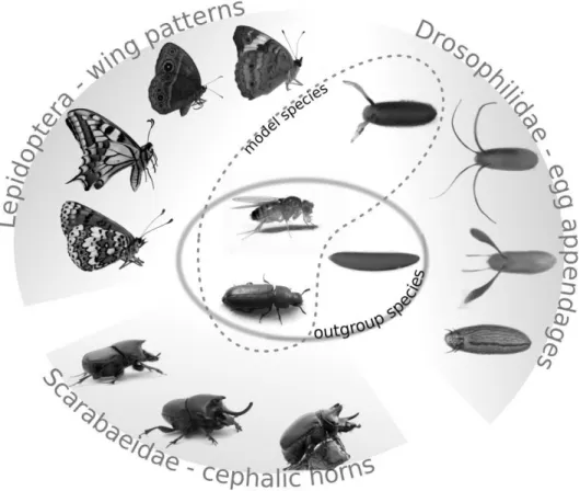
These are no strange concepts to the field of evo-devo. The latter point in particular has been shown to play a role in many current model systems of novelty, as genes or whole networks have been recruited in the development of novel traits. To illustrate this mechanism, we can look at two systems currently on the front line of the evo-devo research programme on evolutionary novelties: beetle horns, and butterfly wing patterns.
1.1.2
Examples of novelty: wing patterns and horns
The butterfly wing pattern is not just a textbook example of a morphological novelty, but a compelling case of a novel trait with a clear adaptive advantage. The immense variety that exists in pigment patterns between different butterfly species is exemplary for the power of novel traits in driving subsequent species radiation (Nijhout, 1991) (Fig. 1.1). The ecological relevance of wing patterns is well understood: elements on the wing may function for example in predator avoidance either by camouflage, or by diverting attention away from the butterfly’s vulnerable body (Brakefield and Reitsma, 1991). Moreover, the wing patterns of Heliconius butterflies provide a classical example of M¨ullerian mimicry (Joron et al., 2006).
be involved in one particular element, the eyespots of the butterflyBicyclus any-nana. Interestingly, a large degree of diversity exists in the genes generating the underlying patterns: no single patterning gene has yet been demonstrated to be required in all species examined thus far (Shirai et al., 2012).
The redeployment of old genes for novel functions is commonly referred to as ‘gene co-option’. This mechanism is widely considered to be responsible for complex change of any kind, but clearly plays an important role in generating novelty. In another example of novelty, beetle horns (Fig. 1.1), the co-option of an appendage-forming genetic programme during prepupal head development has generated an outgrowth on the dorsal head, which can now be used as a weapon in combat (Moczek et al., 2006). Further, a gender-specific regulator has been incorporated in the pathway with the recruitment of the Hox gene Sex combs reduced (Wasik et al., 2010), generating sexual dimorphism in the trait.
These studies are extremely valuable in understanding the genetic and devel-opmental mechanisms generating evolutionary novelty. However, they rely funda-mentally on prior knowledge of genes, pathways, and networks in model systems like Drosophila melanogaster. Genomic tools are increasingly available for both systems(Beldade et al., 2007, 2009; Kijimoto et al., 2009), allowing the work to move beyond a Drosophila-inspired candidate gene approach. Still, the available tools at this time to explore genetic relationships are limited.
Investigating the origin of the novelty in question depends on comparative studies with outgroups, or, as phrased by Shubin et al. (2009): “It is not possible to identify what is new in evolution without understanding the old”. The role of ‘the old’, or, species without the novel trait in question, is usually taken up by the classical models, irrespective of their phylogenetic distance and true basal position (Fig. 1.1). This may be problematic when those model systems are themselves very derived, as the case is with the classical model in development and genetics, Drosophila melanogaster. However, precisely this derived state could prove useful in the research programme on novelty, if we can identify and harness traits within this system that are themselves evolutionary novelties. Indeed, such a novelty can be found, in the dorsal-anterior appendages on theDrosophilaeggshell.
1.2
The
Drosophila
eggshell as a new model
sys-tem for evolutionary novelty
ground for of evolutionary novelties. Indeed, the novel feature central to this the-sis is found in D. melanogaster, and concerns the two appendages found on the dorsal-anterior side of Drosophilid eggs. These are large protrusions of the chorion, and promote the embryo’s access to oxygen in the air, while managing the risk of dessication (Hinton, 1969, 1981). This trait has emerged in a common ancestor of the family Drosophilidae, and the phenotype has since diversified extensively (Okada, 1968). Most importantly for our purposes, the genetic and developmental basis of dorsal appendages is well understood, making it an excellent model system to investigate the generative mechanisms behind this evolutionary novelty.
Analysing the development of any trait is a powerful tool in determining its evolutionary origin, since every phenotype inevitably has to be formed by trans-lating genes through development (Arthur, 2002). In this introductory chapter we will therefore discuss what is known about the ontogeny of dorsal appendages, which takes place during oogenesis. Of specific interest will be the fact that the pathways involved in forming the appendages and those that determine the main body axes of the future embryo are tightly linked. This will provide our research on the developmental basis of this innovation with a useful anchor, as the novel trait is developmentally connected to a highly important, likely conserved ontogenetic process.
To adequately introduce this model system, on the following pages a rough framework will be formed in which to view the processes discussed in this thesis, and with which to interpret our experimental results. The focus will initially be on the morphogenetic aspects of oogenesis, after which the genes and pathways are introduced that play a role in regulating these processes. Finally, we will discuss what is known about the patterning of the dorsal eggshell, which is the formative stage for the appendages we aim to study.
1.3
A framework to
D. melanogaster
oogenesis
1.3.1
Morphogenesis from germarium to eggshell
Drosophila ovaries consist of multiple strands of progressively ordered egg cham-bers, called ovarioles. Multiple developmental stages occur simultaneously in one ovariole: younger stages are located at anterior (Fig. 1.2 A), and the strands ter-minate in a completed egg at the posterior-most end, distally in the fly’s abdomen (Spradling, 1993).
cells and the oocyte are encapsulated in separate follicles but remain connected through a nutritive cord; and polytrophic, where both nurse cells and oocyte form a single capsule as the oocyte matures (Heming, 2003). Drosophilaoogenesis is an example of the latter type.
The egg chamber is formed in the germarium
The polytrophic Drosophila egg chamber is formed by 16 germline cells (one of which is the oocyte), encapsulated by a monolayer of±1000 somatic follicle cells. The ontogenic basis of this structure is in the germarium, the anterior-most struc-ture of the ovariole, from which each egg chamber is released (Fig. 1.2 B). Two germline stem cells are present at the anterior side of the germarium. Presumably, the asymmetric division of a stem cell generates a new stem cell and a cysto-blast. The latter will undergo four consecutive mitoses with incomplete cytokine-sis, forming a cyst with 16 interconnected cells. From this cyst, one cell is selected to become the oocyte. This selection is the first demonstration of asymmetry in the egg chamber, and will lay the foundation for future symmetry-breaking events. The selection of the oocyte and the precise regulation of early egg chamber devel-opment are complex processes that have been excellently reviewed by e.g. Deng and Lin (2001); Huynh and St Johnston (2004); Roth and Lynch (2009).
The oocyte will remain largely, though not entirely, transcriptionally silent (Mahowald and Tiefert, 1970, Raquel A. M. Santos and V´ıtor Barbosa, pers. comm.). Mostly, it is the nurse cells that provide mRNA and proteins through the cytoplasmic bridges (ring canals) that connect the cells, via a cytoplasmic struc-ture called the fusome, and in many cases through transport via the microtubule network (Mahajan-Miklos and Cooley, 1994; Pokrywka and Stephenson, 1995). The minus end of the microtubule network is anchored at the posterior pole of the oocyte, and is vital for the oocyte’s identity (reviewed in Deng and Lin, 2001; Huynh and St Johnston, 2004; Roth and Lynch, 2009).
of processes that shape the follicular epithelium, including the specification of an adjacent group of terminal anterior follicle cells (6-10 in total) known as border cells (Ruohola et al., 1991; Grammont and Irvine, 2001; Torres et al., 2003).
Oogenesis in 14 stages
At its encapsulation, the egg chamber will bud offfrom the germarium, remaining connected only through stalk cells that adhere to the polar cells of the successive egg chambers (Fig. 1.2A, B) (reviewed in Roth and Lynch, 2009). The egg chamber progresses through a total of 14 morphologically distinct stages that can be divided into the pre-vitellogenic stages (1-7) and the post-vitellogenic stages (8-14) (Fig. 1.2A). These stage groups are separated by the onset of vitelline membrane formation, which is the first component of the future eggshell that is synthesized in the egg chamber (Spradling, 1993). During the pre-vitellogenic stages, the egg chamber enlarges rapidly and the follicle cells undergo a number of divisions to enable further growth. Around stage 8 they stop dividing and switch to an endocycle to become polyploid (Brower et al., 1981).
Stage 7 sees a major reorganization of the microtubule network, when the posterior microtubule organizing centre (MTOC) in the oocyte disintegrates (see section 1.3.2), and microtubules now grow from the anterior-lateral cortex of the oocyte. This rearranges the oocyte’s polarity entirely. At this stage, the oocyte nucleus moves from its previous position at the posterior pole to an anterior-lateral location (Guichet et al., 2001), pushed by the polymerizing microtubules (Zhao et al., 2012). This asymmetric localization of the nucleus at the anterior end
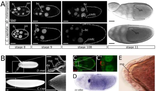
Figure 1.2 (preceding page): An overview of oogenesis in Drosophila
melanogaster. (A) The ovariole consists of eggchambers of progressively later
of the egg chamber constitutes the first event in the specification of the future dorsoventral axis, and the nucleus marks the future dorsal side of the egg (Fig. 1.2 D).
The follicle cell layer at stage 9 is rigourously restructured as most anterior follicle cells move posteriorly to cover the oocyte, and roughly 5% are stretched out into a squamous epithelium overlying the nurse cells. The other 95% form a columnar layer over the oocyte. At the boundary of the nurse cells and the oocyte, a designated group of follicle cells will move centripetally to cover the anterior part of the oocyte (these are referred to as the centripetally migrating follicle cells, or CMFC). This movement starts in early stage 10 egg chambers, and is complete by stage 10B (Spradling, 1993; Deng and Bownes, 1998). Concomitant with the restructuring of the outer follicle cell layer, the border cells (an anterior terminal cluster of specialized follicle cells) travel from the anterior-most end of the egg chamber, through the nurse cell cluster, to the anterior oocyte border (Fig. 1.2A). Subsequently, they move dorsally along the oocyte (Brower et al., 1981; Montell et al., 1992). These cells will later secrete the paracrystalline material that forms the pore of the micropyle, allowing sperm to enter the egg (Spradling, 1993).
Because of the constant content deposition by the nurse cells, the oocyte grows relatively faster than its supporting clones: at stage 10, the oocyte occupies half the egg chamber (Fig. 1.2A). During stages 10B-12 the nurse cells rapidly dump their cytoplasm into the oocyte, and in the final stages (13-14) they shrink and complete apoptosis (Chao and Nagoshi, 1999).
Formation of the vitelline membrane, chorion, and eggshell structures
The eggshellin toto is composed of the following layers: the vitelline membrane, the wax layer, the inner chorionic layer (ICL), the endochorion (itself consist-ing of an inner part, an outer part, and pillars in between), and the exochorion (Fig. 1.2E00) (Margaritis et al., 1980). The various components of the eggshell are secreted by the follicle cell layer, starting with the vitelline membrane: a solid layer that even without the chorion is able to maintain the integrity of the egg and support it during embryonic development. Synthesis of the membrane components starts at stage 8, but the vitelline membrane is not complete before 10B. At this stage, small vesicles containing the vitelline proteins fuse, and a single layer is formed between the oocyte and the follicular epithelium (Spradling, 1993). This breaks the communication between the follicular epithelium and the oocyte.
of the exochorion (Spradling, 1993). This clustering has been conserved in other dipterans (Konsolaki et al., 1990; Tolias et al., 1990).
Secretion of the chorionic proteins is rapid and profuse. Prior to protein syn-thesis, starting at stage 9, the chorion gene clusters undergo rapid amplification, which allows high-level expression of the genes and facilitates fast chorion protein production. Protein secretion occurs between stage 11 and the end of oogenesis, lasting no longer than five hours (Orr-Weaver, 1991; Spradling, 1993).
Some final touches are required to complete the eggshell: interaction between vitelline membrane components and chorionic proteins is necessary to further sta-bilize the eggshell and connect its layers. Final shape and hardness is attained when the shell is hydrated as the egg passes through the oviduct (Spradling, 1993). During the prior eggshell patterning phase, subgroups of follicle cells have been defined that now form several specialized eggshell structures: the CMFC build both the operculum and the micropyle, which relate to hatching and fertilization respectively, while the border cells secrete the micropylar pore (Margaritis et al., 1980; Montell et al., 1992). While the formation of these structures is inextricably connected to the formation of the dorsal appendages, their precise development and morphology is beyond the scope of this thesis. However, an excellent review on micropyle and operculum development was written by Dobens and Raftery (2000).
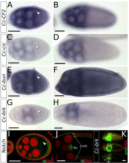
Another subset of anterior-dorsal follicle cells will form the dorsal appendages. On either side of the midline is an appendage primordium (Fig. 1.2E), each con-sisting of two cell groups: floor cells and roof cells, which will form the floor and the roof of the appendage tube, respectively (Fig. 1.2E0) (Ward and Berg, 2005; Boyle et al., 2010).
The roof and floor cells are marked respectively by expression of broad (br), encoding a zinc-finger transcription factor, andrhomboid (rho), encoding a serine protease (Ruohola-Baker et al., 1993; Deng and Bownes, 1997; Ward and Berg, 2005). When forming the tubes, Br-cells constrict apically to form the roof, and Rho-cells elongate forming the floor. The appendage primordia are defined by stage 10B, and reorganization starts at stage 11, finalizing the tubes at stage 14 (Dorman et al., 2004). Within thebr-expressing roof cells, two cell types can be distinguished based on their function during morphogenesis. The ‘leading’ roof consists of those cells adjacent to the floor cells in the epithelium, and is indispensable for the shape change that forms the appendage tube. The ‘trailing’ roof cells, meanwhile, merely follow (Boyle et al., 2010).
(Margaritis et al., 1980). Much more than in other areas of the eggshell, the outer endochorionic and exochorionic layers of the appendages contain pores, through which air can enter the meshwork of the endochorion (Spradling, 1993). This underlines the function of the egg appendages as respiratory structures (Hinton, 1969).
1.3.2
EGF signalling defines the main body axes of the
fu-ture embryo
The eggshell with its dorsal structures is evidently polarized. The origin of this polarity is tightly connected to the establishment of the embryonic main body axes. Determining the anteroposterior and dorsoventral axes is of vital importance in the development of bilaterally symmetric animals, and is generally one of the first events in ontogenesis (Gerhardt and Kirschner, 1997; Gilbert, 2003). In insects, the symmetry-breaking events leading to the definition of the main body axes happen even prior to fertilization, during the development of the egg (Roth and Lynch, 2009). In fact, it is during the very first stages of oogenesis, when in the germarium the oocyte is selected from a group of 16 germ line cells, that the foundation is laid for embryonic polarization inDrosophila melanogaster (Huynh and St Johnston, 2004).
Embryonic anteroposterior axis determinants, specifically: mRNA of bicoid (bcd), oskar (osk), and nanos (nos), localize asymmetrically in the oocyte, and form gradients in the early embryo. Patterning of posterior embryonic regions then occurs downstream ofosk andnos, whilebcd defines anterior structures.
The cellular sublocalization of these mRNAs is microtubule-dependent, and is properly defined after a series of signalling events between the oocyte and the follicular epithelium. This interaction between the oocyte and the somatic follicle cells during oogenesis is essential in establishing polarity (reviewed in e.g. Roth and Sch¨upbach, 1994; Roth and Lynch, 2009).
Anteroposterior polarity
In addition to the location-specific Grk signal, prior differentiation of the follicle cells also determines their response. During stages 6-7, a combination of Notch and JAK/STAT signalling—originating from the germline cyst and polar cells, respectively—specifies a group of cells on either terminus of the follicular epithe-lium to adopt terminal cell fate (Grammont and Irvine, 2002; Xi et al., 2003). These cells are now competent to respond to the first EGF signalling event, which occurs between the oocyte and the terminal follicle cells at stage 7 (Fig. 1.2C). Grk in the oocyte activates EGFr in the adjacent terminal follicle cells, which now take up posterior fate (Peri et al., 1999). The posterior follicle cells now signal back to the oocyte, with a signal of yet unknown nature (Fig. 1.2C0).
Up until this point, microtubules have formed a direct transport system for mRNAs from the nurse cells to the posterior pole at the oocyte MTOC (Cooley and Theurkauf, 1994). This backsignalling event, however, breaks down the pos-terior MTOC, and microtubules rearrange and rebuild in the oocyte, changing mRNA sublocalization with it. The mRNAs required for embryonic anteroposte-rior polarity now take up their final location: bcd at the anterior cortex, andnos and osk at the posterior pole (Becalska and Gavis, 2009). Upon rearrangement of the cytoskeleton the nucleus moves to dorsal-anterior, as does the associated grk (Neuman-Silberberg and Sch¨upbach, 1993, 1996; Cooperstock and Lipshitz, 2001). Both grk mRNA and the Grk protein now border the dorsal-anterior cor-tex of the oocyte, the protein co-localizing with membrane associated F-actin (Neuman-Silberberg and Sch¨upbach, 1996).
Dorsoventral polarity
The second EGF signal, determining dorsoventral polarity, starts when Grk acti-vates EGFr in the overlying dorsal-anterior follicle cells (Fig. 1.2D). Downstream of activated EGFr, expression of mirror (mirr) is up-regulated; a gene in the Iroquois complex that codes for a homeobox-containing transcription factor (Mc-Neill et al., 1997; Jordan et al., 2000; Zhao et al., 2000). Mirr represses the gene pipe (pip), restricting its expression to the ventral side of the egg chamber, which leaves an asymmetric distribution of Pip protein in the perivitelline space by the end of oogenesis (Stein et al., 1991; Nilson and Sch¨upbach, 1998). Precise levels of EGFr activation are required for these patterns to be correctly established, which is achieved by the inhibitory activity of Cbl (Pai et al., 2000).
and Sch¨upbach, 1998). Toll activation then causes the degradation of Cactus (Cact) in a polarized fashion in the embryo. As Cact prevents the transcription factor Dorsal (Dl) from entering the blastoderm nuclei, Toll receptor signalling ultimately results in a gradient of nuclear localization of Dl in the blastoderm stage embryo. This gradient subsequently defines the embryonic germ layers, as the transcription of several key regulators depends on the nuclear concentration of Dl (Gilbert, 2003; Moussian and Roth, 2005).
The EGF signalling cascade in dorsoventral polarity
While the cascade betweenpip expression and embryonic germ layer specification is extremely well known, the link between EGFr activation and pip expression is less straightforward. EGFr is a class I Receptor Tyrosine Kinase (RTK), a family of surface-bound receptors that function via intracellular kinase activity. In EGF signalling, ligand-binding induces phosphorylation of the cytoplasmic domain of the receptor, in turn leading to phosphorylation of Ras1. This initiates a sig-nal transduction cascade via Raf and MEK/MAPKK (Mitogen Activated Protein Kinase Kinase) to MAPK (Mitogen Activated Protein Kinase), which once phos-phorylated can target a number of transcription factors (Alberts et al., 2002, p. 871-879) (Fig. 1.3A).
1.3.3
Dpp signalling establishes anterior eggshell structures
In the follicular epithelium, Dpp signalling is crucial in patterning anterior eggshell domains. Dpp, a BMP ligand of the TGFβ superfamily, binds to a combination of type I and type II receptors (Ruberte et al., 1995), which phosphorylates the receptor-associated Mad (Mothers Against Dpp, a Smad1/Smad5 homolog). The phosphorylated Mad (pMad) now dissociates from the receptor, and binds to its cofactor Medea (Med), a Smad4 homolog (Wisotzkey et al., 1998). The complex moves into the nucleus, and directly drives transcription of target genes (Alberts et al., 2002, p.888) (Fig. 1.3B).
Interference with this signalling cascade can occur in several ways. Firstly, the homolog of the inhibitory Smad7, Daughters against Dpp (Dad), can bind the type I receptor to prevent Mad from associating and being phosphorylated. Secondly, the Chordin homolog Short Gastrulation (Sog) can prevent signalling by binding the Dpp ligand itself (Alberts et al., 2002, p.888). Lastly, Brinker (Brk), an intracellular negative regulator of the pathway, blocks expression of Dpp target genes. Brk has been proposed to be an effector of low-level Dpp signalling, as the gene itself is a negative target of the Dpp signal; and, through repressingbrk, Dpp signalling alleviates this repression on its low-level targets (Jazwinska et al., 1999). At stage 8, Dpp signalling starts with the expression of the ligand dpp in a subset of anterior follicle cells (Twombly et al., 1996). Dpp protein diffuses to more posterior follicle cells, forming a morphogen gradient. It acts through type I receptors Saxophone (Sax) and Thickveins (Tkv) in the follicular epithelium to activate the pathway in a graded manner (Shravage et al., 2007). As the inward movement of the CMFC starts,dpp is expressed in the leading edge of these cells, and disrupted Dpp signalling has been associated with defects in this migration (Twombly et al., 1996). High levels of Dpp pathway activity are associated with operculum formation, thus, high Dpp activity suppresses dorsal appendage (DA) fate (Twombly et al., 1996; Dobens et al., 2000). Importantly, both the micropyle and the operculum are formed by a subset of the CMFC population, emphasizing the importance of Dpp signalling for the establishment of both these anterior structures (Spradling, 1993).
The expression and activity of Dpp persist through stages 9 and 10, and can be detected as late as stage 11, although the pattern is dynamic (Niepielko et al., 2011). Because expression of the receptor gene tkv is up-regulated in the ap-pendage primordia, the dorsal Dpp activity gradient shifts to two patches corre-sponding to the appendage primordia in stages beyond 10B. The suppressive effect of Dpp on DA fate is thought to be involved in temporal regulation of Br, which will be elaborated on next (Yakoby et al., 2008b).
in regulating polarity (EGF), and cell migration (Dpp). How these pathways function in patterning the dorsal-anterior area of the epithelium specifically will be the subject of the following section.
1.4
Specification of the appendage primordia on
the follicular epithelium
Pattern formation, a process that provides individual identities to cells or cell groups within a larger structure, is a classical case in developmental biology, and has played a prime part in theories of development. Who is not familiar with Lewis Wolpert’s famous French flag analogy for the interpretation of a morphogen gradient, or the reaction-diffusion model that marked Alan Turing’s legendary foray into the study of development (Roth, 2011)? Indeed, pattern formation both as a concept and a mechanism in development has been well explored. The many phenomena involved in this process, as well as the interactions between them, are capable of producing an endless variety of forms, which are often the basis for further processes shaping or colouring the organism in question.
The follicular epithelium of the developingDrosophilaegg is no exception. At stage 10B of oogenesis, two groups of cells on either side of the midline have been designated with the dorsal appendage fate. These populations, the appendage primordia, consist of an anterior-midline cell row expressing high levels of rho, and a larger posterior-lateral patch with elevated levels of br expression. They will shape the floor and the roof of the appendage, respectively (Dorman et al.,
2004; Ward and Berg, 2005) (Fig. 1.2E).
In the demarcation of these appendage primordia, several phenomena have been observed to act. In the following section we will describe in detail which genes are involved, and how they interact. Before delving into specifics, however, it is helpful to observe the processes from a distance.
First, global coordination of the epithelium occurs via the signalling activity of two previously described pathways: EGFr and Dpp. We can employ a useful simplification to describe their role in patterning the follicular epithelium: EGF signalling is responsible for providing positional information along the dorsoventral axis, while Dpp defines anteroposterior polarity in the epithelium. Indeed, early attempts at modelling follicle cell patterning have indicated that this is a useful approach of domain specification on the follicular epithelium (chapter 4 of this thesis). Importantly, this rough analysis does not simply stem from patterns of pathway activity, but is reinforced by the mutant phenotypes of elements of each pathway (section 1.4.1, below).
Both pathways also depend on (positive and negative) feedback loops to mod-ulate their signalling activity to the appropriate levels. Here, pathway interactions take place, as the targets of one pathway can be involved in the feedback loop of the other. Moreover, we observe that the initial signals start to combine, as tran-scription factors are targeted by both pathways. Subsequently, these trantran-scription factors will be crucial in specifying those cells that will take part in the appendage primordia. This phase will be discussed in section 1.4.2.
Finally, local organization comes into play. This is an important phenomenon in pattern formation responsible for the tight coordination between domains. In the eggshell patterning network we observe precise coordination between the do-mains of the operculum, the appendage floor cells, and the appendage roof cells. The details of this phenomenon will be elaborated on in section 1.4.3.
1.4.1
Elements of EGFr and Dpp signalling provide global
coordination
Mutation of EGFr pathway elements affects dorsoventral polarity of the eggshell
muta-tion of Ras1, required for EGFr signal transducmuta-tion (Brand and Perrimon, 1994; Schnorr and Berg, 1996). Conversely, ectopic activation of EGFr has dorsalizes the eggshell (Queenan et al., 1997), as does ectopic over-expression of EGFr ligands Grk (Ghiglione et al., 2002) and Spitz (Spi) (Sapir et al., 1998). When Cic, a transcription factor targeted for degradation by EGF signalling, is mutated, the eggshell is dorsalized (Goffet al., 2001). The same is true for three inhibitors of EGF signalling: Kekkon1 (Kek1) (Ghiglione et al., 1999), Sprouty (Sty) (Reich et al., 1999), and Cbl (Pai et al., 2000). Importantly, the respective strengths of the mutant phenotypes differ tremendously. While Sty loss-of-function results in a pronouncedly dorsalized eggshell (Reich et al., 1999), mutation ofkek1 only has a mildly dorsalizing effect, which slightly enlarges the space between the ap-pendages (Ghiglione et al., 1999). Embryos from Kek1 mutant females develop normally (Musacchio and Perrimon, 1996).
Mutations in Dpp signalling affect the anterior portion of the eggshell
Conversely, manipulations of Dpp signalling (Fig. 1.3B) affect the anterior region of the eggshell, which is most clearly visible through its effects on dorsal-anterior structures. Overexpression of thedpp gene encoding the ligand severely enlarges the operculum, and depending on the severity of the phenotype, can either af-fect the shape and number of the dorsal appendages, or remove them altogether (Twombly et al., 1996; Deng and Bownes, 1997; Shravage et al., 2007). Eggs with reduced levels of Dpp receptors Sax and Tkv tend to be shorter, and show reduc-tion in micropyle as well as operculum size (Twombly et al., 1996). Follicle cells mutant for Med produce an eggshell without operculum (Shravage et al., 2007). Loss-of-function of dSno, an antagonist of Dpp signalling, enlarges the operculum, and shifts the dorsal appendages toward posterior. Interestingly, the appendages on dSno mutant eggs are also slightly further apart, likely a testimony to the in-volvement of EGF signalling in the regulation of dSno (Shravage et al., 2007). Another antagonist of Dpp signalling is Brinkerbrk, the mutation of which, again, enlarges the operculum, frequently removing the dorsal appendages completely (Chen and Sch¨upbach, 2006).
1.4.2
Feedback loops amplify, specify, and connect Dpp and
EGF signalling
multiple feedback loops. Several inhibitors of EGF signalling are themselves EGFr targets (Fig. 1.3A), most notably the genes encoding RTK inhibitors Sty and Kek1 (Ghiglione et al., 1999; Reich et al., 1999), and the inhibitory ligand Aos (Golembo et al., 1996; Queenan et al., 1997). Additionally, a positive feedback loop amplifies the EGFr signal by targeting rho, which encodes a protease that cleaves Spi, an activating ligand of the pathway (Queenan et al., 1997). The precise transcription factors used to target the genes in both feedback loops are unknown. In the case ofrho, early studies pointed at CF2, which is degraded after phosphorylation by activated MAPK (Hsu et al., 1996), while later research indicates Cic as a link between EGFr activation andrhoexpression (Astigarraga et al., 2007). The same study also shows the involvement of Cic in the regulation of aos. Additionally, both Mirr and Pnt have been suggested to be upstream ofrhoandaos (Morimoto et al., 1996; Jordan et al., 2000; Chang et al., 2003). However, none of the proposed links have been confirmed as direct regulatory interactions, and indirect evidence deserves to be treated with caution when feedback loops are involved.
Regarding the EGFr feedback loops, while EGFr activity dynamics via Rho (amplification) and Aos (inhibition) have long been the focus of study in eggshell patterning (Wasserman and Freeman, 1998), the importance of these elements has been called into question in the last few years. Most notably, a study by Boisclair Lachance et al. (2009) showed that their respective loss-of-function in follicle cells had no effect on the eggshell phenotype. Conversely, the role of Sty and Kek1 as factors in eggshell patterning was confirmed by the same study, and the importance of their influence on the quality of the EGFr activation gradient was emphasized by computational analyses (Zartman et al., 2011). In chapter 4 of this thesis, the role of these feedback loops will be discussed in more detail.
Similarly, antagonist activity regulates Dpp signalling to the appropriate levels in different cell populations (Fig. 1.3B). brk encodes a repressor of Dpp signalling necessary to facilitatebr expression in the appendage primordia, and restrict op-erculum fate to the anterior-most follicle cells. EGF signalling up-regulates brk expression, while Dpp signalling negatively affectsbrk expression. Thus, the com-bined levels of Dpp and EGF signalling regulate the precise distribution of brk (Chen and Sch¨upbach, 2006). Additionally, EGF signalling targetsdSno, which is another antagonist of the Dpp pathway, to be expressed in the posterior-lateral boundaries of the future appendage primordia (Shravage et al., 2007).
pheno-type (Morimoto et al., 1996). However, they differ in the phenotype they generate when over-expressed: Pnt-P1 over-expression reduces the dorsal appendages in size or abolishes them altogether, while Pnt-P2 over-expression has no effect on the eggshell or the embryo (Morimoto et al., 1996). An explanation for this could be the fact that Pnt-P2 requires activation through phosphorylation by activated MAPK to be functional; thus, solely over-expressing the gene is insufficient for extending its activity over a larger area (O’Neill et al., 1994). A model propos-ing that expression of pnt-p1 is under the control of activated Pnt-P2 (O’Neill et al., 1994; Morimoto et al., 1996) could explain the fact that both proteins have the exact same loss-of-function phenotype while differing in the effect of their over-expression. Furthermore, it would explain how Pnt is targeted by the EGFr pathway.
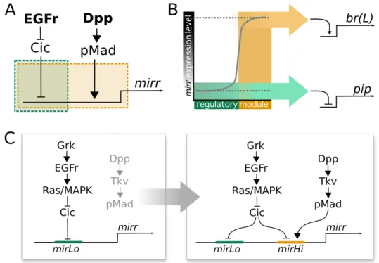
The transcription factor Mirr is a key element of the network. As described before (section 1.3.2),mirr is expressed in a large domain at the dorsal-anterior end of the epithelium, and has been shown to be under the repressive control of Cic (Goffet al., 2001; Atkey et al., 2006). However, up-regulation of mirr in cic mutants is limited to anterior regions, indicating the control of another anterior factor, proposed to be Dpp signalling2, onmirr expression (Go
ffet al., 2001). Ultimately, however, the most relevant outcome of the interactions between Dpp and EGF signalling lies in the localization of the transcription factor Br to the appendage primordia. It has recently been shown that thebr expression pattern is driven by two regulatory elements: brE (‘early’) andbrL(‘late’). brE drives early ubiquitousbr expression, and down-regulatesbr in the dorsal-anterior epithelium starting at stage 10A. By contrast, brL drives the expression of br in the two anterior dorsolateral patches corresponding to the appendage primordia (Fuchs et al., 2012). Both elements respond directly to Mirr: brE is down-regulated, and brLis up-regulated in the dorsal-anterior domain governed by this transcription factor. Additionally,brLis repressed by Pnt in the midline, and by Dpp signalling in the anterior-most cell rows, which generates the characteristic lateral-anterior patches that specify the appendage roof cells (Yakoby et al., 2008b; Fuchs et al., 2012).
1.4.3
Demarcation of domains via local interaction
In the patterning of the follicular epithelium, a clear separation between opercu-lum identity and the appendage primordia has been proven crucial in the proper development of both structures (Dobens and Raftery, 2000). The boundary
be-2
tween operculum and appendage is defined by the expression of bunched (bun), a homolog of the mammalian transcription factor TSC-22. Expression ofbun de-pends on EGFr activation, while high levels of Dpp signalling repressbun(Dobens et al., 1997, 2000). In turn, Bun is necessary to repress operculum fate (Dobens et al., 2000).
The mechanism through which Bun carries out this repression depends on the Notch signalling pathway. Elevated Notch signalling during stage 10 of oogenesis is associated with the centripetal movement of follicle cells belonging to the future operculum (Dobens et al., 2000). Notch is a membrane-bound receptor responding to membrane-bound ligands in adjacent cells, and activity of the pathway in follicle cells up-regulates expression ofnotch, as well as its ligand genesdeltaandserrate. Bun antagonizes Notch activity, likely by repressing ser in a cell-autonomous fashion, down-regulating Notch signalling in adjacent cells (Dobens et al., 2005). Bun is thus responsible for restricting operculum fate and centripetal migration to the anterior-most cells, and maintaining columnar cell fate in the future appendage primordia.
Interestingly, Notch signalling has also been shown to be responsible for the coordination between floor and roof cells. Cells at the floor-roof boundary with high Notch levels express the floor marker rho, whereas cells with lower Notch express br (Ward and Berg, 2005; Ward et al., 2006). Notch is necessary to maintain the boundary between the two cell types: here, the absence of Notch leads to ectopic expression ofbr, at the expense ofrho(Ward et al., 2006). More specifically, however, Notch signalling appears to be responsible for the single row ofrhoexpressing cells bordering the Br domain. Clonal analysis reveals that ectopicrhoexpression occurs in those cells neighbouring a Notch null clone, though only in regions that were not too distant from the wildtyperhohinge (Ward et al., 2006). The precise mechanism for this regulation has not been elucidated.
Finally, reciprocal inhibition between Mirr and Bun after the initiation ofmirr expression may be responsible for the stable demarcation of the posterior border of the appendage primordia. After establishing the border between CMFC and columnar follicle cells, bun expression is down-regulated in the dorsal-anterior follicle cells, and its expression pattern now borders exactly on the mirr domain (Leonard Dobens, pers. comm., Raftery and Dobens, 2012). While these are preliminary and unpublished results, it is interesting to observe again that the gene bun is used in border specification, mimicking its earlier role during the establishment of the anterior border.
which of the steps of eggshell patterning are part of an ancestral property of the epithelium, and which have been co-opted in the evolution of dorsal appendages. Furthermore, we want to explore at which stage(s) in the hierarchy of pattern formation the foundations are laid for phenotypic variation.
1.5
Varying the eggshell phenotype
As described in the previous sections, there is a wealth of data on the genetic and developmental mechanisms underlying dorsal appendage formation. This makes it an exceptionally suitable system for evolutionary developmental research. In this thesis we will address both the putative origin of this feature by comparing oogenesis between species with and without appendages, but also address the many different phenotypes that exist in dorsal appendage morphology between species of Drosophilidae.
1.5.1
Variation in dorsal appendage number, shape, and size
An extensive variety of eggshell phenotypes exists within the family Drosophilidae (Okada, 1968). Drosophila melanogaster belongs to theSophophora subgenus, in which all species studied thus far lay eggs with two appendages, though size and shape may differ. Outside of this subgenus, however, species may be found with one (e.g. Microdrosophila urashimae) three (e.g. D. phalerata), four (e.g. D. virilis), or even up to 12 appendages (e.g. Chymomyzasp.). Some appendages are extraordinarily short (e.g. Chymomyza sp. and D. phalerata), while others can be up to four times as long as the egg (e.g. Microdrosophila urashimae). Another feature can be seen at the tip: D. melanogaster egg appendages are dilated distally to resemble a paddle, while many species’ appendages end in a narrow point (e.g. D. mojavensis). Variation in shape and size can also exist between anterior and posterior appendages of species with more than two egg filaments (e.g. Zaprionus sepsoides) (Okada, 1968). Finally, some Chymomyza species lay eggs that are not entirely symmetrical, and differ in appendage number between left and right (chapter 4).
state (e.g. James and Berg, 2003; Nakamura et al., 2007), though several studies have gone beyond that, and explored epithelial patterning in other species of the Drosophila genus (e.g. Kagesawa et al., 2008). This research has demonstrated the limits of the predictive power of gene expression on the follicular epithelium regarding future phenotypes: indeed, while it is possible to distinguish four ap-pendage primordia in stage 12 patterns of Br, earlier (stage 10B, 11) patterns, where the cells of the appendage primordia are already defined, are only partly predictive of the number of appendages on the future eggshell (James and Berg, 2003).
Chapter 4 of this thesis further explores the developmental basis of this varia-tion. A more detailed review of the literature can also be found in the introduction of that chapter.
1.5.2
Ceratitis capitata
, a tephritid dipteran with
appendage-less eggs
One of the aims of this thesis is to understand the evolutionary origin of a novel morphology—the dorsal egg appendages. To tackle this, we needed to find an ap-propriate model species that could serve as an outgroup of the Drosophilidae, and a representative of an ancestral, appendage-less state. The Tephritid flyCeratitis capitata, perhaps better known as the Mediterranean fruitfly (or Medfly), serves our purpose excellently. Not only does this fly indeed lack eggshell appendages, but it is also a model system for ecological research as it constitutes an agricul-tural pest. The benefits of this status include a genome project (Handler et al., 2012), and the availability and ongoing development of tools for transgenesis (e.g. Loukeris et al., 1995). Not least, the available expertise onC. capitata husbandry greatly facilitates the use of this species as a laboratory model.
1.6
This thesis: objectives and outline
Much is known about the genetics and the development of drosophila oogenesis. Unfortunately, this subject has only marginally been examined in an evolutionary context. So far, the focus has been on the evolution of axis specification (e.g. Lynch et al., 2010) or variation in appendage phenotypes (e.g. Kagesawa et al., 2008). This thesis aims to establish the eggshell structures themselves as a model for morphological innovation, and explore what it can teach us about both the origin and subsequent diversification of novelty.
It is clear that an intimate link exists between the formation of the dorsal eggshell appendages and important developmental events required for the suc-cessful completion of oogenesis. As presented earlier, both the EGF and the Dpp pathways are upstream of both appendage primordia specification, and an array of functions vital for further development, most important of which is the definition of the embryonic main body axes. While it seems likely that the initial signalling events of both pathways are operational elements of oogenesis regardless of the for-mation of eggshell features, and thus precede this innovation evolutionarily, this has not been confirmed experimentally. Further, it is unclear which elements of the targeted network of transcription factors and other genes that eventually establish the appendage primordia are constituents of an ancestral regulatory network, and which have been co-opted concomitant with the evolution of dorsal appendages.
These questions will be addressed in chapter 2, with a comparison of oogenesis between C. capitata and D. melanogaster. This is the first step in analysing which developmental elements correlate with the formation of appendages, and understanding the developmental background from which they evolved. The aim of this chapter is also to identify candidate regulators that have played a role in the evolution of the epithelial patterning network.
The relation between polarity and eggshell patterning will be further explored in chapter 3. Here we will briefly introduce a mutation inD. melanogaster, which partially uncouples the ancestral feature of dorsoventral axis formation from the novel trait of eggshell patterning. Research on this mutant is in full swing at the moment, and this chapter should not be considered anything but prelimi-nary. However, we chose to include this work despite its preliminary state for the simple reason that the severed connection between novel and ancestral traits in this mutant sheds an interesting light on genetic modularity and developmental robustness. Furthermore, our preliminary results diverge from the transcription factor-centred view of gene networks that is so prominent in current evolutionary developmental biology.
in-vestigate which part of the patterning network is responsible for variation between species. This chapter will also contain an elaborate review of current computa-tional models of epithelial patterning during oogenesis, as well as a discussion on one specific element of the network: which genes are responsible for determining the posterior border of the appendage primordia?
Finally, in chapter 5, our results, as well as the available information on dorsal appendage formation, will be discussed in the broader context of novelty. Here, we also look at possibilities for future research, and consider further questions that can be asked about evolutionary novelty with the help of this model system.
Acknowledgements
Abstract
2.1
Introduction
Classically, the concept of evolutionary novelty is that of a new trait, usually an anatomical or morphological one, that opens up the possibility of a wide adaptive radiation into new niches (Mayr, 1960). This definition places its emphasis on adaptation and is thus illustrative of the central role novel traits may have on shaping life on earth. Yet, it is a restrictive definition in that it implies knowledge of the adaptive value of the trait, eliminating traits that have been phylogenetically validated as novelties but lack ecological context.
Moreover, this definition disregards the ontogenic aspects of the new trait par-ticularly of novel morphologies, the most prevalent type of novelty reported. In this light, and in the confined context of this chapter, we will adopt the definition of morphological novelty proposed by M¨uller and Wagner (1991) that to a great extent circumvents the limitations described above by placing the concept in a more fecund ground for an evo-devo research program: “a morphological novelty is a structure that is neither homologous to any structure in the ancestral species nor homonomous to any other structure of the same organism”.
At the mechanistic level, one of the most important contributions of evo-devo to our understanding of the evolutionary process has been the refinement and ex-perimental validation of the gene recruitment concept (co-option). In recent years many examples demonstrate that evolution largely relies on recycling old genes and pathways to generate novel patterns and morphologies (e.g. Brakefield et al., 1996; Moczek and Nagy, 2005). A rewiring of regulatory networks thus seems to be at the core of the dramatic evolutionary changes associated with novelty, and even beyond the novelty concept this has led to an increased effort to under-stand the evolution of whole networks (Abouheif and Wray, 2002). True network evolution, unfortunately, is difficult to analyse, as such an analysis hinges on the understanding of the network as a whole, and beyond the existence of its separate components. Such knowledge is rare in emerging models of evo-devo, but an op-erational standard in many classical genetic models like Drosophila melanogaster (S´anchez et al., 2008).
Most eggs of Drosophilidae bear dorsal appendages, which are thought to have a single origin in their last common ancestor (Hinton, 1969). The appendages are hollow tubes protruding from the dorsal-anterior end of the chorion, and fa-cilitate oxygen supply to the immersed egg (Hinton, 1969, 1981). They display a striking diversity within the Drosophilidae family (Okada, 1968; Nakamura and Matsuno, 2003; Kagesawa et al., 2008), which makes them an interesting sub-ject from an evolutionary perspective. The evolutionary advantage of respira-tory appendages is emphasized by Hinton (1969): they allow the egg to increase its oxygen-absorbing surface without risking desiccation. Indeed, similar eggshell structures have evolved independently at least 11 more times within Diptera, and at seven more instances in other insects (Hinton, 1969, 1981). Nonetheless, and despite their assumed evolutionary advantage, their phylogenetic mapping across Diptera strongly suggests the independent evolution of these structures in different lineages.
In addition to the dorsal appendages, theDrosophilaegg carries an operculum and a micropyle: structures relevant for hatching and fertilization, respectively (Fig. 2.2B). These structures are formed during the last stage of oogenesis by designated cells in the follicular epithelium that change shape prior to the depo-sition of chorionic proteins (Dorman et al., 2004; Berg, 2005). Specification of the appendage primordia occurs chiefly through activity of two main signalling pathways: EGF and Dpp (Peri and Roth, 2000; Berg, 2005).
2.1.1
EGF and Dpp signalling defines appendage primordia
Around stage 8 of Drosophila oogenesis, dorsal patterning is initiated when the TGF-α-like ligand Gurken localizes to the dorsal-anterior corner of the oocyte. Gurken associates with the oocyte nucleus, which is pushed by microtubules to a dorsal-anterior position (Zhao et al., 2012), breaking dorsoventral symmetry in the eggchamber (Neuman-Silberberg and Sch¨upbach, 1993). The Gurken signal then activates EGFr in the adjacent follicle cells, leading (directly and indirectly) to the expression of several transcriptional targets, among which aremirror (mirr) (Jordan et al., 2000; Zhao et al., 2000), rhomboid (rho) (Ruohola-Baker et al., 1993), andpointed (pnt) (Morimoto et al., 1996) (Fig. 2.1).
work by Fuchs et al. (2012) shows how the transcription factor Mirr, regulated by both Dpp and EGF signalling activity, and the ETS domain transcription factor Pnt, expressed in a more narrow stripe along the midline, subsequently establish two groups of cells expressingbroad (br) through two rounds of signalling. First, Mirr represses br, which has been expressed in all follicle cells up to this point, in a wide dorsoanterior region through the brE enhancer. Then, br expression is upregulated again by Mirr, but repressed by Pnt, through the brL enhancer (Fig. 2.1). The two resulting patches of Br-positive cells on either side of the midline are identified as ‘roof cells’: they will later constrict apically and shape the roof of the appendage tube (Ward and Berg, 2005). Adjacent to the Br-positive patches is a single L-shaped row of cells, bordering the anterior and the central edge of the roof domain. These cells express high levels ofrho, and elongate directionally to form the floor of the tube (Ward and Berg, 2005). rhoexpression is regulated mainly by activation of the EGF pathway, which is highly dynamic throughout oogenesis, and shows the same L-shaped pattern at the definition of the floor cells (Nakamura et al., 2007). Rho itself is involved in the dynamic EGFr activation as it cleaves the EGFr ligand Spitz (Spi) into its active form, thereby providing a positive feedback loop for EGF signalling (Ruohola-Baker et al., 1993; Wasserman and Freeman, 1998; Urban et al., 2001) (Fig. 2.1).
Importantly, EGF signalling also determines the dorsoventral axis of the future embryo (Queenan et al., 1997). Via Mirr,pipe(pip) expression is restricted to the ventral side of the egg chamber (Fig. 2.1), leaving an asymmetric distribution of Pip protein in the perivitelline space at the end of oogenesis (Peri et al., 2002; Technau et al., 2011; Andreu et al., 2012; Fuchs et al., 2012). Pip is upstream of a proteolytic cascade in the embryo, leading to the well-known gradient of nuclear Dorsal that regulates the germ layers of the early embryo (Moussian and Roth, 2005).
Dpp, too, is required for processes other than the specification of the appendage primordia. As the inward movement of the centripetally migrating follicle cells starts (Fig. 2.2A),dppis expressed in the leading edge of these cells, and disrupted Dpp signalling has been associated with defects in this migration (Twombly et al., 1996). Dpp is required furthermore for the formation of the operculum (Twombly et al., 1996; Dobens et al., 2000).
Figure 2.1: A simplified representation of the genetic network under-lying dorsoventral polarity (pip) and DA-formation (br) during D.
melanogaster oogenesis. Input comes from two main signalling pathways,
EGF and Dpp, providing dorsoventral and anteroposterior information, respec-tively, and results in the specification of cell domains expressingpipandbr.
2.1.2
Ceratitis capitata
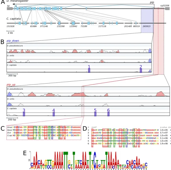
Considering the relatively novel acquisition of elaborate eggshell structures, it is interesting to examine the underlying patterning network in the context of a fly species that does not possess these specialized structures. Tephritidae are esti-mated to be separated by about 65 million years of evolution from Drosophilidae (Wiegmann et al., 2011). For our comparison we chose a Tephritid fly that has been established as a laboratory organism: the Mediterranean fruit fly Ceratitis capitata. C. capitata is an agricultural pest, which has motivated widespread in-ternational research, including a genome project and the development of genetic tools (Loukeris et al., 1995; Zwiebel et al., 1995; Schetelig et al., 2009).
downstream targets in C. capitata oogenesis, in order to understand the ances-tral network patterning the follicular epithelium prior to the evolution of dorsal appendages. Determining which genes differ in their behaviour throughout ooge-nesis of appendage-bearing (D. melanogaster) and appendage-less (C. capitata) eggshells can help generate hypotheses on the co-option of genes and genetic net-work changes in the evolution of this novel feature. Our analysis points to a key role for the transcription factor Mirr, both in its regulation as in its transcriptional targets. Furthermore, the presence of both the EGF and the Dpp pathway inC. capitata oogenesis leads us to hypothesize that the positional information that these pathways provide to the ancestral follicular epithelium could have facilitated further downstream patterning required for developing the dorsal appendages.
2.2
Material and Methods
2.2.1
Fly maintenance
Our initial Ceratitis capitata culture was kindly (and repeatedly) provided by Andrew Jessup (IAEA Seibersdorf, Austria), originating from flies captured in Argentina. Adult flies were maintained on a diet of sugar and hydrolysed yeast protein, and larvae were reared on a mixture of bran, sugar and yeast. All stages were maintained at room temperature. Drosophila melanogaster Oregon R. was maintained on regular fly food at room temperature.
2.2.2
Cloning
Gene-specific sequences were isolated from C. capitata cDNA by PCR using de-generate primers (fordpp,mirr,slbo,tkv, andwind), as well asC. capitata specific primers (forCc-br,Cc-grk, Cc-nud, andCc-pip), designed using contigs from the C. capitata genome project kindly provided by the Medfly Whole Genome Se-quencing Consortium (Handler et al., 2012). ForCc-pip two primer combinations were used, generating two separate probes forin situ hybridization. These probes were (1) against the common part of allpip isoforms, and (2) againstCc-pip-ST2, the homologue ofDm-pip-ST2 (isoform A). Corresponding probes were made for the positive controls inD.melanogaster.