1
Arquivos Brasileiros de Cardiologia - Volume 85, Nº 1, Julho 2005
Case Report
Right Atrial Aneurysm Associated to Fetal Hydrops:
Diagnosis through Fetal Echocardiography
Marcia Ferreira Alves Barberato, Silvio Henrique Barberato, Cláudio Correa Gomes,
Sérgio Luis Costa, Alfred Krawiec
CEMMEF- Centro de Medicina Materno-Fetal - Curitiba, PR - Brazil
Mailing address: Silvio Henrique Barberato - Rua Saint Hilaire, 122/203 - 80240-140 - Curitiba, PR - Brazil
E-mail: silviohb@cardiol.br
Received for publishing on 05/06/2004 Accepted on 02/10/2005
Right atrium aneurysms are entities which are rarely reported in cardiologic practice, especially in intrauterine life, and may be mistaken with pericardial effusion and Ebstein’s anomaly. We show a review of the literature and illustrate with a case of prenatal diagnosis of right atrium aneurysm running through with hydropsy signs.
The right atrial (RA) aneurysm is a cardiac malformation of unknown etiology, rarely found in medical literature, especially when is about prenatal diagnosis. Due to its rareness, it can be easily mistaken with other cardiopathies that lead to RA dilatation, such as Ebstein’s anomaly of tricuspid valve1,2. Besides, its usually
silent intrauterine evolution and the still restricted use of fetal echocardiography in the tracking of congenital cardiopathies in our milieu contribute to the diagnosis is only done, mostly, in adult age3,4. We show a case of intrauterine diagnosis of RA
aneurysm associated to fetal hydrops with syndrome traces.
Case Report
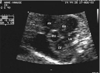
A 26-year-old white patient, 2 pregnancies, 1 delivery, with 22 weeks of pregnancy, sent to the service of materno-fetal medicine due to pericardial effusion, detected in a routine obstetric echography. Fetal echocardiogram dispelled the presence of pericardial effusion and suggested, as initial hypothesis, Ebstein’s anomaly with great RA dilatation and mild tricuspid regurgitation. After 2 weeks, the fetus started to show signs of hydrops (ascitis and skin edema). A new echocardiogram allowed for a more tailed assessment of the anatomy of tricuspid valve, without de-monstrating Ebstein’s typical valvar caudal implantation. The aneurysmatic RA extended cranially to the superior cava vein and ascending aorta and, caudally, to the heart’s apex, bordering the right ventricle (fig. 1). The use of digital and diuretic was dis-cussed, but the assistant team preferred expectant management. With 30 weeks of pregnancy, control echography showed an intrauterine growth retardation and signs of fetal suffering, opting for interruption of pregnancy. The newborn (NB) from operative birth, weighing 2,250 grams, APGAR 4 and 7 at the first and fifth minutes, respectively, with the need for mechanical
ventilation. At the physical examination, generalized edema, syndromic facies (triangular shape, prominent forehead, low auricular implantation) and sturdy consistency hepatomegaly were detected. The NB was transferred to neonatal ICU, where transthoracic echocardiogram confirmed the disease (fig. 2). Spontaneous contrast or thrombus presence was not seen inside the aneurysm. Discreet tricuspid regurgitation was confirmed and the presence of small interatrial communication was verified. The NB had cardiorespiratory arrest and death with only 10 hours of life, having not been assessed by a geneticist. The necropsy showed a giant aneurysm of free wall of RA with paper-thin aspect, measuring 7x2.8 cm and taking almost the whole mediastinum. Atrioventricular and semilunar valves of normal macroscopic aspect, ventricles with preserved dimensions and pericardium without changes. Oval foramen-type interatrial communication with diameter of 1.6 mm was verified. Liver with increased size, of hardened consistency and nodular aspect. Hepatic biopsy showed atresia of biliary ways. Karyotype was not performed.
Discussion
Reports on aneurysm of RA have been carried out at childhood5,
adult age3,4 and, very rarely in intrauterine life6,7. The greatest review
published on congenital malformations of RA, comprising from 1955 to 19998, listed 60 cases of global congenital dilatation of RA
among 105 reports, which made it the most common type of malformation of this chamber. After a thoroughly search in the related literature, we found 4 cases of intrauterine dilatation of RA diagnosed by means of fetal ultrasound. In only two of them there was the correct initial diagnosis of RA aneurysm. Ebstein’s anomaly was suspected of in the first case, after the finding of RA dilatation associated to tricuspid insufficiency. Soon after the birth, a trans-thoracic echocardiogram confirmed the correct diagnosis1. In the
second case, RA aneurysm was diagnosed in a fetus with 17 weeks of pregnancy, without signs of heart failure. The option was the interruption of the pregnancy and the study of necropsy was confir-matory6. In the third case, the Ebstein’s anomaly diagnosis was
done in 35 weeks and confirmed after birth. At 10 months of life, the infant showed signs of refractory right heart failure and was submitted to surgery. In the operative act, the tricuspid valve was normally positioned and the RA had its free wall partially dissected. The evolution was asymptomatic for 4 years of follow-up2. In the
2
Arquivos Brasileiros de Cardiologia - Volume 85, Nº 1, Julho 2005
Right Atrial Aneurysm Associated to Fetal Hydrops: Diagnosis through Fetal Echocardiography
without intercurrences. The infant evolved asymptomatically up to one year of age when we serial echocardiograms showed a progressive increase of the aneurysm (generating vascular compression) and appearance of intracardiac thrombus. The option was surgical treatment that went by without complications. After an one year follow-up, the infant stayed healthy7.
The present case, although initially have also been mistaken with Ebstein’s anomaly, shows some peculiarities. The severe evolu-tion with fetal hydrops and suffering, needing to interrupt the pregnan-cy, have not been reported yet. All cases were found of exam in systematic obstetric echography. In none of them the fetus showed signs of heart failure, despite of large atrial dilatation. In most cases, the RA aneurysm diagnosis is only done at a more advanced age, either through exam finding5, atrial arrhythmias4 and embolic
phenomena9. It is possible to infer that its evolution in the fetal
period is usually benign and silent. In the present case, two possibi-lities may have led to hydrops: the probable syndrome the fetus carried (without relation to the aneurysm itself) or, alternatively, due to the compression of vascular structures and right ventricle, obstructing the venous return and impairing the cardiac output. The large dilatation of RA and of tricuspid ring, causing a
pseudo-1. Silva AM, Witsemburg M, Elzenza N, Stewart P. Idiopathic dilatation of the right atrium diagnosed in utero. Rev Port Cardiol 1992; 11: 161-3.
2. Reinhardt-Owlya L, Sekarski N, Hurni M, Laurini R, Payot M. Idiopathic dilatation of the right atrium simulating Ebstein’s anomaly. Apropos of a case diagnosed in utero. Arch Mal Coeur Vaiss 1998; 91: 645-9.
3. Zeebregts CJ, Hensens AG, Lacquet LK. Asymptomatic right atrial aneurysm: for-tuitous finding and resection. Eur J Cardiothorac Surg 1997; 11: 591-3. 4. Barberato SH, Barberato MF, Avila BM, Perretto S, Blume Ld Ldo R, Chamma Neto
M. Aneurysm of the right atrial appendage. Arq Bras Cardiol 2002; 78: 236-41. 5. Chatrath R, Turek O, Quivers ES, Driscoll DJ, Edwards WD, Danielson GK.
Asymp-tomatic giant right atrial aneurysm. Tex Heart Inst J 2001; 28: 301-3. 6. Gross B, Petrikovsky B, Challenger M. Prenatal diagnosis of an aneurysm of the
right atrium. Prenat Diagn 1996; 16: 1043-5.
References
7. Haut Cilly FB, Schleich JM, Lacour-Gayet F, Almange C. Right atrial compressive aneurysm with favorable outcome after surgery at the age of 1 month. Arch Mal Coeur Vaiss 2002; 95: 487-90.
8. Binder TM, Rosenhek R, Frank H, Gwechenberger M, Maurer G, Baumgartner H. Congenital malformations of the right atrium and the coronary sinus: an analysis based on 103 cases reported in the literature and two additional cases. Chest 2000; 117: 1740-8.
9. Staubach P. Large right atrial aneurysm: rare cause of recurrent pulmonary embo-lism. Z Kardiol 1998; 87: 894-9.
10. McElhinney DB, Krantz ID, Bason L et al. Analysis of cardiovascular phenotype and genotype-phenotype correlation in individuals with a JAG1 mutation and/or Alagille syndrome. Circulation 2002; 106: 2567-74.
Fig. 2 - Transthoracic echocardiogram: asterisks mark the collum of the aneurysm of right atrium (AN ). RA - right atrium; LA - left atrium; RV - right ventricle; LV - left ventricle.
AN
LV
RV
LA
RA AN
*
Fig. 1 - Fetal echocardiogram: cross-section of 4 chambers showing aneurysm of right atrium (AN). RA - right atrium; LA - left atrium; RV - right ventricle; LV - left ventricle.
AN
RV
LV RA
LA
displacement of the septal leaflet of the valve, led to the mistaken initial diagnostic impression of Ebstein’s anomaly. The syndromic facies associated to intrahepatic biliary disease and right heart malformation made us include the syndrome of Alagille10 in the
differential diagnosis. However, through the classic criteria for the definition of the syndrome we cannot ascertain that it is a typical case. Although the right heart malformations are the most common in that disease, we did not find any report of RA aneurysm. Unfor-tunately, the non-performance of the karyotype, a mandatory ma-nagement in cases of severe fetal cardiopathy with suspicion of syndrome, limited the correct etiological clarifying.
