3 1 4
Pêgo-Fernandes et al
Ligation of the thoracic duct for the treatment of chylothorax
Arq Bras Cardiol 2003; 81: 314-7.
Instituto do Coração do Hospital das Clínicas - FMUSP.
Mailing address: Paulo M. Pêgo-Fernandes – InCor - Av. Dr. Enéas C. Aguiar,44 – 05403-000 – São Paulo, SP, Brazil - E-mail: [email protected]
Arq Bras Cardiol, volume 81 (nº 3), 314-317, 2003
Paulo M. Pêgo-Fernandes, Fábio B. Jatene, Clayton Cesar Tokunaga, Danielle Tiemi Simão, Ricardo Beirutty, Eliza Rumiko Iwahashi, Sérgio Almeida de O liveira
São Paulo, SP - Brazil
Ligation of the Thoracic Duct for the Treatment of
Chylothorax in Heart Diseases
Brief Report
If conservative treatment is not effective, surgical treatment should be considered 8. Studies indicate that
liga-tion of the thoracic duct and pleurodesis are efficient alter-natives for resolution of chylothorax 9.
Recently, thoracic duct ligation through video-assis-ted thoracic surgery has been used in patients who did not respond to conservative treatment. Kirby et al 11 describe
this conduct as less painful and with fewer postoperative complications.
Pereira et al 12 describe the use of lymphatic ligation for
the treatment of postoperative chylopericardium. However, in our country, ligation of the thoracic duct for the treatment of postoperative chylothorax is still a rare procedure, and at what point the conservative treatment should be replaced by postsurgical treatment is still controversial. Thus, our study evaluates the initial results of ligation of the thoracic tract in 4 patients with chylothorax unresponsive to conser-vative treatment.
Case Reports
Case 1 - A four-year-old-male patient with congenital cardiopathy and cyanosis at birth with a previous history of pulmonary atresia without interventricular communication, single left ventricle, hypoplastic right ventricle with sinu-soids, left pulmonary artery stenosis, and coronary artery cavitary fistulas. At 5 days of life, he underwent atrial septo-tomy with the Rashkind procedure, and after 2 days, he underwent a modified left Blalock Taussig shunt procedure. At 4 years old, he underwent a bidirectional Glenn operation with maintenance of the previous Blalock shunt, evolving, in the postoperative period with low cardiac output maintai-ned with vasoactive drugs. He was discharged on the 5th postoperative day, when minor pleural effusion was sus-pected. A chest X-ray was indicated.
After chylothorax was diagnosed, pleural drainage was started. Parenteral nutrition and fasting were introdu-ced after 1 week. The patient evolved with significant per-sistence of pleural drainage. Nineteen days after the initial
In children, chylothorax occurs mainly after cardiac and thoracic surgeries. One of the recommended postsur-gery treatments is ligation of the thoracic tract, when all other conservative treatments have failed. We report 4 cases of chylothorax in patients who were successfully treated with this approach, which resulted in a decrease in pleural drainage without recurrent chylothorax.
Chylothorax refers to the presence of lymphatic fluid in one or both pleural spaces, secondary to leakage in the thora-cic duct or one of its main tributaries 1. Quinke 2 first described
traumatic chylothorax in 1875, after rupture of the thoracic duct. When chylothorax is not treated, the risk of death is high, with rates above 45% 3, requiring, in the majority of the
cases, aggressive treatment.
According to DeMeester 4, chylothorax may be
accor-ding to its cause, either congenital, traumatic, neoplastic, or multiple causes. Among the traumatic causes surgical pro-cedures with a thoracic approach may cause thoracic duct lesions, leading to the development of chylothorax. Post-operative chylothorax occurs in less than 1% of thoracic surgeries, with a prevalence of 0.5% to 2% 5,and may cause
nutritional deficiencies, respiratory system involvement, dehydration, and immunosuppression, making the patient more vulnerable to infections.
Postoperative chylothorax is initially treated conser-vatively 6.Treatment consists of extracting chyle from the
Arq Bras Cardiol 2003; 81: 314-7.
Pêgo-Fernandes et al Ligation of the thoracic duct for the treatment of chylothorax
3 1 5
clinical picture, one more drainage procedure and surgical cleansing were performed. On the 49th admission day, due to maintenance of chylothorax, the patient underwent liga-tion of the thoracic duct through a right antero-lateral thora-cotomy. A small amount of chyle, multiple pleuro-pulmona-ry adherences, and a bulge in the pericardial sac were found.
After precise identification of the thoracic duct, liga-tion was carried out, pericardiocentesis was performed, and 100 mL of chyle was drained under pressure. Drains were maintained in the 6th intercostal space. The patient evolved well, with withdrawal of right and left drains on the 16th and 18th postoperative days, respectively.
Case 2 - A one-year-old female patient presenting with a history of viral myocarditis (positive test for Cox sackie virus) evolving to dilated myocardiopathy with congestive heart failure and acute renal failure. She was sent to our ser-vice to stabilize the clinical picture and to discuss the possi-bility of cardiac transplantation. After 1 month, a thoracic X-ray showed an image suggestive of left pleural effusion from a posterior puncture, with leakage of a milky liquid, which was diagnosed through biochemical analysis as chyle. We suspect that the cause of chylothorax was the lesion in the thoracic duct during the insertion of an intraca-theter days before. Treatment was initiated with placement of a drain in the left side and fasting maintenance and admi-nistration of parenteral nutrition.
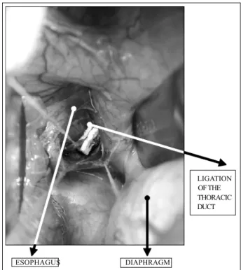
After 19 days, due to the persistence of increased pleu-ral drainage and the poor condition of the patient, with great weight loss, the thoracic duct was identified and liga-ted (fig. 1) through a postero-lateral thoracotomy in the 7th right intercostal space. The patient evolved well, with a decrease in pleural drainage. Oral food consumption was initiated with maintenance of parenteral nutrition until the 8th postoperative day. The patient showed progressive clinical improvement and underwent cardiac transplantation 25 days after the thoracic duct ligation.
Case 3 – A 2-year-old male patient underwent surgical closure of patent ductus arteriosus, evolving with pleural drai-nage above normal limits. As of the 4th postoperative day, the appearance of the pleural drainage fluid, which remained elevated, became milkier and was sent for biochemical analysis, and the diagnosis of chylothorax was made. The patient was treated for 5 days with a fat-restricted oral diet; however, the amount of liquid drained did not decrease. Fasting and parenteral nutrition were used for 10 days, with persistent significant drainage and significant weight loss.
Due to the persistence of chylothorax, we opted for ligation of the thoracic duct through a video-assisted thora-cic surgery. The patient evolved well with drain withdrawal on the 8th postoperative day.
Case 4 – A 70-year-old male patient underwent myo-cardial revascularization. On the 3rd postoperative day, he evolved with leakage of a milky secretion through the left pleural drain, which later revealed high levels of triglyceri-des. On the 7th postoperative day, parenteral nutrition was introduced. (BNJ5). On the 14th postoperative, the patient
underwent ligation of the thoracic duct through a right thoracoscopy. Left and right pleural drainage was maintai-ned. The right drain was removed 3 days after ligation. On the 19th postoperative day after revascularization, a liquid diet was reintroduced. Parenteral nutrition was interrupted on the 21st postoperative day and a light nonfat enteral diet was established, and complemented with medium-chain triglycerides.
On the 24th postoperative day, the patient underwent a chest X-ray that demonstrated bilateral pleural effusion with septation, more evident in the left side, and discreet in the right side. After 4 days, the patient underwent a left thoracotomy with pleural decortication, pleurocentesis, with placement of anterior and posterior drains. A pleural fluid culture indicated the presence of Pseudomonas
aeru-ginosa. On the 5th postoperative day (33 days after
revas-cularization), a general oral diet was instituted. On the 7th postoperative day, the drains were withdrawn. The patient was discharged on the 9th postoperative day (37 days after revascularization).
Discussion
Chylothorax is characterized by the effusion of lym-phatic fluid in the pleural space. Among other causes, it may originate as a secondary complication of surgical interven-tions 13, especially in thoracic approaches, when it is called
postoperative chylothorax.
In the 3 pediatric patients in this study, the chylothorax was due to surgical procedures, 2 of them directly related to the cardiac approach and one possibly to the placement of an intracatheter. These data are similar to other studies that
Fig. 1 – Ligation of the thoracic duct.
ESOPHAGUS DIAPHRAGM
3 1 6
Pêgo-Fernandes et al
Ligation of the thoracic duct for the treatment of chylothorax
Arq Bras Cardiol 2003; 81: 314-7.
show that in pediatric patients, postoperative chylothorax occurs, mainly as a thoracic and cardiac postoperative com-plication 14.
The 70-year-old patient developed chylothorax after myocardial revascularization with the internal thoracic arte-ry, a rare complication of this procedure due to the location of the thoracic duct 15.
Postoperative chylothorax may develop in 2 to 4 weeks after surgery and can be mild or severe, according to the volume and the amount of chyle loss 14. Although it is a
rare condition, when not treated, postoperative chylothorax has a high mortality rate, reaching 50% 6.
Leakage of chyle in the pleural cavity may compress the ipsilateral lung and, when the secretion is intense, it can cause mediastinum elevation involving the other lung and cardiac function 16. Additionally, the loss of chyle causes a
decrease in the number of T lymphocytes, involving the immunologic system and predisposing patients to infec-tions 5. Metabolic alterations are due to depletion of
electro-lytes, and chylomicrons with long-chain fatty acids determi-ne the metabolic alterations.
Most patients with chylothorax experience dyspnea and pleural secretion. Diagnosis may be confirmed by bio-chemical and microscopic examinations of the drained liquid, checking for the presence of triglycerides and chylomi-crons. The cause can be defined by investigation of the thoracic wall, pleura, lungs, intrathoracic lymphonodus, pulmonary lymphatic or thoracic duct 13.
Chylothorax treatment aims at a) reducing chyle pro-duction, b) treating the underlying causes of intrapleural drainage with fluid replacement, and d) obliteration of the pleural space 13.
Postoperative chylothorax is initially treated conser-vatively, based on the drainage of the pleural space and nutritional support, with the use of medium-chain triglyceri-des 7, low-fat oral diet or enteral rest by giving parenteral
nutrition 17.However, the use of medium-chain triglycerides
is avoided by some physicians, after the Peitersen and Jacob-sen 18 study demonstrated the substantial increase in
trigly-ceride content in pleural effusion. Additionally, repeated or continuous drainage of the pleural secretion is inefficient and promotes fluid, plasma protein and electrolyte loss.
If conservative treatment fails, it should be suspended and surgery should be performed. The timing of surgical management is controversial. Some studies recommend surgical intervention if drainage lasts for 1 to 3 weeks 17, or
when daily leakage exceeds 200mL to 500mL per day. Surgical treatment of chylothorax includes pleurode-sis 19, ligation of the lymphatic vessels, and ligation of the
thoracic duct 17.
Ligation of the thoracic duct, performed for the first time in 1948 by Lampson 3 was very efficient low mortality rate.
Evidence exists that thoracic duct ligation increases col-lateral lymphatic circulation regardless of the level of duct ligation. Thus, even with the withdrawal of the thoracic duct and the cisterna chyli, adverse effects are not triggered 10.
Ligation of the thoracic tract through video-assisted
thoracic surgery in the treatment of spontaneous and pos-toperative chylothorax has been widely described in recent years 6. It is a very efficient procedure, enabling the
identifi-cation of the thoracic duct and its selective ligation, and can be indicated when conservative treatment fails, although the presence of extensive pleural adhesions may determine the need for open surgery 10. Another important factor is
that video-assisted surgery causes less postoperative pain and decreases the risk of pulmonary dysfunction 11.
Accor-ding to Fahimi et al 6, video-assisted ligation of the thoracic
duct is a great alternative for early treatment of persistent postoperative chylothorax, due to the low cost and facility of its use associated with a lower mortality rate.
For the use of video-assisted thoracic surgery in the treatment of chylothorax, Sachs et al 20 proposed
lymphan-giography as a useful method of preoperative localization of the leakage site and reported the value of computed tomo-graphy as an additional but not essential method. Fahimi et al 6 proposed the administration of a cream meal mixed with
Sudan black that enables stabilization of the spot of leakage in the majority of cases, with a simple, efficient, and non-invasive method, in comparison with lymphangiography and computed tomography.
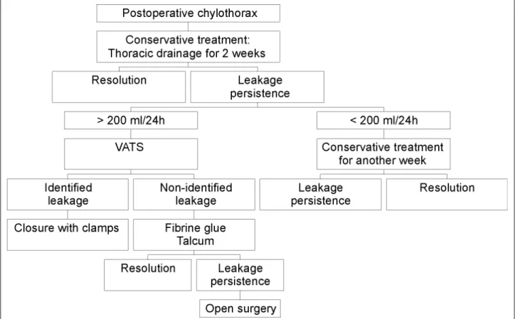
According to Fahimi et al 6, postoperative chylothorax
must follow this conduct: (fig. 2) the use of video-assisted thoracic surgery is indicated when daily leakage exceeds 200 mL after 2 weeks of conservative treatment.
Some studies demonstrate that the ligation of the thoracic duct by using video-assisted thoracic surgery or thoracotomy for the treatment of chylothorax in children may fail due to the anatomic variations of the thoracic duct or due to the impossibility of identifying the leakage sites 21.
Such difficulty in identifying and isolating structures with the use of video-assisted thoracic surgery is, in part, related to the duration of chylothorax which, if long-lasting, may cause the development of pleural adherences, as men-tioned above 10, hindering the identification of mediastinal
structures. In this case, ligation of the thoracic duct may be performed through open surgery as that performed in patients 1 and 2.
In other conditions, pleurodesis may be associated with ligation of the thoracic duct in the treatment of chylo-thorax, as in patient 4 in whom, due to an infectious process, chylothorax persisted even after duct ligation.
Thoracic duct ligation is an efficient method to treat chylothorax. Thus, the possibility of prompt surgical inter-vention is recommended. At one time, shorter periods of conservative treatments, which are often unsuccessful, could minimize cost and damages to the patients, such as weight loss due to a restrictive diet and the pain caused by the permanence of the chylothorax. However, Sieczka and Harvey 5 reported that the conservative treatment may
Arq Bras Cardiol 2003; 81: 314-7.
Pêgo-Fernandes et al Ligation of the thoracic duct for the treatment of chylothorax
3 1 7
In conclusion, our 4 patients had significant improve-ment of in the clinical features of chylothorax after thoracic duct ligation. The treatment was characterized as a good therapeutic alternative for postoperative chylothorax when
Fig. 2 - Postoperative chylothorax treatment according to Fahimi et al 6.VATS: video-assisted thoracic surgery.
no response occurs to conservative treatment. Early liga-tion of the thoracic duct may eliminate the possible failure of conservative treatment, which could be aggressive, causing unnecessary suffering for patients.
References
1. Miller JI. Chylothorax and anatomy of the thoracic duct. In: Shields TW, ed. General Thoracic Surgery. Philadelphia: Lea & Febiger, 1989: 625. 2. Wurnig PN, Hollaus PH, et al. Thoracoscopic direct clipping of the thoracic
duct for chylopericardium and chylothorax. Ann Thorac Surg 2000; 70: 1662-5.
3. Fahimi H, Casselman FP, et al. Current management of postoperative chylothorax. Ann Thorac Surg 2001; 71: 448-51.
4. Demeester TR. The pleura. In: Sabiston DCaS, EC, ed. Surgery of the Chest. Philadelphia: Lea & Febiger, 1983.
5. Sieczka EM, Harvey JC. Early thoracic duct ligation for postoperative chylotho-rax. J Surg Oncol 1996; 61: 56-60.
6. Fahimi H, Casselman FP, Mariani MA, et al. Current management of postoperative chylothorax. Ann Thorac Surg 2001; 71: 448-51.
7. Hashim SA, Roholt HB, Babayan VK, Itallie TB. Treatment of chyluria and chylo-thorax with medium-chain triglyceride. N Engl J Med 1964; 270: 756-61. 8. Graham DD, McGahren ED, Tribble CG, et al. Use of video-assisted thoracic
surgery in the treatment of chylothorax. Ann Thorac Surg 1994; 57: 1507-12. 9. Allen EM, van Heeckeren DW, Spector ML, Blumer JL. Management of
nutritio-nal and infectious complications of postoperative chylothorax in children. J Pediatr Surg 1991; 26: 1169-74.
10. Wurnig PN, Hollaus PH, Ohtsuka T, et al. Thoracoscopic direct clipping of the thoracic duct for chylopericardium and chylothorax. Ann Thorac Surg 2000; 70: 1662-5.
11. Kirby TJ, Mack MJ, Landreneau RJ, Rice TW. Lobectomy-video-assisted
thora-cic surgery versus muscle-sparing thoracotomy.: a randomized trial. J Thorac Cardiovasc Surg 1995; 109: 997-1002.
12. Pereira WM, Kalil RAK, Prates PR, Nesralla IA. Cardiac tamponade due to chy-lopericardium after cardiac surgery. Ann Thorac Surg 1988; 46: 572-3. 13. Browse NL, Allen DR, Wilson NM. Management of chylothorax. Br J Surg 1997;
84: 1711-6.
14. Stringel G, Mercer S, Bass J. Surgical management of persistent postoperative chylothorax in children. Can J Surg 1984; 27: 543-6.
15. Pego-Fernandes PM, Ebaid GX, Nouer GH, et al. Chylothorax after myocardial revascularization with the left internal thoracic artery. Arq Bras Cardiol 1999; 73: 383-90.
16. Wolff AB, Silen ML, Kokoska ER, Rodgers BM. Treatment of refractory chylotho-rax with externalized pleuroperitoneal shunts in children. Ann Thorac Surg 1999; 68: 1053-7.
17. Bond SJ, Guzzetta PC, Snyder ML, Randolph JG. Management of pediatric pos-toperative chylothorax. Ann Thorac Surg 1993; 56: 469-73.
18. Peitersen B, Jacobsen B. Medium chain triglycerides for treatment of sponta-neous, neonatal chylothorax: lipid analysis of the chyle. Acta Paediatr Scand 1977; 66: 121-5.
19. Adler RH, Levinsky L. Persistent chylothorax: treatment by talc pleurodesis. J Thorac Cardiovasc Surg 1978; 76: 859-64.
20. Sachs PB, Zelch MG, Rice TW, et al. Diagnosis and localization of laceration of the thoracic duct: usefulness of lymphangiography and CT. Am J Roentgenol 1991; 157: 703-5.