Case Report
3 3 0 Arq Bras Oftalmol. 2014;77(5):330-3 http://dx.doi.org/10.5935/0004-2749.20140083
INTRODUCTION
Dengue fever (DF) is a viral infection disease endemic in tropical and subtropical areas; it is caused by four different virus serotypes. Its clinical features vary from asymptomatic infection to critical systemic disease. One of the more severe forms of the disease is dengue he-morrhagic fever, characterized by fever, evidence of a hehe-morrhagic event or a positive tourniquet test, low platelets levels (≤100,000 cells/ mm3) and evidence of disseminated plasma leakage such as pleural effusion, ascites and hypoalbuminemia(1).
Acute visual loss in a patient with DF is an unusual occurrence, which generally arises due to hemorrhagic complications in the retina(2) or to virus-related optic neuritis(3,4). Visual loss from intracranial compli-cations of diseases involving the optic pathways is extremely rare, but was recently described in two patients with hemorrhage in pituitary adenomas(1,4).
In this paper we document, to our knowledge, the first reported case of DF and bilateral acute visual loss caused by chiasmal com-pression from Rathke’s cleft cyst (RCC) apoplexy.
CASE REPORT
A 47-year-old man was referred for evaluation of acute bilateral visual loss, most severe in the left eye (OS). One month prior to exa-mination, the patient had experienced rapidly progressing blurred vi-sion over a period of three days, associated with headache, retro-ocular pain, fever and prostration. A medical evaluation at another facility suggested the diagnosis of DF, which was subsequently confirmed by positive serologic testing for dengue virus-specific IgM antibodies. The visual complaints were apparently considered unspecific symptoms because of generalized malaise. In addition, a positive tourniquet test
indicated a hemorrhagic tendency. As there were no signs of dengue shock syndrome, and the patient was advised to hydrate, rest, and watch for warning signs of complications of DF.
The systemic symptoms gradually resolved, but the blurred vision improved only slightly and the patient was referred for ophthalmic evaluation. His past medical history was unrevealing, except for the use of glasses for the preceding 30 years and slightly decreased vision due to anisometropic amblyopia in the right eye (OD) since childhood. On ophthalmic examination, the best-corrected visual acuity was 20/60 in OD (+3.75 sph X -0.75 cyl axis 80) and 20/300 in OS (+0.25 sph). The pupils were equal in size but a slight relative afferent pupillary defect was observed on the left side. The slit lamp findings and intraocular pressure measurements were within normal limits. Ophthalmoscopy was normal except for mild temporal optic disc pallor in OS. The visual field (VF) examination demonstrated loss of the temporal field in both eyes and constriction of the inferior nasal field in OS (Figure 1). Optical coherence tomography revealed peripapillary retinal nerve fiber layers of normal thickness in both eyes. Magnetic resonance imaging (MRI) revealed a tumor in the sellar region with extension to the suprasellar cistern and compres-sing the optic chiasm. Heterogeneous areas in the tumor suggested intralesional bleeding. The lesion showed a hyperintense signal on T1-weighted images and a hypointense signal on T2-weighted images, indicating hemorrhage inside the cystic tumor (Figure 2). Subsequently, the patient developed nausea and vomiting. A repeat laboratory investigation revealed a marked decrease in cortical and adrenocorticotrophin (ACTH) hormone levels, indicating adrenal in-su fficiency. The patient was in-submitted to urgent transsphenoidal surgery. The histological characteristics of the tumor were consistent with RCC (Figure 3).
Bilateral acute visual loss from Rathke’s cleft cyst apoplexy in a patient with dengue fever
Perda visual aguda secundária a apoplexia de cisto de Rathke em paciente com dengue
AnA CláudiA de FrAnCo Suzuki1, rAFAel BArBoSAde ArAújo1, eduArdo CunhAde SouzA1, Mário luiz riBeiro Monteiro1
Submitted for publication: October 10, 2013 Accepted for publication: January 3, 2014
Study conducted at the Department of Ophthalmology and Otolaringology, University of São Paulo Medical School.
1 Division of Ophthalmology, University of São Paulo Medical School, São Paulo, Brazil.
Funding: No specific financial support was available for this study.
Disclosure of potential conflicts of interest: None of the authors have any potential conflicts of interest to disclose.
Corresponding author: Ana Claudia De Franco Suzuki. Av. Dr. Eneas de Carvalho Aguiar, 255 - 6o an dar - sala 6121 - São Paulo - SP - 05403-000 - Brazil - Email: [email protected] ABSTRACT
Hemorrhagic complications of optic pathway diseases are extremely rare causes of acute visual loss associated with dengue fever. In this paper we report a patient presenting with dengue fever and bilateral acute visual loss caused by chiasmal compression due to Rathke’s cleft cyst apoplexy. Considering the importance of early diagnosis and treatment to visual recovery, apoplexy of sellar and suprasellar tumors should be considered in the differential diagnosis of patients with acute visual loss and dengue fever.
Keywords: Visual disorders/etiology; Dengue/complications; Retinal hemorrhage; Pituitary apoplexy; Optic chiasm; Humans; Case reports
RESUMO
Complicações hemorrágicas de doenças da via óptica são causas extremamente raras de perda aguda de visão em pacientes com dengue. Nesse trabalho, documentamos um caso de paciente com dengue apresentando perda de visão bilateral aguda secundária a compressão quiasmática por quadro hemorrágico em cisto de Rathke. Considerando a importância do diagnóstico e tratamento precoces para um bom prognóstico visual, a apoplexia de tumores da região selar e suprasselar deve ser incluída como um raro, porém importante, diagnóstico diferencial de perda visual aguda nesses pacientes.
Suzuki ACF, et al.
331
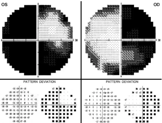
Arq Bras Oftalmol. 2014;77(5):330-3 Figure 1. Automated static perimetry (Humphrey 24-2 SITA-Standard test), gray scale (above) and pattern deviation (below),
showing complete temporal ield loss in both eyes, some nasal ield constriction in the right eye (OD) and severe inferior and nasal loss in the left eye (OS).
Bilateral acute visual loss from Rathke’s cleft cyst apoplexy in a patient with dengue fever
332 Arq Bras Oftalmol. 2014;77(5):330-3
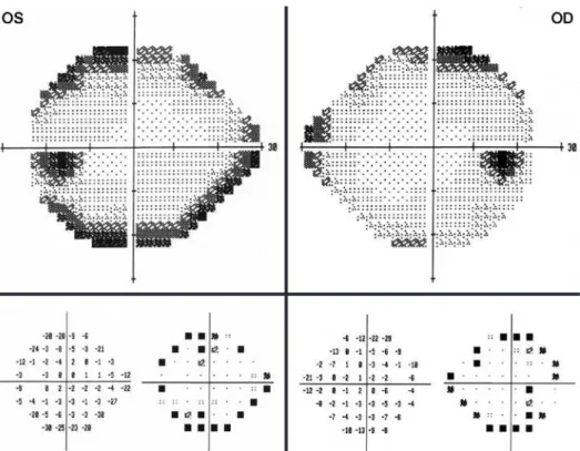
Figure 4. Automated static perimetry (Humphrey 24-2 SITA-Standard test), gray scale (above), and pattern deviation (below), showing 2 weeks after surgery, with only a peripheral, predominantly upper temporal depression in the right eye (OD) as well as in the left eye (OS).
Figure 3. Histopathological study indicative of Rathke’s cleft cyst. Above: a thick, colla genous wall and areas of squamous metaplasia (H&E, x 50). Below: Simple surface epithelium of Rathke’s cleft cyst, composed of well-diferentiated, columnar, and nondescriptive cells (H&E, x200).
Shortly after surgery, vision and systemic symptoms improved mar kedly. Two weeks after surgery, visual acuity improved to 20/30 in OD and 20/20 in OS. A repeat VF examination revealed only mild depression, predominantly upper temporal, in both eyes (Figure 4). A repeat MRI scan revealed marked reduction of the suprasellar mass and complete decompression of the visual pathway.
DISCUSSION
RCC is a benign and usually asymptomatic epithelial sellar lesion that develops as a result of incomplete obliteration of Rathke’s pouch. Histologically, RCCs present a single layer of ciliated cuboidal or co-lumnar epithelium and often contain goblet, squamous, and basal cells. Histopathological confirmation is required for diagnosis. RCCs are found in 13-22% of autopsies of patients with normal pituitary. In-frequently, when the cysts become larger and compress the pituitary
Suzuki ACF, et al.
3 3 3 Arq Bras Oftalmol. 2014;77(5):330-3 clots. Yet, hemorrhage into the cyst most often appears as an area of
signal hyperintensity on T1-weighted noncontrast images. Without the aid of surgical and histopathological findings, this condition is very difficult to distinguish from pituitary tumor apoplexy (unless the patient is known to have a RCC)(6,7). In cases of RCC apoplexy with visual impairment or hypopituitarism, transsphenoidal surgery is the treatment of choice. Long-term follow up is required to monitor pitui-tary function and because of the potential for recurrence of apoplexy. Our case draws attention to the need to broaden the differential diagnosis of causes of visual loss in patients with DF. While the pos-sibility of visual loss from virus-related hemorrhagic or inflammatory retinal and optic nerve conditions is generally appreciated, hemor-rhagic complications in other parts of the visual pathway are uncom-mon and not widely recognized. The condition should, however, be promptly recognized to prevent further permanent visual loss. While our patient fortunately experienced dramatic visual improvement after surgery, a late diagnosis would most likely have led to perma-nent optic pathway damage. The pathogenesis of RCC apoplexy is not completely understood, but arterial ruptures in its single-layer epithelium or in compressed portal veins due to possible blood pres-sure fluctuations are the suggested mechanisms(8). However, in our case, the precipitating mechanism was likely dengue-related blood dyscrasia in a predisposed individual. Dengue hemorrhagic fever is one important cause of low platelet count that may lead to sponta-neous bleeding from mucosal surfaces. Although our patient did not have other systemic hemorrhagic complications, the positive
tourni-quet test clearly indicated that he had the hemorrhagic form of the disease that was manifested solely by bleeding inside the cyst and subsequent hypopituitarism from secondary endocrine dysfunction.
In conclusion, this report emphasizes that visual loss in patients with DF may occur not only from ocular complications but also from acute enlargement of suprasellar tumors, including not only pituitary apoplexy but also cleft cyst apoplexy.
REFERENCES
1. Wildemberg LE, Neto LV, Niemeyer P, Gasparetto EL, Chimelli L, Gadelha MR. Associa-tion of dengue hemorrhagic fever with multiple risk factors for pituitary apoplexy. Endocr Pract. 2012;18(5):e97-e101.
2. Preechawat P, Poonyathalang A. Bilateral optic neuritis after dengue viral infection. J Neuroophthalmol. 2005;25(1):51-2.
3. Aragao RE, Barreira IM, Lima LN, Rabelo LP, Pereira FB. [Bilateral optic neuritis after dengue viral infection: case report]. Arq Bras Oftalmol. 2010;73(2):175-8. Portuguese. 4. Kumar V, Kataria R, Mehta VS. Dengue hemorrhagic fever: a rare cause of pituitary
tumor hemorrhage and reversible vision loss. Indian J Ophthalmol. 2011;59(4):311-2. 5. Haritoglou C, Dotse SD, Rudolph G, Stephan CM, Thurau SR, Klauss V. A tourist with
dengue fever and visual loss. Lancet, 2002;360(9339):1070. Comment in: Lancet. 2003; 361(9352):181-2.
6. Chaiban JT, Abdelmannan D, Cohen M, Selman WR, Arafah BM. Rathke cleft cyst apo-plexy: a newly characterized distinct clinical entity. J Neurosurg. 2011;114(2):318-24. 7. Binning MJ, Liu JK, Gannon J, Osborn OG, Couldwell WT. Hemorrhagic and nonhe-morrhagic Rathke cleft cysts mimicking pituitary apoplexy. J Neurosurg. 2008;108(1):3-8. 8. Kim E. A Rathke’s cleft cyst presenting with apoplexy. J Korean Neurosurg Soc. 2012;
52(4):404-6.