American Journal of Applied Sciences 11 (5): 857-859, 2014 ISSN: 1546-9239
©2014 Science Publication
doi:10.3844/ajassp.2014.857.859 Published Online 11 (5) 2014 (http://www.thescipub.com/ajas.toc)
857
Science Publications AJAS
THYROID HORMONES (T3 AND T4) AS A MARKER OF
UTERINE CANCER RISK FACTOR AFTER MENOPAUSE
Zena A.M. Al-Jawadi
Department of Chemistry, College of Science, Mosul University, Mosul, Iraq
Received 2014-01-13; Revised 2014-03-12; Accepted 2014-03-15
ABSTRACT
Due to limited available information of the correlation between T3 and T4 on uterine cancer. This study focused on the following: Possibility of T3 and T4 as a marker of uterine cancer by studying the relationship between T3 and T4 and uterine cancer through a hormonal parameters. The study included collection of samples attending the Hospitals in Mosul, divided to two groups (40 healthy women and 35 women diagnosed with uterine cancer only). Each patient was evaluated clinically and by a biochemical laboratory tests, which include estrogen, progesterone, T3, T4, adiponectin and BMI in serum. The results showed that T3 and T4 may increases the incidence of uterine cancer especially after menopause because its increase significantly BMI and estrogen and decreased adiponectin lead to disorder in hormonal imbalance in women body. Novel of this research showed that thyroid hormones are a new risk factor for uterine cancer especially after menopause. Accordingly, women should pay attention for the level of thyroid hormones during menopausal age to reduce the risk of uterine cancer.
Keywords: T3, T4, Estrogen, Progesterone, Adiponectin
1. INTRODUCTION
The concept that hormones can cause increase the incidence of human cancer is mainly seen in for the four hormone-related cancers which are numerically the most important, namely, breast, prostate, uterus and ovary. Even for these sites, large gaps remain in our knowledge of the responsible hormones and the conditions which create the optimal opportunity for carcinogenesis (Schottenfeld and Fraumeni, 2006). The thyroid hormones, Triiodothyronine (T3) and Thyroxine (T4), are tyrosine-based hormones produced by the thyroid gland. Thyroid disease is commonly affecting women more than men in Iraq (Aljawadi and Altalib, 2000), thyroid dysfunction is associated with significant morbidity and mortality (Singh et al., 2008) The thyroid hormones are essential to proper development and differentiation of all cells of the human body (Fierabracci, 2011). Thyroid function may vary in obese women in association with body weight and fat mass (Reinehr, 2010), thus obesity can exacerbate the age-related decline in physical function and
lead to frailty (Villareal et al., 2011). Monotonically increasing risks for excess body weight were also observed for deaths from cancer (Jiemin et al., 2011). Aim of the study is the possibility T3 and T4 as a marker of uterine cancer by studying the relationship between T3 and T4 and uterine cancer through a hormonal parameters and study the effect according to the age.
2. MATERIALS AND METHODS
The study included collection of samples attending the Hospitals in Mosul, from 75 women divided to two groups (40 healthy women and 35 women diagnosed with uterine cancer only). Each patient was evaluated clinically and by a biochemical laboratory tests, which include estrogen, progesterone, T3,T4 and
adiponectin in serum by using commercial kits (BioMerieux Vitek, Inc., UAS), also BMI (weight (kg)/height (m)2) was done.
Zena A.M. Al-Jawadi / American Journal of Applied Sciences 11 (5): 857-859, 2014
858
Science Publications AJAS
maximum, while T-test and Anova analysis was used to compare between total control and total patients according to the occupation at p≤0.05, p≤0.01 and p≤0.001 (Kirkpatrick and Feeney, 2012).
3. RESULTS
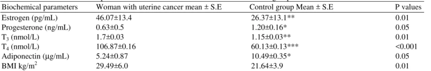
Result in Table 1 showed a significant differs at p = 0.01, p = 0.05, p<0.001, p = 0.05 and p = 0.01 of biochemical parameters (Estrogen, Progesterone, T3, T4,
adiponectin and BMI) in uterine cancer women compared with control group.
4. DISCUSSION
Although the exact cause of uterine cancer is unknown, increased levels of estrogen appear to play a role. Estrogen helps to stimulate the buildup of the lining tissue of the uterus. Study have showed that high levels of estrogen at p = 0.01 result to excessive endometrial growth and cancer as seen in Table 1 and it was showed high correlation with thyriod hormones and utrrine cancer at 0.78. thus T3 and T4 was associated with uterine cancer at p = 0.01 and p<0.001, respectively. Thyroid disorders are common cause of abnormal bleeding thus increasing the risk of uterine cancer (Sweet et al., 2012) and lead to increase lipids, because adipose tissue is working on the production large quantities of estrogen hormone (Wright et al., 2012), thus increase BMI, The International Classification BMI according to WHO showed the normal range: 18.50-24.99 kg/m2, pre-obese:
25.00-29.99 kg/m2 and obese ≥30.00 kg/m2 (WHO, 2004), also thyroid hormones decreased production of adiponectin. Circulating levels of adiponectin, a hormone with insulin-sensitizing properties, are decreased in conditions related to obesity which are recognized risk factors for endometrial cancer, because its play an important role not only in glucose and lipid metabolism but also in the development and progression of several obesity-related malignancies (Maso et al., 2004; Kelesidis et al., 2006). as the result in Table 1 Low level of progesterone reduces the immunity of woman's body, because progesterone plays a critical role in suppressing the inflammatory signals (Singh et al., 2013). Accordingly, thyroid hormones may be serve as a novel and independent prognostic parameter for patients with uterine cancer.
Most cases of uterine cancer occur between the ages of 60 and ≤70 years, but a few cases may occur before age 40 as showed in Table 2 and this corresponding with other study which found increased uterine cancer after menopause (Park et al., 2008; Barlin et al., 2010). Because high levels of thyroid hormones lead to obesity and low levels of adiponectin, adiponectin found to be significantly associated with an increased risk for endometrial cancer postmenopausal in women (Luhn et al., 2013; Erdogan et al., 2013).
Therefore the primary prevention of cancer will probably depend on modification of the factors which affect the secretion and metabolism of the responsible hormones rather than on control of exposure to classical exogenous initiators.
Table 1. Effect of biochemical parameters in women with uterine cancer and control group
Biochemical parameters Woman with uterine cancer mean ± S.E Control group Mean ± S.E P values
Estrogen (pg/mL) 46.07±13.4 26.37±13.1** 0.01
Progesterone (ng/mL) 0.63±0.5 1.20±0.16* 0.05
T3 (nmol/L) 1.7±0.03 1.15±0.03** 0.01
T4 (nmol/L) 106.87±0.16 60.13±0.13*** <0.001
Adiponectin (µg/mL) 5.24±0.87 10.49±0.35* 0.05
BMI kg/m2 29.49±6.0 21.64±3.9 0.01
*Significant differences at p = 0.05, ** Significant differences at p = 0.01, ***Significant differences at p<0.001
Table 2. Effect of biochemical parameters in women with uterine cancer according to the age
Biochemical parameters (30-39)No.= 4 (40-49) No. = 6 (50-59)No.= 6 (60-69) No.= 10 ≥70 No.= 9
Estrogen (pg/mL) 34.22±14.90 19.76±5.43* 13.50±3.82 88.21±12.93* 110.13±1.10
Progesterone (ng/mL) 0.40±0.5 1.22±0.83* 0.17±0.15 0.20±0.10* 0.16±0.9
T3 (nmol/L) 2.05±0.60 1.64±0.61 2.09±0.52* 1.86±0.70* 0.96±0.61
T4 (nmol/L) 92.27±7.16 97.30±9.16 116.68±10.22 124.43±11.20 72.61±7.17
Adiponectin (ug/mL) 5.94±0.77 4.71±0.57* 4.22±0.87* 4.27±0.57* 5.26±0.87
BMI kg/m2 28.40±1.34 28.14±1.60 28.49±4.32 29.49±6.0* 32.58±5.9*
Zena A.M. Al-Jawadi / American Journal of Applied Sciences 11 (5): 857-859, 2014
859
Science Publications AJAS
5. CONCLUSION
Novel of this research showed that high concentration of T3 and T4 especially after menopause is a risk factor for uterine cancer. Accordingly, women should pay attention for the level of thyroid hormones during menopause age and early diagnosis and treatment of thyroid disorders to reduce the risk of uterine cancer.
6. REFERENCES
Aljawadi, Z.AM. and N. Altalib, 2000. Clinical study of thyroid disease in mosul and dohuk provinces. J. Educ. Sci., 21: 15-20.
Barlin, J.N., I. Puri and R.E. Bristow, 2010. Cytoreductive surgery for advanced or recurrent endometrial cancer: A meta-analysis. Gynecol.
Oncol., 118: 14-18. DOI:
10.1016/j.ygyno.2010.04.005
Erdogan, S., S. Sezer, E. Baser, O. Gun-Eryilmaz and T. Gungor et al., 2013. Evaluating vaspin and adiponectin in postmenopausal women with endometrial cancer. Endocr. Relat. Cancer, 20: 669-675. DOI:10.1530/ERC-13-0280
Fierabracci, A., 2011. Identifying thyroid stem/progenitor cells: Advances and limitations. J Endocrinol., 213: 1-13. PMID: 22028390
Jiemin, M., W.D. Flanders, E.M. Ward and A. Jemal, 2011. Body mass index in young adulthood and premature death: Analyses of the US national health interview survey linked mortality files. Am. J. Epidemiol., 174: 934-944. DOI: 10.1093/aje/kwr169
Kelesidis, I., T. Kelesidis and C.S. Mantzoros, 2006. Adiponectin and cancer: A systematic review. Brit.
J. Cancer, 94: 1221-1225. DOI:
10.1038/sj.bjc.6603051
Kirkpatrick, L.A. and B.C. Feeney, 2012. A Simple Guide to IBM SPSS Statistics for Versions 18.0 and 19.0. 11th Edn., Wadsworth Cengage Learning, Belmont, ISBN-10: 1111352550, pp: 115.
Luhn, P., C.M. Dallal, J.M. Weiss, A. Black and W. Huang et al., 2013. Circulating adipokine levels and endometrial cancer risk in the prostate, lung, colorectal and ovarian cancer screening trial. Cancer Epidemiol. Biomarkers Prev., 22: 1304-1312. DOI:10.1158/1055-9965.EPI-13-0258
Maso, L.D., L. Dal, L.S.A. Augustin, A Karalis and R Talamini et al., 2004. Circulating adiponectin and endometrial cancer risk. J. Clin. Endocrinol. Metabolism, 89: 1160-1163. DOI: 10.1210/jc.2003-031716
Park, C.K., S. Apte, G. Acs and E. Harris, 2008. Cancer of the Endometrium. In: Abeloff’s Clinical Oncology, Abeloff, M.D., J.O. Armitage, J.E. Niederhuber and M.B. Kastan et al., (Eds)., Philadelphia, pp:
Reinehr, T., 2010. Obesity and thyroid function. Mol. Cell Endocrinol, 316: 165-71. PMID: 19540303 Schottenfeld, D. and J.F. Fraumeni, 2006. Cancer
Epidemiology and Prevention. 3rd Edn., Oxford University Press, New York, ISBN-10: 0199747970, pp: 1416.
Singh, S., J. Duggal, J. Molnar, F. Maldonado and C.P. Barsano et al., 2008. Impact of subclinical thyroid disorders on coronary heart disease, cardiovascular and all-cause mortality: A meta-analysis. Int. J. Cardiol. , 125: 41-48. PMID: 17434631
Singh, A.K., R. Chattopadhyay, B. Chakravarty and K. Chaudhury, 2013. Altered circulating levels of matrix metalloproteinases 2 and 9 and their inhibitors and effect of progesterone supplementation in women with endometriosis undergoing in vitro fertilization. Fertility Sterility, 100: 127-134. DOI:10.1016/j.fertnstert.2013.03.006 Sweet, M.G., T.A.S. Dalton, P.M. Weiss and K.P.
Madsen, 2012. Evaluation and management of abnormal uterine bleeding in premenopausal women. Am. Fam. Physician., 85: 35-43. PMID: 22230306
Villareal, D.T., G.I. Smith, D.R. Sinacore, K. Shah and B. Mittendorfer, 2011. Regular multicomponent exercise increases physical fitness and muscle protein anabolism in frail, obese, older adults. Obesity, 19: 312-318. PMID: 20489691
WHO, 2004. Appropriate body-mass index for Asian populations and its implications for policy and intervention strategies. Lancet, 363: 157-163. PMID: 14726171