Primary Cerebrocortical Cells: Role of Thyroid Hormone
Receptor Subtypes and Interactions with Retinoic Acid
and Glucocorticoids
Pilar Gil-Iba´n˜ez1,2,3, Juan Bernal1,2,3*, Beatriz Morte1,2,3*
1Instituto de Investigaciones Biome´dicas, Consejo Superior de Investigaciones Cientı´ficas (CSIC), Madrid, Spain,2Universidad Auto´noma de Madrid (UAM), Madrid, Spain, 3Center for Biomedical Research On Rare Diseases (Ciberer), Instituto de Salud Carlos III, Madrid, Spain
Abstract
The effects of thyroid hormone on brain development and function are largely mediated by the binding of 3,5,39 -triiodo-L-thyronine (T3) to its nuclear receptors (TR) to regulate positively or negatively gene expression. We have analyzed by quantitative polymerase chain reaction the effect of T3 on primary cultured cells from the embryonic mouse cerebral cortex, on the expression ofHr,Klf9,Shh,Dio3,Aldh1a1, andAldh1a3. In particular we focused on T3 receptor specificity, and on the crosstalk between T3, retinoic acid and dexamethasone. To check for receptor subtype specificity we used cerebrocortical cells derived from wild type mice and from mice deficient in thyroid hormone receptor subtypes. Receptor subtype specificity was found forDio3 andAldh1a1, which were induced by T3 only in cells expressing the T3 receptor alpha 1 subtype. Interactions of T3 with retinoic acid signaling through the control of retinoic acid metabolism are likely to be important during development. T3 had opposing influences on retinoic acid synthesizing enzymes, increasing the expression of Aldh1a1, and decreasing Aldh1a3, while increasing the retinoic acid degrading enzyme Cyp26b1. Dexamethasone increasedKlf9 and Aldh1a1 expression. The effects of T3 and dexamethasone on Aldh1a1were highly synergistic, with mRNA increments of up to 20 fold. The results provide new data on thyroid hormone regulation of gene expression and underscore the importance of thyroid hormone interactions with retinoic acid and glucocorticoids during neural development.
Citation:Gil-Iba´n˜ez P, Bernal J, Morte B (2014) Thyroid Hormone Regulation of Gene Expression in Primary Cerebrocortical Cells: Role of Thyroid Hormone Receptor Subtypes and Interactions with Retinoic Acid and Glucocorticoids. PLoS ONE 9(3): e91692. doi:10.1371/journal.pone.0091692
Editor:Michael Schubert, Laboratoire de Biologie du De´veloppement de Villefranche-sur-Mer, France
ReceivedDecember 18, 2013;AcceptedFebruary 14, 2014;PublishedMarch 11, 2014
Copyright:ß2014 Gil-Iba´n˜ez et al. This is an open-access article distributed under the terms of the Creative Commons Attribution License, which permits unrestricted use, distribution, and reproduction in any medium, provided the original author and source are credited.
Funding:Supported with grants from Plan Nacional de I+D SAF2011-25608, the Ramon Areces Foundation, and the Center for Biomedical Research on Rare Diseases (CIBERER), Instituto Carlos III, Spain. PG-I is recipient of a Junta de Ampliacio´n de Estudios (JAE) predoctoral contract from the Consejo Superior de Investigaciones Cientı´ficas (CSIC). The funders had no role in study design, data collection and analysis, decision to publish, or preparation of the manuscript.
Competing Interests:The authors have declared that no competing interests exist.
* E-mail: [email protected] (JB); [email protected] (BM)
Introduction
Thyroid hormone action on mammalian brain development is exerted through regulation of gene expression mediated by binding of the active hormone T3 to the nuclear receptors encoded by the THRA/Thra and THRB/Thrb genes [1,2]. The
THRA/Thra gene encodes the T3 binding isoform TRa1, and several non T3-binding splicing products. TheTHRB/Thrbgene encodes the T3 biding isoforms TRb1 and TRb2. The TR subtypes and isoforms have different but overlapping distribution in tissues, with distinct roles in development and physiology [3].
Hypothyroidism in rodents during the fetal and neonatal period, as well as in adult animals leads to increased or decreased expression of many genes in the cerebral cortex and other brain regions [4–6]. Expression changes of some of these genes have been used as sensitive indicators of the impact on the brain of situations leading to altered metabolism and/or transport of thyroid hormones, such as inactivation of the deiodinase genes
Dio2 and Dio3, thyroid hormone transporter genes Mct8 and
Oatp1c1, and the receptor genesThraandThrb, in addition to hypo or hyperthyroidism [5–9]. These studies have produced data that
in some cases are difficult to explain, and reflect the complexity of the physiological mechanisms regulating gene expression in vivo. For example, inactivation of the Mct8 gene which leads to restricted entry of T3 through the blood-brain barrier has a limited effect on cerebral cortex gene expression postnatally. This is due to up regulation of Dio2 and increased local T3 formation. Consequently, compound inactivation ofMct8and Dio2leads to a situation more similar to hypothyroidism, but surprisingly some genes profoundly affected in the hypothyroid brain such asAldh1a1
are not affected by concomitantMct8and Dio2inactivation [5]. Single inactivation of the Dio2 gene in mice leads to brain T3 concentrations similar to hypothyroidism, but relatively minor effects on gene expression [10]. More recently it was found that
Dio2 inactivation preferentially affects expression of the genes regulated negatively by T3 rather than the positively regulated genes. The latter observation suggests that gene expression is also dependent on the route of entry of T3 to the brain [5]. Paradoxical effects ofDio3inactivation and T3 administration on cortex gene expression were also found [9]. Positive gene such asHr,Kcnj10,
genes such asCirbp,Fxyd6,Gpc3, and others were sensitive toDio3
inactivation but resistant to T3 administration. These results could not be explained by a simple mechanism of neuronal T3 increase by decreased degradation in theDio3KOmice, and indicate that the source of T3 is also important.
On the other hand, single or compound inactivation of the mouse Thraand Thrb genes leads to mild perturbations of gene expression compared to hypothyroidism. In a recent study [6] we found that from 27 cortex and striatal genes which expression was reduced by hypothyroidism only around 10 were reduced by TRa1 deficiency and 6 were increased by TRb deficiency. Inactivation of both receptor genes increased the number of affected genes to 17, and there was no correlation between the effects of hypothyroidism and the effects of receptor gene inactivation. In general the conclusion of these studies was that in the absence of TRa1 thyroid hormone-dependent gene expression can be largely maintained by TRb, and that the effects of hypothyroidism are largely due to the transcriptional effects of unliganded TR.
It is likely that thyroid hormone action on the brainin vivo is modulated by many interacting physiological factors acting on micro domains. The bulk effect observedin vivoin individual brain regions would be an aggregate result of the interaction of T3 with additional factors that may be modulated differently in each micro domain, depending on the cellular composition, expression of receptors, transporters, deiodinases, and the crosstalk with other signaling pathways. To better understand the role of different factors in the action of thyroid hormone on neural gene expression we have analyzed the expression of some selected genes in primary cultures of mouse cerebrocortical cells. Specifically we studied the effects of TR knockdown in vivo on the effects of T3 to better define the relative roles of TRa1 and TRb. In addition we studied the crosstalk between thyroid hormone and retinoic acid, or glucocorticoids, as it is known that the effects of these hormonal systems are frequently interrelated. For example, retinoic acid and thyroid hormone may regulate the expression of some genes through similar responsive elements [11–13]. Examples of genes regulated by T3 and glucocorticoids at the transcriptional level include GH [14–17], cholesterol 7a-hydrolase [18], liver cytosolic PEPCK [19], FAS [20], Pdk4[21], andKlf9 [22], among other genes.
Materials and Methods
Ethics statement: All experimental procedures involving animals were performed following the European Union Council guidelines (directive 2010/63/UE) and Spanish regulations (R.D.1201/2005, and Law 32/2007). They were approved by the ethics committee of our institution (Consejo Superior de Investigaciones Cientı´ficas, CSIC; approval number SAF2011–25608).
Animals were housed in temperature (2262uC) and light (12:12 light-dark cycle; lights on at 7 a.m.) controlled conditions and had free access to food and water. Experiments involving comparisons between genotypes were from the strains of mice described elsewhere [6]. For the effects of dexamethasone (DEX) and retinoic acid (RA) we used cultures derived from mice of the C57BL/6J strain.
Pregnant dams were euthanized with CO2 on gestation day 17.5, and the fetuses were extracted and euthanized by decapitation. The cerebral cortices were dissected at in PBS containing 1% BSA and 0.1% Glucose. The tissue was disaggre-gated by enzymatic digestion with 0.4 mg/ml Papain (Roche) and passing through a 0.9-mm syringe in the presence of DNAse I. The homogenate was centrifuged and the cells were resuspended
in serum free culture medium (Neurobasal Medium supplemented with 2% B27, Glutamax, 10 U/ml Penicillin, and 10 U/ml Streptomycin). The cells were seeded on poly-L-ornithine-coated 12-well multiwells (Sigma). The wells were previously preincubat-ed with a mixture of Horse Serum and culture mpreincubat-edium (1:1), and Laminin (1mg/ml) (Sigma). Cells were added to this media in culture medium (66105cells per well). After 9 days, the cells were incubated 24 h in the same medium without B27 supplement before adding the different treatments: 1 nM T3, 1mM RA, or 10 nM DEX. Incubation times are indicated in the figure legends. Control cultures without treatment were incubated in parallel. To examine the response to T3 in the presence of inhibition of protein synthesis, cycloheximide (CHX, Sigma) was added to the cultures at a concentration of 8mg/ml 30 min before T3 (10 nM) and the cells were harvested 6 hours after T3 addition.
The composition of the primary cultures was analyzed by immunofluorescence as follows: Cells plated on glass coverslips were fixed with absolute acetone for 7 min at 220uC. After permeabilization for 5 min in 0.5% Triton X-100 in PBS, the cells were incubated for 1 h in 1% BSA 2% cold fish skin gelatin (Sigma) 0.05% Triton X-100 in PBS to block non-specific antibody binding sites. For immunofluorescence labeling, the cells were stained by overnight incubation at 4 C in the blocking buffer with the following combination of primary antibodies dilut-ed1:500: monoclonal anti glial fibrillary acidic protein (Clone G-A-5, Sigma) for astrocytes and rabbit polyclonal anti-NeuN [23] (Millipore) for neurons. The secondary antibodies were donkey anti mouse Alexa 488 (green) and donkey anti rabbit Alexa 555 (red) and were used at 1:500 dilution. Cells were then washed in PBS and incubated for 10 min with 49 ,6-diamidino-2-phenylin-dole (DAPI, GibcoHLife Technologies) 0.1mg/ml in PBS to label the nucleus. Omitting the first antibodies in the incubation reaction gave no signal. Confocal images were acquired using an inverted Zeiss LSM 710 laser scanning microscope with a plan-apochromatic objective 636/N.A 1.3. Sequential scanning mode was used to avoid crosstalk between channels. All images shown correspond to the maximum intensity projection of a z-stack. Images were processed with Zen 2009 software and Adobe Photoshop (Figure S1). To calculate the relative abundance of neurons and astrocytes in the different cultures antibody-labeled cells and DAPI-stained nuclei were counted in photographs taken using a 406objective. A total number of 100–200 cells were counted in sextuplicate for each culture. The relative number of neurons and astrocytes were calculated as a percentage of DAPI-stained nuclei. The results are presented in Table S1. These cultures are enriched in neurons and there were no differences in the % cells stained as neurons in the three cultures. The % astrocytes were slightly higher in the TRa12/2and the TRb2/2
than in the Wt, although only statistically significant when comparing Wt and TRa12/2. As in other studies [24] less than 10% of the cells were not stained as astrocytes or neurons.
Total RNA was isolated using the Trizol procedure (Invitrogen, Carlsbad, CA). 250 ng RNA was used as template for the cDNA synthesis with the high-capacity cDNA reverse transcription kit (Applied Biosystems, Foster City, CA). PCR assays were performed using SYBR Green or TaqMan assays (Applied Biosystems) on a 7900 HT fast real-time PCR system. The PCR program consisted in a hot start of 95uC for 10 min, followed by 40 cycles of 15 s at 95uC and 1 min at 60uC. Expression ofRARb,
59CTGTTAGCACCCTGTCAAAG39; Cyp26b1 forward 59
TA-CAGGGTTCCGGCTTCCAG39; Cyp26b1 reverse
59GCACCGGTCACACGAATCAG3. For analysis we used the 2-Ct method. For internal control RNAs we compared 18S RNA and Cyclophylin (Ppia) mRNA. Either control gave similar results (Figure S2). For the final calculations the data were corrected for 18S RNA and expressed relative to the value obtained for the control cells, which was given a value of 1.0.
To calculate significance of differences between means we used one- or two-way ANOVA, or two-tailed, unpaired Student’st-test depending on the experiments. Calculations and graphics were performed using the GraphPad Prism software (www.graphpad. com/prism/).
Results
The goal of this work was to analyze the effect of T3 on gene expression in cerebrocortical primary cultures. As T3 targets we selected genes previously known to be thyroid hormone dependent
in brain, and that likely play a prominent role in mediating the effects of thyroid hormones on neural development, as follows: 1) the transcription factor and T3 receptor co-repressorHr(Hairless), which was originally shown to be under transcriptional regulation by T3 in the cerebellum [25].Hr has since then been a widely studied T3 target to analyze the effects of thyroid hormone on the brain. 2) The developmental morphogen Shh (Sonic hedgehog), which is regulated by thyroid hormone in the rat and mouse embryonic and adult brain, presumably at the transcriptional level [26]. 3)The transcription factor Klf9 (Kru¨pfel-like transcription factor 9, also known as Basic Transcription Element Binding Protein or BTEB), which is regulated by thyroid hormone at the transcriptional level during tadpole metamorphosis, oligodendro-cyte differentiation, and in the rodent brain [27]. 4)Dio3, which is regulated specifically by TRa1 [28]. 5) Aldh1a1 (Aldehyde dehydrogenase 1a1), a gene sensitive to hypothyroidism produced by blocking thyroid hormone formation, but not by brain hypothyroidism resulting from Mct8 and Dio2 inactivation, as explained in the Introduction [5]. 6)Aldh1a3, a gene negatively Figure 1. Effect of T3 on gene expression in primary mouse cerebrocortical cell cultures in the presence or absence of TRa1 or TRb. Cells (n = 6) from wild type (Wt), TRa1 KO (aKO), or TRbKO (bKO) mice were incubated for 24 hours in the absence (open bars) or in the presence of 1 nM T3 (filled bars). Statistical analysis was by two-way ANOVA; ns = P.0.05; * = P,0.05; ** = P,0.01; *** = P,0.001.
regulated by T3 [29], which participates in the same retinoic acid synthesis pathway asAldh1a1[30]. To search for clues that may explain the puzzling behaviors mentioned above we analyzed the thyroid hormone receptors subtypes specificity and also the effects of other factors such as retinoic acid, and glucocorticoids and their interactions with T3.
The relative role of receptor subtypes in the gene responses to T3 is illustrated in Figure 1. For this experiment we isolated E17.5 cerebrocortical cells from Wt mice, TRa1 KO mice, and TRb
KO mice. Gene expression was measured in the absence of T3, and 24 h after addition of 1 nM T3 to the cultures. In previous experiments (not shown) we found that increasing the T3 concentration up to 100 nM did not increase the response above that attained with 1 nM T3. As an experimental control of the effect of receptor inactivation we measured the expression ofDio3
(encoding the type 3 deiodinase), which is regulated by T3 in a TRa1 isoform-specific way.Dio3expression in cells lacking TRa1 or TRbwas similar to Wt cells in the absence of T3. Addition of T3 increasedDio3expression by 4-fold in the Wt cells, and had no effect on the TRa1-deficient cells. TRbdeficiency potentiated the effect of T3, with an 8-fold induction, to twice the level obtained in the Wt cells. From the rest of the genes studied, onlyAldh1a1was unresponsive in cells deficient of TRa1. T3 increased Aldh1a1
expression in the Wt cells, but had no effect on the TRa1-deficient cells. In the TRb-deficient cells the basal expression ofAldh1a1in the absence of T3 was increased with respect to the untreated Wt cells, and was further increased by T3.
Hr,Klf9, andShhshowed no TR isoform specificity, and were induced by T3 in the three culture types.HrandShhwere induced by 5-fold and 6-fold, respectively in Wt cells and about 2-fold in the TRa1-deficient cells. SimilarlyKlf9increased 2-fold after T3 addition to the Wt cells and 1.3-fold in the TRa1-deficient cells. In the TRb-deficient cells T3 induced a similar effect as in the Wt cells onShh, but the effects onHrandKlf9were increased.
The Aldh1a1 enzyme is involved in RA synthesis, catalyzing conversion of all-trans-retinal to all-trans-retinoic acid. Another aldehyde dehydrogenase Aldh1a3 is also involved in this pathway. In contrast to Aldh1a1, Aldh1a3 is negatively regulated by T3 (Figure 1), with a 40% reduction after T3 addition to the Wt cells. TRa1-deficient cells behave exactly as the Wt cells. In the absence of TRb the basal expression increased by about 60% and T3 induced a similar effect as in the Wt cells.
To check whether the effects of T3 were due to a direct action on gene transcription, or mediated through increased expression of T3-dependent auxiliary proteins or transcription factors, we analyzed the effect of T3 in a shorter time, in the presence of the protein synthesis inhibitor CHX (Figure 2). After 6 hours in the presence of T3 an increased expression ofHr,Klf9, andShh was already observed. The effect on any of these genes was not blocked by CHX pretreatment, indicating a direct effect of T3 on these genes. In contrast T3 had no significant effects onDio3, Aldh1a1
and Aldh1a3 at 6 hours. Therefore no conclusions about the mechanism of induction could be made for Dio3 and Aldh1a1. CHX had a strong stabilizing effect onAldh1a3mRNA, and in the cells pretreated with CHX T3 was able to reduce Aldh1a3
expression, suggesting that also for this gene the effect was direct. Since Aldh1a1 and Aldh1a3 are involved in RA synthesis we analyzed the effect of RA and a possible synergism with T3. As controls for the effects of RA we measured the expression of two RA-target genes, the RA receptorb,RARband the RA degrading enzymeCyp26b1(Figure 3) [31].RARbwas increased by RA but not by T3. When added together the presence of T3 inhibited the effect of RA. Interestingly we found that Cyp26b1was positively regulated by T3, with an effect quantitatively similar to that
obtained with RA. There was no additive effect when both agents were added together. The relative effects of RA and T3 onAldh1a1
were a mirror image of those onRARb.Aldh1a1was increased by T3 but without effect of RA, but RA inhibited the effect of T3 when added together. Aldh1a3 was down regulated by T3 and there was no effect of RA.
Many thyroid hormone regulated genes are also influenced by glucocorticoids, and in some instances there is synergism between the two hormones. Among the genes analyzed in this work, it is known thatKlf9in tadpoles is synergistically regulated by T3 and glucocorticoids at the transcriptional level. Indeed we found that also on Wt cerebrocortical cells (Figure 4) DEX stimulatedKlf9
expression, similarly to T3, and there was a synergistic effect when both hormones were added together.Aldh1a1expression was also stimulated by dexamethasone, in addition to T3. In the presence of both hormones there was a strong synergistic effect of T3 and DEX andAldh1a1 expression was increased 20-fold. To explore possible mechanisms for the interaction between T3 and DEX we measured the effect of DEX on the TR heterodimeric partner RXRa, as it has been reported that DEX increases the expression of RXRa [32]. Indeed we found that DEX increased RXRa
expression (Figure 4), but not RXRc (not shown). In TRa 1-deficient cells T3 and DEX also stimulatedKlf9expression, as was expected due to lack of receptor specificity. However and in agreement with the previous finding, TRa1 was needed for the effect of T3 onAldh1a1expression, since the response to T3 in cells derived from TRa1 KO mice was suppressed, whereas the response to DEX was maintained. Although T3 alone had no Figure 2. Effect of T3 in the presence or absence of cycloheximide (CHX) on gene expression in primary mouse cerebrocortical cell cultures from wild type mice.The cells (n = 4) were incubated for 30 min with or without CHX (8mg/ml) before adding T3 (10 nM), and then incubated for 6 hours. Statistical analysis was by one-way ANOVA; ns = P.0.05; * = P,0.05; ** = P,0.01; *** = P, 0.001.
doi:10.1371/journal.pone.0091692.g002
effect in the absence of TRa1, it increased the response to dexamethasone.
Among the possible mechanisms by which T3 potentiates the effect of DEX in the absence of TRa1, is an increased expression of the glucocorticoid receptor. To test this hypothesis we measured the effect of T3 on the glucocorticoid receptor mRNANr3c1in Wt cells and in TRa1-deficient cells (Figure 4). In both types of cells T3 increasedNr3c1expression by 30–50%. DEX alone decreased
Nr3c1expression by 25%, but not when added together with T3. The effect of T3 was not observed at shorter times of incubation, i.e., 6 hours, and we cannot conclude that it is a direct action of the hormone (not shown).
Discussion
In this work we have analyzed the contribution of factors others than T3 itself, that might be involved in the regulation of gene expression and that may play diverse roles in vivo. This experi-mental set up allowed us to confirm that genes which expression in the mouse cerebral cortex [6] is strongly dependent on thyroid hormones are indeed cellular targets of T3. The altered expression previously observedin vivoafter induction of hypothyroidism was not due to a distal effect of the hypothyroid condition, but reflected a cellular effect of the hormone. The cellular system employed in this work is based on the culture of primary cells isolated from the embryonic mouse brain. These cultures are enriched in neurons but also contain astrocytes and unidentified cells (Table S1, Figure S1 and [24]). Therefore any effect observed by hormonal manipulations of these cells could have its origin in neurons or in astrocytes, depending among other factors on the relative expression of the target gene in each of these cell types.
First we analyzed the specific contribution of the TRa1 and TRb receptor subtypes by measuring the response to T3 of primary cells derived from the cerebral cortex of TRa1 and TRb
KO mice. The results confirm [6] that at the cellular level both TRa1 and TRb mediate the effects of T3, with two exceptions,
Dio3 and Aldh1a1. Dio3 was already shown to be regulated specifically by TRa1 [28], and we confirm this fact in the cultured neurons. Also Aldh1a1 appears to be regulated specifically by TRa1, since no induction by T3 was observed in cells derived from TRa1 KO mice.Klf9and Aldh1a3show no preference for each of the TR subtypes. A recent global analysis of TR specificity in HeLa cells expressing exogenous TRa1 or TRb1 [33] concluded that there are no complete TR subtype specificity, although TRa1 or TRb1 showed some gene preferences, depending on the time of exposure and the dose of T3. In established neural cell lines expression TRa1 or TRb1 lead to substantial differences in the gene network regulated by T3, without correlation with differential chromatin occupancy [34].
Direct transcriptional regulation by T3, has previously been shown for Hr [25], Klf9 [27,35] Dio3 [28], and Shh [26]. Chromatin occupancy by the TR has been shown for Hr and
Klf9in established neural cell lines [34]. Here we confirm that also in cerebrocortical primary cells Hr, Klf9, and Shh are direct transcriptional targets of T3, given that the effect of the hormone was not blocked by previous treatment with CHX to inhibit protein synthesis. We could not determine whetherDio3was also transcriptionally regulated by T3 because it was not stimulated by T3 at 6 hours of incubation, preventing to test the effect of CHX. However, a specific TRa1 binding site has been demonstrated in the upstream region of the gene [28]. The fact that we could not determine whether the effect of T3 was direct, suggest the possibility that the full effect of T3 requires interaction with other intermediate proteins.Dio3is a transcriptional target ofShh [36] which, as mentioned above is a direct T3 target. This raises the possibility that rapid and full induction ofDio3by T3 is the result Figure 3. Effects of T3 and all-trans retinoic acid (RA) on gene expression in primary mouse cerebrocortical cell cultures from wild type mice.The cells (n = 4) were incubated for 48 hours in the absence or presence of T3 (1 nM), RA (1mM) or both. Statistical analysis was by one-way ANOVA; ns = P.0.05; *** = P,0.001.
of a direct transcriptional effect of TRa1 potentiated by the T3-dependent accumulation of Shh protein.
A relevant new finding is the influence of T3 on the expression of genes involved in the synthesis and degradation of RA. We had previously observed a reduction ofAldh1a1expression in the brain of hypothyroid mice. By stimulatingAldh1a1expression T3 would increase RA synthesis. However, this effect would be counteracted by down regulation ofAldh1a3acting in the same pathway, and by up regulation of Cyp26b1, a gene encoding a RA degrading enzyme. During development Aldh1a3 expression is maximal at E14, and decreases to very low levels near birth, whereasAldh1a1
rises postnatally in the telencephalon [37]. It is likely that these changes of expression are influenced by the thyroid hormone concentrations and TR expression changes occurring during development. In addition to this bulk effect, the net effect of thyroid hormone on RA production should depend on the relative expression of each enzyme in specific brain regions. Since the effect of T3 on Aldh1a1 expression is TRa1 dependent, the net effect of T3 in specific cells would depend on the relative concentrations of TRa1 and TRb. Aldh1a1 is involved in the regulation of the total number of dopaminergic neurons [38] and in the metabolism of dopamine and norepinephrine [39].Aldh1a3
is expressed in the ganglionic eminence where RA and thyroid hormone influence tangential neuronal migrations leading to the generation of cerebral cortex interneurons [40,41]. These effects are likely to be important in the regulation of developmental events by T3.
The effect of T3 onAldh1a1expression is also sensitive to RA. Although at the doses used RA in isolation does not influence
Aldhla1expression, it inhibits the effect of T3. On the contrary, the RARbresponse to RA is reduced by the presence of T3.Cyp26b1, which is sensitive to RA and T3, does not show an additive effect in the presence of both compounds, which might also be due to mutual interference. In this respect it is likely that the combined responses to RA and T3 are modulated by COUP-TF1, which is widely expressed in the nucleus of primary neurons (unpublished results from our laboratory). In neurons derived from embryonic stem cells COUP-TF1 represses T3 induction of the T3 target gene Camk4 and confers RA sensitivity. This effect is mediated through a regulatory element containing overlapping binding sites for TR, RAR, and COUP-TF1 [13].
In our primary cultures Klf9 and Aldh1a1 expression were increased by DEX which also potentiated the effects of T3.Klf9is induced by corticosterone in the tadpole brain and in theXenopus Figure 4. Effects of T3 and dexamethasone (DEX) on gene expression in primary mouse cerebrocortical cell cultures from wild type mice or from TRa1 KO mice.The cells (n = 4) were incubated for 48 hours in the absence or presence of T3 (1 nM), DEX (10 nM) or both. Statistical analysis was by one-way ANOVA; ns = P.0.05; * = P,0.05; ** = P,0.01; *** = P,0.001.
doi:10.1371/journal.pone.0091692.g004
laevisfibroblast cell line XTC-2, and is proposed to mediate effects of glucocorticoids on neuronal structure and function [22]. To the best of our knowledge,Aldh1a1was not known to be induced by glucocorticoids. DEX had an effect comparable to that of T3. But when added together there was a synergistic effect and Aldh1a1
expression increased by 20-fold, the largest increment effected by T3 that we have knowledge on any gene in neural cells.
A summary of the crosstalk between T3, RA and glucocorticoid signaling, as described in the present work is illustrated in Figure 5. These interactions are likely to be operating in different cell population micro domains at different stages of development. The net result would be a function of the expression of the different components present in the figure as the cells undergo their differentiation program.
Supporting Information
Figure S1 Confocal images of the cerebrocortical cul-tures from Wt, TRa1KO and TRbKo mice.Upper panels: Cells stained with antibodies against GFAP and NeuN. Lower panels: Nuclei stained with DAPI. Scale bar = 25mm.
(TIF)
Figure S2 Correlation between qPCR measurements using as internal control 18S RNA or Cyclophylin RNA (Ppia).Correlation coefficient was 0.95, P,0.0001.
(TIF)
Table S1 Cellular composition of the cerebrocortical primary cultures. Shown are the percentage of neurons and astrocytes relative to the total number of DAPI-stained nuclei. Data are mean6 SD (n = 6), and 95% Confidence Interval (CI). (*): P, 0.05 compared to Wt. Other comparisons were not significant. (DOCX)
Acknowledgments
We acknowledge the technical assistance of Irene Cuevas, Diana Perez, Diego Navarro and Lucia Sa´nchez.
Author Contributions
Conceived and designed the experiments: PG-I JB BM. Performed the experiments: PG-I. Analyzed the data: PG-I JB BM. Wrote the paper: JB BM.
References
1. Bernal J (2007) Thyroid hormone receptors in brain development and function. Nat Clin Pract Endocrinol Metab 3: 249–259.
2. Cheng SY, Leonard JL, Davis PJ (2010) Molecular aspects of thyroid hormone actions. Endocr Rev 31: 139–170.
3. Flamant F, Gauthier K (2013) Thyroid hormone receptors: the challenge of elucidating isotype-specific functions and cell-specific response. Biochim Biophys Acta 1830: 3900–3907.
4. Diez D, Grijota-Martinez C, Agretti P, De Marco G, Tonacchera M, et al. (2008) Thyroid hormone action in the adult brain: gene expression profiling of the effects of single and multiple doses of triiodo-L-thyronine in the rat striatum. Endocrinology 149: 3989–4000.
5. Morte B, Ceballos A, Diez D, Grijota-Martinez C, Dumitrescu AM, et al. (2010) Thyroid hormone-regulated mouse cerebral cortex genes are differentially dependent on the source of the hormone: a study in monocarboxylate transporter-8- and deiodinase-2-deficient mice. Endocrinology 151: 2381–2387. 6. Gil-Ibanez P, Morte B, Bernal J (2013) Role of thyroid hormone receptor subtypes alpha and beta on gene expression in the cerebral cortex and striatum of postnatal mice. Endocrinology 154: 1940–1947.
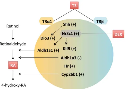
7. Liao XH, Di Cosmo C, Dumitrescu AM, Hernandez A, Van Sande J, et al. (2011) Distinct roles of deiodinases on the phenotype of Mct8 defect: a comparison of eight different mouse genotypes. Endocrinology 152: 1180–1191. Figure 5. A summary of the interactions between T3 receptor subtype, and retinoic acid and glucocorticoid signaling in the T3-mediated control of gene expression described in this work.T3 induces up regulation (+) or down regulation (2) of gene expression. Specificity of TR subtypes is depicted by a color gradient.Dio3andAldh1a1require TRa1, but no clear preferences were detected for other genes. Transcriptionally direct T3 responses includeShh, Hr, andKlf9. Induction ofShhis probably involved inDio3regulation by T3. T3 regulates the expression of enzymes involved in the synthesis and degradation of retinoic acid (RA). The effect of T3 onKlf9and onAldh1a1is potentiated by glucocorticoids, in part by up regulation of the glucocorticoid receptorNr3c1.
8. Mayerl S, Visser TJ, Darras VM, Horn S, Heuer H (2012) Impact of Oatp1c1 deficiency on thyroid hormone metabolism and action in the mouse brain. Endocrinology 153: 1528–1537.
9. Hernandez A, Morte B, Belinchon MM, Ceballos A, Bernal J (2012) Critical role of types 2 and 3 deiodinases in the negative regulation of gene expression by T(3)in the mouse cerebral cortex. Endocrinology 153: 2919–2928.
10. Galton VA, Wood ET, St Germain EA, Withrow CA, Aldrich G, et al. (2007) Thyroid hormone homeostasis and action in the type 2 deiodinase-deficient rodent brain during development. Endocrinology 148: 3080–3088.
11. Bedo G, Santisteban P, Aranda A (1989) Retinoic acid regulates growth hormone gene expression. Nature 339: 231–234.
12. Castillo AI, Sanchez-Martinez R, Moreno JL, Martinez-Iglesias OA, Palacios D, et al. (2004) A permissive retinoid X receptor/thyroid hormone receptor heterodimer allows stimulation of prolactin gene transcription by thyroid hormone and 9-cis-retinoic acid. Mol Cell Biol 24: 502–513.
13. Liu YY, Brent GA (2002) A complex deoxyribonucleic acid response element in the rat Ca(2+)/calmodulin-dependent protein kinase IV gene 59-flanking region mediates thyroid hormone induction and chicken ovalbumin upstream promoter transcription factor 1 repression. Mol Endocrinol 16: 2439–2451.
14. Barinaga M, Yamonoto G, Rivier C, Vale W, Evans R, et al. (1983) Transcriptional regulation of growth hormone gene expression by growth hormone-releasing factor. Nature 306: 84–85.
15. Evans RM, Birnberg NC, Rosenfeld MG (1982) Glucocorticoid and thyroid hormones transcriptionally regulate growth hormone gene expression. Proc Natl Acad Sci U S A 79: 7659–7663.
16. Yaffe BM, Samuels HH (1984) Hormonal regulation of the growth hormone gene. Relationship of the rate of transcription to the level of nuclear thyroid hormone-receptor complexes. J Biol Chem 259: 6284–6291.
17. Diamond DJ, Goodman HM (1985) Regulation of growth hormone messenger RNA synthesis by dexamethasone and triiodothyronine. Transcriptional rate and mRNA stability changes in pituitary tumor cells. J Mol Biol 181: 41–62. 18. Pandak WM, Heuman DM, Redford K, Stravitz RT, Chiang JY, et al. (1997)
Hormonal regulation of cholesterol 7alpha-hydroxylase specific activity, mRNA levels, and transcriptional activity in vivo in the rat. J Lipid Res 38: 2483–2491. 19. Hanson RW, Reshef L (1997) Regulation of phosphoenolpyruvate
carboxyki-nase (GTP) gene expression. Annu Rev Biochem 66: 581–611.
20. Lu Z, Gu Y, Rooney SA (2001) Transcriptional regulation of the lung fatty acid synthase gene by glucocorticoid, thyroid hormone and transforming growth factor-beta 1. Biochim Biophys Acta 1532: 213–222.
21. Attia RR, Connnaughton S, Boone LR, Wang F, Elam MB, et al. (2010) Regulation of pyruvate dehydrogenase kinase 4 (PDK4) by thyroid hormone: role of the peroxisome proliferator-activated receptor gamma coactivator (PGC-1 alpha). J Biol Chem 285: 2375–2385.
22. Bonett RM, Hu F, Bagamasbad P, Denver RJ (2009) Stressor and glucocorticoid-dependent induction of the immediate early gene kruppel-like factor 9: implications for neural development and plasticity. Endocrinology 150: 1757–1765.
23. Mullen RJ, Buck CR, Smith AM (1992) NeuN, a neuronal specific nuclear protein in vertebrates. Development 116: 201–211.
24. Ruiz F, Alvarez G, Ramos M, Hernandez M, Bogonez E, et al. (2000) Cyclosporin A targets involved in protection against glutamate excitotoxicity. Eur J Pharmacol 404: 29–39.
25. Thompson CC (1996) Thyroid hormone-responsive genes in developing cerebellum include a novel synaptotagmin and a hairless homolog. J Neurosci 16: 7832–7840.
26. Desouza LA, Sathanoori M, Kapoor R, Rajadhyaksha N, Gonzalez LE, et al. (2011) Thyroid hormone regulates the expression of the sonic hedgehog signaling pathway in the embryonic and adult Mammalian brain. Endocrinology 152: 1989–2000.
27. Denver RJ, Williamson KE (2009) Identification of a thyroid hormone response element in the mouse Kruppel-like factor 9 gene to explain its postnatal expression in the brain. Endocrinology 150: 3935–3943.
28. Barca-Mayo O, Liao XH, Alonso M, Di Cosmo C, Hernandez A, et al. (2011) Thyroid hormone receptor alpha and regulation of type 3 deiodinase. Mol Endocrinol 25: 575–583.
29. Gil-Iban˜ez P, Bernal J, Morte B (2012) Thyroid hormone action on cerebrocortical neurons in primary culture. 94th Annual Meeting of the Endocrine Society, Abstract#Houston, Texas, June 23–26.
30. Kumar S, Sandell LL, Trainor PA, Koentgen F, Duester G (2012) Alcohol and aldehyde dehydrogenases: retinoid metabolic effects in mouse knockout models. Biochim Biophys Acta 1821: 198–205.
31. Samarut E, Rochette-Egly C (2012) Nuclear retinoic acid receptors: conductors of the retinoic acid symphony during development. Mol Cell Endocrinol 348: 348–360.
32. Steineger HH, Arntsen BM, Spydevold O, Sorensen HN (1997) Retinoid X receptor (RXR alpha) gene expression is regulated by fatty acids and dexamethasone in hepatic cells. Biochimie 79: 107–110.
33. Lin JZ, Sieglaff DH, Yuan C, Su J, Arumanayagam AS, et al. (2013) Gene specific actions of thyroid hormone receptor subtypes. PLoS One 8: e52407. 34. Chatonnet F, Guyot R, Benoit G, Flamant F (2013) Genome-wide analysis of
thyroid hormone receptors shared and specific functions in neural cells. Proc Natl Acad Sci U S A 110: E766–775.
35. Furlow JD, Kanamori A (2002) The transcription factor basic transcription element-binding protein 1 is a direct thyroid hormone response gene in the frog Xenopus laevis. Endocrinology 143: 3295–3305.
36. Dentice M, Luongo C, Huang S, Ambrosio R, Elefante A, et al. (2007) Sonic hedgehog-induced type 3 deiodinase blocks thyroid hormone action enhancing proliferation of normal and malignant keratinocytes. Proc Natl Acad Sci U S A 104: 14466–14471.
37. Smith D, Wagner E, Koul O, McCaffery P, Drager UC (2001) Retinoic acid synthesis for the developing telencephalon. Cereb Cortex 11: 894–905. 38. Jacobs FM, Smits SM, Noorlander CW, von Oerthel L, van der Linden AJ, et al.
(2007) Retinoic acid counteracts developmental defects in the substantia nigra caused by Pitx3 deficiency. Development 134: 2673–2684.
39. Anderson DW, Schray RC, Duester G, Schneider JS (2011) Functional significance of aldehyde dehydrogenase ALDH1A1 to the nigrostriatal dopamine system. Brain Res 1408: 81–87.
40. Crandall JE, Goodman T, McCarthy DM, Duester G, Bhide PG, et al. (2011) Retinoic acid influences neuronal migration from the ganglionic eminence to the cerebral cortex. J Neurochem 119: 723–735.
41. Cuevas E, Auso E, Telefont M, Morreale de Escobar G, Sotelo C, et al. (2005) Transient maternal hypothyroxinemia at onset of corticogenesis alters tangential migration of medial ganglionic eminence-derived neurons. Eur J Neurosci 22: 541–551.