Marker of Circulating Tumor DNA in Cervical Cancer
Patients
Maura Campitelli1, Emmanuelle Jeannot2, Martine Peter2, Emmanuelle Lappartient2, Ste´phanie Saada2, Anne de la Rochefordie`re1, Virginie Fourchotte3, Se´verine Alran3, Peter Petrow4, Paul Cottu5, Jean-Yves Pierga5, Olivier Lantz2, Je´roˆme Couturier2, Xavier Sastre-Garau2*
1Department of Radiation Oncology, Institut Curie, Hospital, Paris, France,2Department of Biopathology, Institut Curie, Hospital, Paris, France,3Department of Surgical Oncology, Institut Curie, Hospital, Paris, France,4Department of Radiology, Institut Curie, Hospital, Paris, France,5Department of Medical Oncology, Institut Curie, Hospital, Paris, France
Abstract
Introduction:In most cases of cervical cancers, HPV DNA is integrated into the genome of carcinoma cells. This mutational insertion constitutes a highly specific molecular marker of tumor DNA for every patient. Circulating tumor DNA (ctDNA) is an emerging marker of tumor dynamics which detection requires specific molecular motif. To determine whether the sequence of the cell-viral junction could be used in clinical practice as a specific marker of ctDNA, we analyzed a series of cervical cancer patient serums.
Methods and Findings:Serum specimens of 16 patients diagnosed with HPV16/18-associated cervical cancer, and for which the viral integration locus had been previously localized, were analyzed. Sequential serum specimens, taken at different times during the course of the disease, were also available for two of these cases. ctDNA was found in 11 out of 13 patients with tumor size greater than 20 mm at diagnosis, and analysis of sequential serum specimens showed that ctDNA concentration in patients serum was related to tumor dynamics.
Conclusions: We report that HPV mutational insertion constitutes a highly specific molecular marker of ctDNA in HPV-associated tumor patients. Using this original approach, ctDNA was detected in most cervical cancer patients over stage I and ctDNA concentration was found to reflect tumor burden. In addition to its potential prognostic and predictive value, HPV mutation insertion is likely to constitute a new molecular surrogate of minimal residual disease and of subclinical relapse in HPV-associated tumor. This is of major importance in the perspective of specific anti-HPV therapy.
Citation:Campitelli M, Jeannot E, Peter M, Lappartient E, Saada S, et al. (2012) Human Papillomavirus Mutational Insertion: Specific Marker of Circulating Tumor DNA in Cervical Cancer Patients. PLoS ONE 7(8): e43393. doi:10.1371/journal.pone.0043393
Editor:Rui Medeiros, IPO, Inst Port Oncology, Portugal
ReceivedFebruary 23, 2012;AcceptedJuly 20, 2012;PublishedAugust 24, 2012
Copyright:ß2012 Campitelli et al. This is an open-access article distributed under the terms of the Creative Commons Attribution License, which permits unrestricted use, distribution, and reproduction in any medium, provided the original author and source are credited.
Funding:This work was supported in part by a partnership with La Maison Goyard and by a generous gift from Ms. A. Mauresmo. The funders had no role in study design, data collection and analysis, decision to publish, or preparation of the manuscript.
Competing Interests:This work was supported in part by the partnership of La Maison Goyard, luxury luggage-maker, through the event ‘Un Bagage pour Curie’. This does not alter the authors’ adherence to all the PLOS ONE policies on sharing data and materials.
* E-mail: [email protected]
Introduction
Specific types of human papillomaviruses (HPV) are recognized as the major etiological factor in cervical neoplasia [1]. In pre-invasive lesions, the viral genomes are present as episomal molecules in the nucleus of infected cells. In most cases of invasive carcinoma, however, HPV DNA sequences are integrated into the cellular genome [2,3,4]. Viral genome integration thus represents a crucial step in tumorigenesis [5]. A single unique viral integration site has been found in more than 80% of cervical cancers [6].
The characteristic clonality, specificity and stability over time of HPV DNA insertion into the genome of carcinoma cells constitute a highly specific genetic marker of tumor DNA. Recent data have shown that mutant DNA could be detected in the blood of patients with tumors and that the amount of circulating tumor DNA
sequences, we also looked for c-HPV DNA using primers located in the E7 HPV16/18 gene.
Materials and Methods
Patients and tumors
From a series of HPV16 (14 cases) or HPV18 (2 cases) cervical carcinoma accumulated between 2001 and 2011, we determined the viral insertion locus using the DIPS-PCR method [15]. In the 16 cases, serum specimens taken at diagnosis before treatment were available. In two of these cases, additional sequential serum specimens taken during the course of the disease were also available. Fourteen tumors were invasive squamous carcinoma and two were glandular. In accordance with French regulation, all patients were informed and did not object to their biological specimens being used for research. The viral load, in tumor biopsy specimens was assessed by quantitative PCR (qPCR) using primers designed in the E7 HPV16/18 gene, as previously described [6]. Since integrated HPV DNA sequences may present amplification at the insertion locus, mutational insertion copy number was also assessed by qPCR using primers and reagents provided in Table S1.KLK3gene status was taken as a two copies reference. DNA serum analyses
DNA was isolated from 200ml of serum with QIAamp Min
Elute Virus Spin Kit (QiagenH, Courtaboeuf, France) following the manufacturer’s instructions. The elution step was performed with 25ml of supplied buffer. Isolated DNA concentrations were measured using a Real-Time quantitative PCR (RT-qPCR) system based on Long Interspaced Nuclear Elements (LINE) sequence detection [16]. Dilutions of normal human DNA were used as standards and PCR was performed in 25ml with SybrH Green
PCR Master Mix (Applied Biosystems, Villebon-sur-Yvette, France), primers mix (1mM) and 2ml of eluted DNA with the following cycling conditions: 15 min at 95uC and 45 cycles (15 s, 95uC; 15 s, 61uC; 1 min, 72uC) followed by a dissociation stage (15 s, 95uC; 15 s, 60uC; 15 s, 95uC). RT-qPCR assay using SybrHGreen (Applied Biosystems) was designed to specifically amplify cell-virus junction DNA sequences (eluted DNA 2ml) or
HPV16 E7 DNA (eluted DNA 2ml) with 400 nM of each primer in a final volume of 25ml. Each set of primers was tested on serial
dilutions (from 50 ng/ml to 0.5 pg/ml) of matching tumor DNA in
Tris-EDTA Buffer with thermocycler conditions of 15 min at 95uC and 45 cycles (15 s, 95uC; 1 min, 62uC) followed by a dissociation stage (15 s, 95uC; 1 min, 60uC; 15 s, 95uC). Primer sequences and MgCl2concentrations are given in the Table S1.
The sensitivity of the PCR assay had to be equal or greater than 5 pg/ml to validate the primer sets. In order to increase the
sensitivity of the detection, 10 replicates of RT-qPCR assay were performed on each serum specimen for the detection of ctDNA and of c-HPV DNA. The amount of ctDNA in 200ml of serum
corresponded to the sum of the ctDNA detected in each positive replicate. The concentration of ctDNA was finally expressed in copies/ml serum.
Results
Clinically, FIGO stages were from Ib to IVa and one case was a pelvic relapse of cervical SCC (Table 1). Tumor size ranged from 10 to 120 mm. The available squamous cell carcinoma antigen (SCC) values ranged from 1.1 ng/ml to 17.4 ng/ml (threshold of positivity: 1.5 ng/ml). HPV DNA was integrated at different loci (Table 1). The viral load in tumor specimens varied from 0.5 to 160 HPV16 genomes per cell (0.8 106to 24 106HPV16 copies/mg
DNA). The concentration of circulating host DNA ranged from 6 101to 77 103copies/ml serum (Table 1). We were able to detect ctDNA in 11 out of 16 patients diagnosed with invasive cervical cancer. Three negative cases corresponded to the early stage (Ib) carcinoma with a size ranging from 10 to 20 mm (Table 1). The serum concentration of ctDNA ranged from 5 to 890 copies/ml serum, representing from 0.01% to 25% of circulating host DNA. c-HPV DNA was detected in 13/16 cases, the three early stage cases remaining negative. Values ranged from 5 to 8.5 103copies/ ml serum (Table 1). The three highest values (8.5, 3.5 and 3.4 103 copies/ml) corresponded to cases with high viral load in tumor biopsy specimens, suggesting that DNA derived from episomal molecules was also detected by the assay.
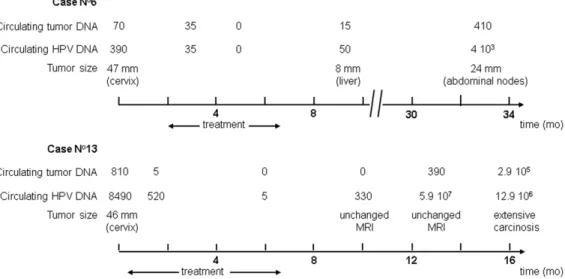
ctDNA dynamics were analyzed on sequential serum specimens in two patients. The first patient received combined chemor-adiotherapy followed by utero-vaginal brachytherapy and surgery (Figure 1, case nu6). The ctDNA value decreased during treatment and was null by the end of therapy at which time a complete histological response was observed on the hysterectomy specimen. Two months later, the patient developed a 8 mm liver metastasis and ctDNA became detectable again. Increasing levels of ctDNA were later found when the patient developed abdominal node recurrence. The same dynamics were observed in the second patient treated exclusively with chemoradiotherapy (Figure 1, case nu13). Of interest during the follow-up, while the pelvic MRI showed no significant modification, a rise of ctDNA was observed. Soon afterwards the patient presented an abdominal relapse associated with high levels of ctDNA. The same dynamics were observed with c-HPV DNA.
Discussion
We report here that, using HPV integration mutation as a molecular marker, ctDNA could be detected in the serum of most patients diagnosed with invasive cervical carcinoma over stage Ib and that the amount of ctDNA reflected tumor dynamics. Since the viral integration site is unique for each patient, this marker is highly specific, much more so than the SSC marker [17]. Regarding sensitivity, we could detect ctDNA in 11 out 13 (85%) patients with stage II–IV tumors, a rate largely higher than the 12% [9], 18% [13], 20% [14] and 50% [11] of positivity reported so far for c-HPV DNA. The practice of 10 replicates for the detection of ctDNA in each serum specimen may account for the increased sensitivity of our assay. For instance, in most of our positive cases, less than 50% of the replicates provided evidence of ctDNA. However, in 2 of the 13 cases with a tumour size
.20 mm, no ctDNA was found whereas c-HPV DNA was detected. Both cases corresponded to tumors with low HPV insertion load (#1 copy/cell). In contrast, cervical carcinoma cells frequently harbour free HPV genomes which are also released in the general circulation, thus increasing the rate of detection of circulating viral DNA. Nevertheless this asset may be balanced by a risk of false positivity. For instance, assays based on the detection on HPV DNA provided false positive rates of 13.5% in normal individuals and 33% in CIN3 patients [11]. In addition, frequent discrepancies between HPV genotypes found in cervical cancer and in the serum of the same patients were also reported [13]. These difficulties should be overcome by adequate controls of specificity and, for clinical purpose, c-HPV DNA analysis may represent a useful alternative where the identification or the use of viral-cell junction sequences is difficult.
Analysis of sequential serum specimens showed the dynamics of the results: the concentration of ctDNA decreased under treatment and increased at the time of relapse. The positivity of ctDNA
Circulating Tumor DNA in Cervical Cancer Patients
Cases
Tumor stage (FIGO)
Tumor size
(mm) HPV type
SCC marker
(ng/ml) HPV integration sites
HPV-E7 load in tumor (copies/cell)
HPV insertion load in
tumor (copies/cell) Circulating DNA (copies/ml serum)
Host DNA HPV DNA Tumor DNA
Nu1 Ib 10 16 NA Xq21.31 17 1 19 103 0 0
Nu2 Ib 15 16 1.1 3q26.32 1 1 21 103 0 0
Nu3 Ib 20 18 NA 17q23.1 0.6 0.6 41 103 0 0
Nu4 IIa 35 16 NA 14q32.2 55 25 17 103 770 790
Nu5 IIb 42 16 1.9 15q23 5 1 40 103 5 5
Nu6 IIb 47 16 NA 3q21.3 88 12 7 103 390 70
Nu7 IIb 53 16 6.4 5p31 27 0.7 7 103 5 5
Nu8 IIb 54 16 15.4 17q25.2 160 79 5 103 3.4 103 890
Nu9 IIb 55 16 2.8 18q21.33 2 2 6 101 30 15
Nu10 IIb 56 16 12.9 4q13 58 1 55 103 3.5 103 0
Nu11 IIIb 55 16 11.6 1p22.1 2 3 21 103 45 20
Nu12 IIIb 74 16 17.4 2q22.1 1 1 32 103 10 20
Nu13 IVa 46 16 NA 17q21.31 94 51 66 103 8.5 103 810
Nu14 IVa 62 16 7.3 Xq22.3 0.5 0.5 77 103 15 0
Nu15 IVa 120 18 NA 1p31.1 5 11 3 103 500 680
Nu16 relapse 27 16 3.9 Repeated sequences* 22 18 5 103 30 25
Abbreviations: FIGO: International Federation of Gynecology and Obstetrics; SCC: squamous cell carcinoma associated antigen; NA: not available; *Repeated sequences: Homology with the centromeric sequences of various chromosomes.
doi:10.1371/journal.pone.0043393.t001
Circulatin
g
Tumor
DNA
in
Cervical
Cancer
ONE
|
www.ploson
e.org
3
August
2012
|
Volume
7
|
Issue
preceded radiological relapse in one case and was associated with an 8 mm liver metastasis in the other one. Tumor DNA might be more easily released from tumor tissue corresponding to relapse or metastasis than to primary lesion. It might also derive from circulating tumor cells. A specific and quantitative molecular marker of ctDNA may be used to follow the course of cervical cancer. During initial treatment, ctDNA may help to assess disease prognosis and tumor sensitivity to therapy. Larger prospective studies will be necessary to validate this utility. During follow-up, ctDNA concentration could be a highly specific surrogate marker of minimal residual disease and subclinical relapse. This is of major importance in the perspective of specific anti-HPV therapy [18] whose potential efficiency is largely dependent on the tumor mass. These approaches could be easily extended to other types of HPV-associated tumors, anal canal and head and neck carcinoma, for example. Localization of the HPV integration site is not currently assessed in clinical practice. New technologies such as Next Generation Sequencing [19], however, should facilitate this determination and provide optimal molecular characterization of tumors to personalize the handling of patients. In addition, the sensitivity of the detection of ctDNA can be easily increased, merely by analyzing a larger amount of serum and/or by
increasing the efficiency of the PCR assay, for instance using digital PCR [20].These advances will facilitate the assessment of the use of ctDNA in clinical oncology and will improve the biological follow-up of patients treated with cervical cancer.
Supporting Information
Table S1 Primers sequences and reagents for detection of ct-DNA.
(DOC)
Acknowledgments
We thank Mr F Radvanyi for fruitful discussions and Ms A. Le Cunff and Z. Maciorowski for their help in the preparation of the manuscript.
Author Contributions
Conceived and designed the experiments: XSG JC OL EL EJ. Performed the experiments: MC EJ MP EL. Analyzed the data: ADLR VF SA PC JYP EL EJ MC. Contributed reagents/materials/analysis tools: SS PP. Wrote the paper: MC EJ XSG OL.
References
1. Walboomers JM, Jacobs MV, Manos MM, Bosch FX, Kummer JA, et al. (1999) Human papillomavirus is a necessary cause of invasive cervical cancer worldwide. J Pathol 189: 12–19.
2. Cullen AP, Reid R, Campion M, Lorincz AT (1991) Analysis of the physical state of different human papillomavirus DNAs in intraepithelial and invasive cervical neoplasm. J Virol 65: 606–612.
3. Kraus I, Driesch C, Vinokurova S, Hovig E, Schneider A, et al. (2008) The majority of viral-cellular fusion transcripts in cervical carcinomas cotranscribe cellular sequences of known or predicted genes. Cancer Res 68: 2514–2522. 4. Wentzensen N, Ridder R, Klaes R, Vinokurova S, Schaefer U, et al. (2002)
Characterization of viral-cellular fusion transcripts in a large series of HPV16 and 18 positive anogenital lesions. Oncogene 21: 419–426.
5. Hopman AH, Smedts F, Dignef W, Ummelen M, Sonke G, et al. (2004) Transition of high-grade cervical intraepithelial neoplasia to micro-invasive carcinoma is characterized by integration of HPV 16/18 and numerical chromosome abnormalities. J Pathol 202: 23–33.
6. Peter M, Stransky N, Couturier J, Hupe P, Barillot E, et al. (2010) Frequent genomic structural alterations at HPV insertion sites in cervical carcinoma. J Pathol 221: 320–330.
7. Diehl F, Schmidt K, Choti MA, Romans K, Goodman S, et al. (2008) Circulating mutant DNA to assess tumor dynamics. Nat Med 14: 985–990.
8. Schwarzenbach H, Hoon DS, Pantel K (2011) Cell-free nucleic acids as biomarkers in cancer patients. Nat Rev Cancer 11: 426–437.
9. Pornthanakasem W, Shotelersuk K, Termrungruanglert W, Voravud N, Niruthisard S, et al. (2001) Human papillomavirus DNA in plasma of patients with cervical cancer. BMC Cancer 1: 2.
10. Shimada T, Yamaguchi N, Nishida N, Yamasaki K, Miura K, et al. (2010) Human papillomavirus DNA in plasma of patients with HPV16 DNA-positive uterine cervical cancer. Jpn J Clin Oncol 40: 420–424.
11. Yang HJ, Liu VW, Tsang PC, Yip AM, Tam KF, et al. (2004) Quantification of human papillomavirus DNA in the plasma of patients with cervical cancer. Int J Gynecol Cancer 14: 903–910.
12. Tseng CJ, Pao CC, Lin JD, Soong YK, Hong JH, et al. (1999) Detection of human papillomavirus types 16 and 18 mRNA in peripheral blood of advanced cervical cancer patients and its association with prognosis. J Clin Oncol 17: 1391–1396.
13. Dong SM, Pai SI, Rha SH, Hildesheim A, Kurman RJ, et al. (2002) Detection and quantitation of human papillomavirus DNA in the plasma of patients with cervical carcinoma. Cancer Epidemiol Biomarkers Prev 11: 3–6.
14. Liu VW, Tsang P, Yip A, Ng TY, Wong LC, et al. (2001) Low incidence of HPV DNA in sera of pretreatment cervical cancer patients. Gynecol Oncol 82: 269– 272.
Figure 1. Circulating tumor DNA related to tumor dynamics in cervical cancer patients.
doi:10.1371/journal.pone.0043393.g001
Circulating Tumor DNA in Cervical Cancer Patients
15. Luft F, Klaes R, Nees M, Durst M, Heilmann V, et al. (2001) Detection of integrated papillomavirus sequences by ligation-mediated PCR (DIPS-PCR) and molecular characterization in cervical cancer cells. Int J Cancer 92: 9–17. 16. Rago C, Huso DL, Diehl F, Karim B, Liu G, et al. (2007) Serial assessment of
human tumor burdens in mice by the analysis of circulating DNA. Cancer Res 67: 9364–9370.
17. Gadducci A, Tana R, Cosio S, Genazzani AR (2008) The serum assay of tumour markers in the prognostic evaluation, treatment monitoring and follow-up of
patients with cervical cancer: a review of the literature. Crit Rev Oncol Hematol 66: 10–20.
18. Kenter GG, Welters MJ, Valentijn AR, Lowik MJ, Berends-van der Meer DM, et al. (2009) Vaccination against HPV-16 oncoproteins for vulvar intraepithelial neoplasia. N Engl J Med 361: 1838–1847.