This clinical case report describes the orthodontic treatment of an 8-year and 9-month old female patient with An-gle Class I malocclusion, anterior crossbite and canine Class III relationship. Orthodontic treatment was carried out in two stages. The first one was orthopedic, while the second one included the use of a fixed appliance and the need for space gain for reshaping of maxillary lateral incisors. The two-stage treatment combined with multidisciplinary Restorative Cosmetic Dentistry allowed excellent esthetic and functional outcomes to be achieved. This case was presented to the Brazilian Board of Orthodontics and Dentofacial Orthopedics (BBO) as a requirement for the title of certified by the BBO.
Keywords:Angle Class I malocclusion. Orthopedics. Corrective Orthodontics.
INTRODUCTION
A Caucasian, 8-year and 9-month old, female pa-tient in good general oral health was referred for treat-ment by her legal guardians, with the major esthetic complaint of anterior negative overjet. She had no functional complaint. The patient’s mother and two sisters had Class III skeletal pattern, which revealed a strong possibility of her having the same unfavorable
DIAGNOSIS
Facial analysis in frontal view revealed the presence of passive lip seal. At smiling, she had little maxillary inci-sors exposure, with discreet asymmetry of the mandible to the right. In lateral view, the patient presented with a deiciency in the premaxilla and a concave proile with a tendency to become worse overtime.1,2,3 There was
more lower lip protrusion in comparison to the upper O presente caso clínico relata o tratamento ortodôntico de uma paciente com oito anos e nove meses de idade, portadora de má oclusão de Classe I de Angle, com mordida cruzada anterior e relação de classe III entre os caninos. O tratamen-to ortratamen-todôntico foi realizado em duas etapas, sendo a primeira ortratamen-topédica e a segunda constando de aparatratamen-tologia fixa, havendo necessidade de ganho de espaço para reanatomização dos incisivos laterais superiores. O tratamento em duas etapas, aliado à multidisciplinariedade com a Dentística Restauradora, permitiu a obtenção de excelente resultado final estético e funcional. O presente caso clínico foi apresentado à Diretoria do Board Brasileiro de Ortodontia e Ortopedia Facial (BBO) como parte dos requisitos para obtenção do título de Diplomado pelo BBO.
Souza PA BBO case report
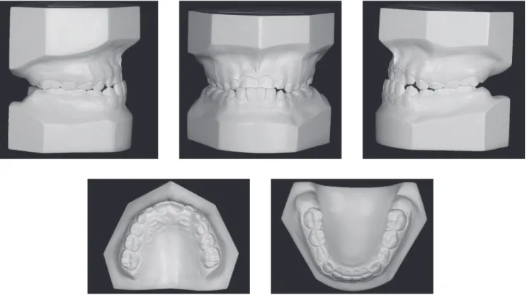
features are shown in Figure 1. Dental assessment (Figs 1, 2) revealed molars in Class I relationship4 and canines
in Class III relationship, with crossbite in the anterior region. Patient’s mandibular midline had a 1-mm shit to the right, while maxillary lateral incisors had crowns signiicantly reduced in size.
Panoramic radiograph (Fig 3) revealed the presence of all permanent teeth at diferent odontogenic stages, with teeth #18, #38 and #48 found to be at the initial stages of crown formation.
Figure 2 - Initial casts.
Souza PA BBO case report
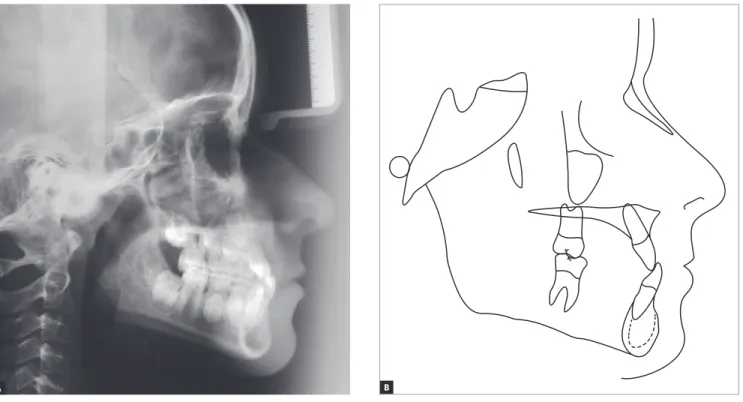
Figure 4 - Initial lateral cephalogram (A) and cephalometric tracing (B).
A B
TREATMENT PLAN
In view of patient’s conditions, a two-stage treat-ment plan was established: the irst stage would include orthopedic intervention, while the second one would include the use of a mandibular ixed tongue crib fol-lowed by conventional orthodontic treatment.
Initially, a progenic appliance with digital springs would be placed in order to protrude maxillary incisors, which would correct anterior crossbite. Subsequently, the patient would be followed-up, so as to have the devel-opment of her dentition monitored. Immediately before her mandibular deciduous second molars were lost, she would have a mandibular ixed tongue crib placed in or-der to have the leeway space5 preserved and the action of
the tongue minimized, thereby leading to a physiological
retraction of mandibular incisors. Thereater, orthodontic bands would be installed on teeth #16 and #26 and brack-ets (MBT Straight Wire slot 0.022 x 0.028-in) would be bonded to all other teeth in both upper and lower arches.
For alignment and leveling, stainless steel Twist Flex 0.015-in, 0.0175-in and 0.020-in archwires, fol-lowed by 0.016-in and 0.018-in smooth archwires and 0.018 x 0.025-in rectangular wires would be used. In the maxilla, the spaces between mesial and distal surfaces of lat-eral incisors would be preserved, so as to allow resin to be placed in those areas; thus, improving the anatomical traits.
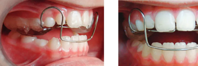
Figure 5 - Protraction archwire.
TREATMENT PROGRESS
Treatment was carried out as planned without changes in the planned sequence. Initially, an orthope-dic progenic appliance with digital springs was installed in the anterior region, aiming at protruding maxillary incisors (Fig 5). Ater anterior crossbite had been cor-rected, a maxillary removable bite plate was used to favor extrusion of posterior mandibular teeth, thus correcting posterior open bite caused during crossbite correction.
The patient was followed-up and before exfoliation of mandibular deciduous second molars, a ixed tongue crib was installed, supported by teeth #36 and #46. At 11 years and nine months of age, ater mandibular second premolars had fully erupted, and the patient had complete permanent dentition, except for third molars, new examination was required with a view to initiating conventional orthodontic treatment (Figs 6 to 9).
Orthodontic bands were placed around maxillary permanent irst molars and brackets bonded to all other maxillary and mandibular teeth. Metal brackets (MBT Straight Wire, slot 0.022 x 0.018-in) were used.
Alignment and leveling archwires were placed in the maxilla and mandible in the following sequence: Twist Flex stainless steel 0.015-in, 0.0175-in and 0.020-in
wires, stainless steel smooth 0.016-in and 0.018-in round wires, and stainless steel 0.018 x 0.025-in rectan-gular wires. With a view to gaining space adjacent to the mesial and distal surfaces of lateral incisors in the upper arch, open springs were compressed between central in-cisors and canines. The space was used for reshaping of lateral incisors carried out by means of placing resin in the proximal surface of those teeth.
Once the 0.018-in steel wire had been installed, an elastomeric chain was placed from tooth #36 to #46 in the lower arch, combined with intermaxillary Class III elastics, with a view to not only closing residual spaces resulting from leeway space, from posterior to anterior direction up to the region of incisors, but also to en-hance retraction of those teeth. At this stage, the ixed tongue crib was removed and spaces fully closed.
Souza PA BBO case report
Souza PA BBO case report
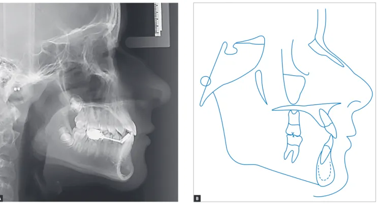
Figure 9 - Intermediate lateral cephalogram (A) and cephalometric tracing (B).
A B
Once all treatment goals had been achieved, the orthodontic ixed appliance was removed and the reten-tion phase started. A removable appliance manufactured with a 1.5-mm acetate sheet was used in the maxilla, while an intercanine bar manufactured with stainless steel Twist Flex 0.032-in wire was used in the mandible.
RESULTS
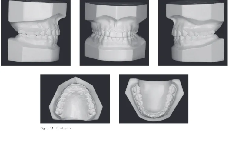
Patient’s inal records (Figs 10 to 13) assessment revealed that all treatment goals were achieved. There was vertical gain in patient’s lower third of the face, in addition to signiicant upper lip protrusion, there-by improving patient’s proile signiicantly. Howev-er, passive lip seal was preserved. MoreovHowev-er, patient’s smile was signiicantly improved, with greater
maxil-was excellent for occlusion in protrusive excursion and right as well as let lateral guidance, with centric relation coinciding with maximal intercuspation. It is worth noting that, as shown by panoramic radiograph taken at treatment completion (Fig 12), changes were achieved without radiographically noticeable apical root remodeling.
As planned, cephalometric examination revealed that patient’s skeletal pattern was preserved, with the ANB6 angle increasing from 1° to 2°, and Wits value
Souza PA BBO case report
Figure 11 - Final casts.
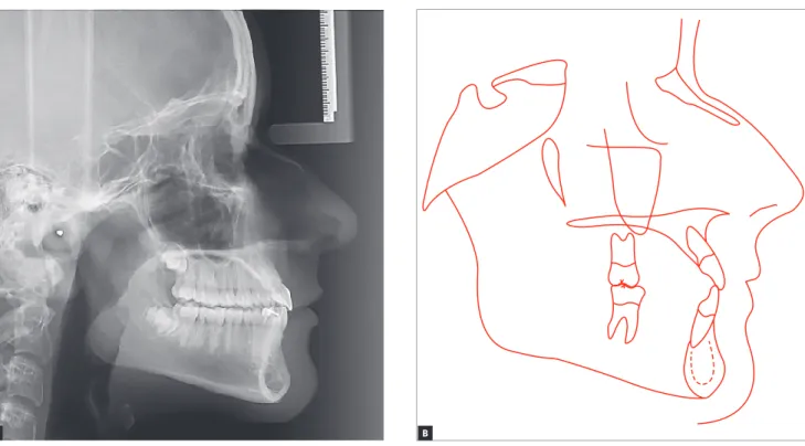
Figure 13 - Final lateral cephalogram (A) and cephalometric tracing (B).
Souza PA BBO case report
Figure 15 - Total (A) and partial (B) cephalometric superimpositions of initial (black) and final (red) tracings. A
A B
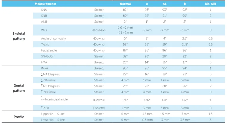
Table 1 - Initial cephalometric values (A) at the beginning of the second treatment phase (A1) and final (B) cephalometric values.
Measurements Normal A A1 B Dif. A/B
Skeletal pattern
SNA (Steiner) 82° 93° 93° 92° 1
SNB (Steiner) 80° 92° 91° 90° 2
ANB (Steiner) 2° 1° 2° 2° 1
Wits (Jacobson) ♀ 0 ±2 mm
♂ 1 ±2 mm -2 mm -3 mm -2 mm 0
Angle of convexity (Downs) 0° 3° 4° 2.5° 0.5
Y-axis (Downs) 59° 55° 59° 61.5° 6.5
Facial angle (Downs) 87° 95° 96° 96° 1
SN-GoGn (Steiner) 32° 20° 20° 22° 2
FMA (Tweed) 25° 14° 16° 17° 3
Dental pattern
IMPA (Tweed) 90° 95° 95° 94° 1
1.NA (degrees) (Steiner) 22° 16° 19° 21° 5
1-NA (mm) (Steiner) 4 mm 1 mm 4 mm 5 mm 4
1.NB (degrees) (Steiner) 25° 28° 28° 26° 2
1-NB (mm) (Steiner) 4 mm 4 mm 4 mm 4 mm 0
1
1. Ritter DE. Class I maloclusion with anterior crossbite and severe crowding. Dental Press J Orthod 2014 Mar-Apr;19(2):115-25.
REFERENCES success achieved by conventional therapy,8 which
war-rants early treatment of such malocclusion.
As it has been previously reported, there was some concern about proportionality between max-illary central and lateral incisors dimensions. Since lateral incisors were rather small, there was a need for space gain in their mesial and distal surfaces, for