DOI: 10.1590/2317-1782/20162015048
CoDAS 2016;28(4):486-488
Case Report
Relato de Caso
Swallowing endoscopy findings in
Huntington’s disease: a case report
Thaís Coelho Alves1
Paula Cristina Cola1
Rarissa Rúbia Dallaqua dos Santos1
Suely Mayumi Motonaga1
Roberta Gonçalves da Silva1
Keywords
Deglutition Disorders Huntington’s Disease Neurodegenerative Diseases Chorea Endoscopy
Correspondence address:
Roberta Gonçalves da Silva Universidade Estadual Paulista – UNESP
Av. Hygino Muzzi Filho, 737, Marília (SP), Brazil, CEP: 17515-901. E-mail: [email protected]
Received: February 26, 2015
Accepted: August 04, 2015
Study carried out at Dysphagia Research Center of the Universidade Estadual Paulista – UNESP - Marília (SP), Brazil.
1 Universidade Estadual Paulista – UNESP - Marília (SP), Brazil. Financial support: none.
Conlict of interests: nothing to declare.
ABSTRACT
Huntington’s disease (HD) is a degenerative genetic disorder with autosomal-dominant transmission. The triad of symptoms of this disease consists of psychiatric disorders, jerky movements, and dementia. Oropharyngeal dysphagia, which is more evident with disease progression, is also present. Few studies have addressed the swallowing characteristics using objective analysis in this population. The purpose of this research was to
describe the swallowing endoscopic indings of the pharyngeal phase in HD. This is a cross-sectional study
addressing a clinical case which included two individuals of the same family, male, 32 and 63 years old,
designated as individual A and individual B, with progression of the disease for ive and 13 years, respectively.
CoDAS 2016;28(4):486-488
Huntington’s disease and swallowing 487
INTRODUCTION
Huntington’s disease (HD) is a degenerative genetic disorder with autosomal-dominant transmission1-3. The triad of symptoms of this disease consists of psychiatric disorders, jerky movements, and dementia. Oropharyngeal dysphagia is more evident with disease progression and compromises the swallowing function.
In the descriptions of this disease in the literature, there are studies on swallowing which investigate the characteristics of
the oral and pharyngeal phases using videoluoroscopy (VFD) and iberoptic endoscopic evaluation of swallowing (FEES), as
well as the risks of aspiration associated with bronchopulmonary infections, airway obstruction, dehydration, and malnutrition4,5.
Although the clinical assessments of swallowing provide relevant information on the condition of oral feeding in patients with oropharyngeal dysphagia, the variability found in their accuracy shows the importance of conducting supplemental exams to objectively assess swallowing in the diagnosis of dysphagia. Also, there are few studies addressing this diagnosis in this
population. They veriied the swallowing of individuals using videoluoroscopy, and identiied postural changes, inadequate
movement of the tongue, multiple swallowing, pharyngeal residue after swallowing, and residues in the valleculae and pyriform sinuses6,7. The presence of penetration and/or laryngotracheal aspiration was observed only in the advanced stages of the disease4.
In this paper we used Fiberoptic Endoscopic Evaluation of Swallowing (FEES)8, which is an excellent method to analyze the pharyngeal phase of swallowing, identifying posterior oral spillage, pharyngeal residue, penetration, and aspiration. The main purpose of this study was to describe the swallowing
endoscopic indings of the pharyngeal phase in HD.
CASE REPORT
This descriptive study addresses the clinical case of two individuals of the same family, male, 32 and 63 years old, designated as individual A and individual B, respectively. The individuals were referred to the Dysphagia Center due to complaints of speech and swallowing. Both of them signed the Informed Consent Form. The individuals were diagnosed with
HD conirmed by genetic testing, with progression of the disease for ive and 13 years, respectively. They presented weight loss
and gagging. Functional activities such as dressing, brushing teeth, and bathing were compromised. During the evaluation, both individuals presented hyperkinetic dysarthria, postural instability, and involuntary movements of the head and upper and lower limbs. These signals were worse in the A individual, who is younger than the B individual. In the clinical assessment
of swallowing, the individuals presented dificulties in labial
sealing, oral incoordination, compensatory maneuvers of the head, and disorder in oral transit time, but no clinical signs suggestive of penetration and/or laryngotracheal aspiration. The individuals were referred to FEES, which was performed by an Ear, Nose and Throat Physician (ENT) and accompanied by a speech-language pathologist. The patients were positioned with support to minimize involuntary movement. During the
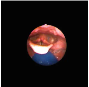
endoscopy examination, no structural disorder was found in the nasopharynx, oropharynx, and hipopharynx. Disorders in the vocal folds were also not found. We used consistent liquid, nectar, and puree in volumes of 3-10 mL. The results show that both individuals presented laryngeal and pharyngeal sensitivity, posterior oral spillage for liquid and nectar (Figures 1 and 2), absence of salivary stasis, pharyngeal residues in small quantity, presence of pharyngeal clearance, and absence of laryngeal penetration and/or laryngotracheal aspiration.
Figure 1. Posterior oral spillage in the A individual
CoDAS 2016;28(4):486-488
Alves TC, Cola PC, Santos RRD, Motonaga SM, Silva RG 488
DISCUSSION
According to few authors, dysphagia is a common complication of Huntington’s disease, which is often responsible for episodes of respiratory impairment9. Clinical assessment provides information on the presence, nature, and severity of dysphagia4,10, but supplementary exams through objective assessment of swallowing are needed for its diagnosis8.
In this study, we used the FEES method because it provides easy visualization of the pharyngeal phase of swallowing11,12 in
agreement with videoluoroscopy indings. It is known that the
oral phase in HD can become poor and compromise the oral transit time. However, the presence of posterior oral spillage can also provide us with some information on the coordination
of the oral phase, and this parameter can be identiied in the FEES and in the videoluoroscopy13. In both tests, the presence of involuntary movement present in HD further complicates the oral control and the presence of posterior oral spillage.
Other studies using swallowing videoluoroscopy in liquid and puree consistencies veriied involuntary movements of
the tongue, presence of posterior oral spillage, and pharyngeal
residue, corroborating the indings of this study6,14.
These results on posterior oral spillage and pharyngeal
residue found in both individuals were also identiied by another
authors7 as the most prevalent signs in HD. The FEES is an excellent method for the research of these symptoms because it can clearly identify the presence of pharyngeal residue, penetration, and aspiration.
Penetration and/or tracheal aspiration were not observed in either of the individuals, showing once again agreement with
other studies that used videoluoroscopy15. Also, the literature reports the presence of these signs only in advanced stages of the disease. In addition to the progression of the disease, other aspects that should be considered and that can increase the risk of tracheal aspiration are also related to the amount of pharyngeal residue4.
There are no studies in the literature describing oropharyngeal dysphagia by the progression of the disease in this population due to the particular manifestations of each individual. New studies with larger samples and different periods of the disease should be conducted to provide additional information on the characteristics and management of swallowing in this population. This demonstrates that there is little knowledge on
dysphagia, its most frequent indings during the course of the
disease, and its prevalence in HD7.
FINAL COMMENTS
The use of FEES in assessing the pharyngeal phase of
swallowing in HD identiied indings consistent with those of VFD for this population. This does not preclude the use of VFD
as an important tool to assessment oral phase of swallowing in this population. However, because it provides the possibility
to conirm or exclude the presence of pharyngeal residue,
laryngeal penetration, and aspiration in this population, the
FEES is a more accessible evaluation method in present context
of the country. This study identiied the presence of posterior
oral spillage and small amount of pharyngeal residues, and no presence of penetration or aspiration in individuals with HD.
REFERENCES
1. Gusella JF, MacDonald ME. Huntington’s disease. Semin Cell Biol. 1995;6(1):21-8. http://dx.doi.org/10.1016/1043-4682(95)90011-X. PMid:7620118.
2. Yorkston KM, Miller RM, Strand EA. Management of speech and swallowing in degenerative diseases. Tucson: Communication Skill Builders; 1995. 3. Pringsheim T, Wiltshire K, Day L, Dykeman J, Steeves T, Jette N. The
incidence and prevalence of Huntington’s disease: a systematic review and meta-analysis. Mov Disord. 2012;27(9):1083-91. http://dx.doi.org/10.1002/ mds.25075. PMid:22692795.
4. Hamilton A, Heemskerk AW, Loucas M, Twiston-Davies R, Matheson KY, Simpson SA, et al. Oral feeding in Huntington’s disease: a guideline document for speech and language therapists. Neurodegener Dis Manag. 2012;2(1):45-53. http://dx.doi.org/10.2217/nmt.11.77.
5. Hamakawa S, Koda C, Umeno H, Yoshida Y, Nakashima T, Asaoka K, et al. Oropharyngeal dysphagia in a case of Huntington’s disease. Auris Nasus Larynx. 2004;31(2):171-6. http://dx.doi.org/10.1016/j.anl.2003.11.001. PMid:15121228.
6. Heemskerk AW, Roos RA. Dysphagia in Huntington’s disease: a review. Dysphagia. 2011;26(1):62-6. http://dx.doi.org/10.1007/s00455-010-9302-4. PMid:20838817.
7. Kagel MC, Leopold NA. Dysphagia in Huntington’s disease: a 16-year retrospective. Dysphagia. 1992;7(2):106-14. http://dx.doi.org/10.1007/ BF02493441. PMid:1533361.
8. Marinque D. Avaliação otorrinolaringológica da deglutição. In: Furkin AM, Santini CS. Disfagias orofaríngeas. Carapicuiba: Pró-fono; 1999. p. 49-66.
9. Leopold NA, Kagel MC. Dysphagia in Huntington’s disease. Arch Neurol. 1985;42(1):57-60. http://dx.doi.org/10.1001/archneur.1985.04060010063017. PMid:3155611.
10. Langmore SE. Issues in the management of dysphagia. Folia Phoniatr. 1999;51(1):220-30.
11. Langmore SE, Schatz K, Olson N. Endoscopic and videofluoroscopic evaluations of swallowing and aspiration. Ann Otol Rhinol Laryngol. 1991;100(8):678-81. http://dx.doi.org/10.1177/000348949110000815. PMid:1872520.
12. Kelly AM, Drinnan MJ, Leslie P. Assessing penetration and aspiration: how do videofluoroscopy and fiberoptic endoscopic evaluation of swallowing compare? Laryngoscope. 2007;117(10):1723-7. http://dx.doi.org/10.1097/ MLG.0b013e318123ee6a. PMid:17906496.
13. Dodds WJ, Logemann JA, Stewart ET. Radiologic assessment of abnormal oral and pharyngeal phases of swallowing. AJR Am J Roentgenol. 1990;154(5):965-74. http://dx.doi.org/10.2214/ajr.154.5.2108570. PMid:2108570.
14. Barsottini OGP. Doença de Huntington: o que é preciso saber. Einstein. 2007;5(3):83-8.
15. Lee TH, Lee JS, Kim WJ. High resolution impedance manometric findings in dysphagia of Huntington’s disease. World journal of gastroenterology. WJG. 2012;18(14):1695-9. http://dx.doi.org/10.3748/wjg.v18.i14.1695. PMid:22529701.
Author contributions