Published: October 01, 2009 by CRC Press - 464 Pages
Editor(s): João Manuel R.S. Tavares, Faculty of Engineering, University of Porto, Portugal; R.M. Natal Jorge, Faculty of Engineering, University of Porto, Portugal
Share Gosto 0
Computational Vision and Medical Image Processing:
VipIMAGE 2009
» Recommend to Librarian
Hardback £140.00
ISBN 9780415570411
Cat# K11337
Purchasing Options
Description Table of Contents
Table of Contents
Preface
Acknowledgements Invited lectures Thematic sessions
Home » Medicine » General Reference » Computational Vision and Medical Image Processing: VipIMAGE 2009
Region: PORTUGAL
Search Advanced Search
All Subjects Textbooks Only
(Login/Register My Account
Contact Us |
Items: 0 Total: £0.00
Home About Us Resources Textbooks CRCnetBASE Featured Authors
Computa(onal Vision and Medical Image Processing -‐ CRC Press Book h<p://www.crcpress.com/product/isbn/9780415570411
On building photographic portraits using statistical techniques, R. Roque, A. Puga, E. Carrapatoso & F. Barbosa Geometric analysis of female pelvic floor muscles by using manual segmentation, T.H. Da Roza, R.M. Natal Jorge, M. Parente, João Manuel R.S. Tavares, C. Saleme, M.P. Barbosa, A.L.S. Filho, T. Mascarenhas & J. Loureiro
Analysis of the fibers direction in the tympanic membrane, C. Garbe, F. Gentil, M.P.L. Parente, P. Martins, R.M. Natal Jorge & S. Santos
Construction of a 3D model for the female pelvic organs, A.J.C. Arteiro, M.P.L. Parente, R.M. Natal Jorge, A.A. Fernandes & T. Mascarenhas
A versatile matching algorithm based on dynamic programming with circular order preserving, Francisco P.M. Oliveira, João Manuel R.S. Tavares & Todd C. Pataky
Construction of a 3D model from medical images: Application to knee joint, H. Mata, R.M. Natal Jorge, S. Santos, A. Sousa & F. Raposo
A novel method to segment the levator ani muscles in MR images, Zhen Ma, João Manuel R.S. Tavares & R.M. Natal Jorge
3D reconstruction of human temporomandibular structure, V.L. Trindade, R.M. Natal Jorge & S. Santos Spectral imaging of retinal tissue oxygenation, E. Ohel, S. Rotman, D.G. Blumberg & N. Belfair
Automatic segmentation of the optic radiation using DTI in glaucoma patients, A. El-Rafei, J. Hornegger, T. Engelhorn, A. Dörfler, S. Wärntges & G. Michelson
Comparison between Kalman and unscented Kalman filters in tracking applications of computational vision, Raquel R. Pinho & João Manuel R.S. Tavares
Thematic session on computer vision in robotics
Model based localization for a mobile robot in an indoor environment, F. Valente & M. Ramalho Obtaining the distance map for perspective vision systems, A.J.R. Neves, D.A. Martins & A.J. Pinho
Real-time vision in the RoboCup—Robotic Soccer international competitions, L.P. Reis, A.J.R. Neves & A. Sousa Thematic session on imaging of biological flows trends and challenges
Diffusion in concentrated suspensions of biological cells, T. Ishikawa X-ray micro-imaging of various bio-fluid flows, S.J. Lee
Patient-specific blood flow analysis of pulmonary artery affected by severe deformations of the lung 335
J.-J. Christophe, N. Matsuki, T. Yamaguchi, T. Ishikawa, Y. Imai, M. Thiriet & K. Takase
Motions of particles and red blood cells in a bifurcation: Comparison between experiments and numerical simulations, M. Lagoela, B. Oliveira, D. Cidre, C. Fernandes, C. Balsa, R. Lima, R. Dias, T. Ishikawa, Y. Imai & T. Yamaguchi In silico multiscale study of the hemodynamics in a human carotid bifurcation model, D. Gallo, D. Massai, F. Consolo, A. Satriano, F.M. Montevecchi, U. Morbiducci, R. Ponzini, L. Antiga & A. Redaelli
Influence of blood rheology on bulk flow in carotid bifurcation: A numerical study, D. Massai, D. Gallo, A. Satriano,
Computa(onal Vision and Medical Image Processing -‐ CRC Press Book h<p://www.crcpress.com/product/isbn/9780415570411
1 INTRODUCTION
Several studies have revealed that the information obtained on the rheological properties of blood in straight glass microtubes differs from the in vivo situation (Pries et al. 1994, Suzuki et al. 1996). The main potential causes for these discrepancies may be due to the endothelial surface layer and microvascu-lar networks composed with short irregumicrovascu-lar vessel segments which are linked by numerous bifurcations (Maeda 1996, Pries and Secomb 2003).
The main purpose of this paper is to analyse the non-Newtonian flow characteristics of blood flowing in microvascular network models. To accomplish it experimental flow studies performed with a micro-PIV/PTV system will be complemented by hemody-namics computational models (commercial finite element software package POLYFLOW®). By using this combination we expect to gain understanding about several important parameters that affect the blood flow through a diverging microvessel bifurca-tion.
2 MATERIALS AND METHODS
2.1. Working fluids and microchannel
Two working fluids were used in this study: pure wa-ter and dextran 40 (Dx40) containing about 14% (14Hct) of human red blood cells (RBCs). The blood was col-lected from a healthy adult volunteer, where ethyl-enediaminetetraacetic acid (EDTA) was added to prevent coagulation. The RBCs were separated from the bulk
blood by centrifugation and aspiration and then washed twice with physiological saline (PS). The washed RBCs were labeled with a fluorescent cell tracker (CM-Dil, C-7000, Molecular Probes) and then diluted with Dx40 to make up the required RBCs concentration by volume. All blood samples were stored hermetical at 4ºC until the ex-periment was performed at controlled temperature of about 37ºC (Lima et al. 2009).
The microchannel used in this study was a symmetric PDMS bifurcation (150 µm wide, 50 µm deep for parent vessel; 75 µm wide, 50 µm deep for daughter vessel) fab-ricated by a soft lithography technique (Lima et al. 2008).
Fig. 1 Symmetrical bifurcation geometry used in this study: Q0
= 0.18 l/min, W0 = 150 µm, W1 = 75 µm, W2 = 75 µm, θ =
60º, depth = 50 µm.
Motions of particles and red blood cells in a bifurcation: comparison between
experiments and numerical simulations
M. Lagoela, B. Oliveira, D. Cidre, C. Fernandes, C. Balsa
ESTiG, IPB, C. Sta. Apolonia, 5301-857 Braganca, Portugal.
R. Lima, R. Dias
ESTiG, IPB, C. Sta. Apolonia, 5301-857 Braganca, Portugal. CEFT, FEUP, R. Dr. Roberto Frias, 4200-465 Porto, Portugal.
T. Ishikawa, Y. Imai
Dept. Bioeng. & Robotics, Grad. Sch. Eng., Tohoku Univ., 6-6-01 Aoba, 980-8579 Sendai, Japan
T. Yamaguchi
Dept. Biomedical Eng., Grad. Sch. Eng., Tohoku Univ., 6-6-01 Aoba, 980-8579 Sendai, Japan.
The blood flow dynamics in microcirculation depends strongly on the microvascular networks composed with short ir-regular vessel segments which are linked by numerous bifurcations. This paper presents the application of a confocal micro-PTV system to track RBCs through a rectangular polydimethysiloxane (PDMS) microchannel with a bifurcation. By using a confocal micro-PTV system, we have measured the effect of bifurcation on the flow behaviour of both fluo-rescent particles diluted in pure water and RBCs in concentrated suspensions. After performing simulations with the
commercial finite element software package POLYFLOW®, some experimental results were compared with the
2.2.Confocal micro-PTV experimental set-up
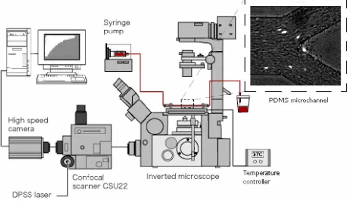
The confocal micro-PTV system used in our ex-periment consists of an inverted microscope (IX71, Olympus, Japan) combined with a confocal scanning unit (CSU22, Yokogawa) and a diode-pumped solid state (DPSS) laser (Laser Quantum Ltd) with an ex-citation wavelength of 532 nm. Moreover, a high-speed camera (Phantom v7.1) was connected into the outlet port of the CSU22. The microchannel was placed on the stage of the inverted microscope where the flow rate of the working fluids was kept constant (Re = 0.007) by means of a syringe pump (KD Sci-entific Inc.). The Reynolds number (Re) and associ-ated experimental parameters are summarized in Ta-ble 1. A thermo plate controller (Tokai Hit) was set to 37ºC. All the confocal images were captured in the middle of the microchannels with a resolution of 640×480 pixels, at a rate of 100 frames/s with an ex-posure time of 9.4 ms. The recorded images were transferred to the computer and then evaluated in the Image J (NIH) (Abramoff et al. 2004) by using the manual tracking MtrackJ (Meijering et al. 2006) plugin. As a result it was possible to track single RBCs through the middle plane of the microchannel.
Fig. 2 Experimental set-up.
Table 1 - Experimental parameters used to calculate the Re.
Density (kg/m3) 1046
Mean velocity (m/s) 3.8×10⁻⁴ Hydraulic diameter (m)
7.5×10⁻⁵ Viscosity of Dx-40 (Ns/m2)
4.5×10⁻³
Re 0.007
2.3. Simulation method
The numerical calculations for the laminar iso-thermal flow of pure water were performed using the finite-element computational fluid dynamics (CFD) program POLYFLOW®. The simulations were car-ried out in a 3D geometry representing the micro-channel. The mesh used in the simulations was mainly constituted by quadrilateral elements, the discretization of the walls of the channel. The size of
the elements was fixed after a grid independence test. The grids were successively refined and the ve-locity obtained with the different meshes were com-pared, the results being considered independent of the mesh when a difference bellow 1 % was achieved (Fernandes et al. 2007, 2008).
The equations solved were the conservation of mass and momentum equations for laminar incom-pressible flow of water. The problem is a non-linear problem, so it was necessary to use an iterative method to solve the referred equations. In order to evaluate the convergence of this process, a test based on the relative error in the velocity field was per-formed and the convergence test value was set to 10-4 (Fernandes et al. 2007, 2008).
The boundary conditions were established in or-der to reproduce the experimental conditions. The flow rate at the inlet of the microchannel was 0.18 l/min and slip at the walls of the channel was assumed to be non-existent.
3 RESULTS AND DISCUSSION
The confocal micro-PIV system was first evalu-ated by comparing the experimental results not only with a well established analytical solution for steady flow in a rectangular microchannel (Lima et al. 2006) but also with a reliable numerical method that was used in past investigations to study the flow be-haviour of Newtonian or non-Newtonian fluids at low Reynolds numbers (Fernandes et al. 2007, 2008).
The numerical, experimental and analytical re-sults of the present work were obtained for the mid-dle plane (25 m height) of the rectangular micro-channel. The averaged velocity data obtained from the confocal micro-PTV measurements, analytical solution and numerical simulation were in close agreement. A more detail description of these results can be found elsewhere (Oliveira et al. 2009).
By using a confocal micro-PTV system it was possible to obtain series of successive images at the middle of the bifurcation. Figures 3a and 4 show im-ages with both fluorescent particles and labeled RBCs (laser-emitted light) flowing through a sym-metric bifurcation, together with the correspondent time position tracking of both particles and individ-ual RBCs.
a)
b)
Fig. 3 a) Paths displacement of fluorescent particles flowing in pure water; b) Numerical trajectories using pure water.
Fig. 4 Paths displacement of labeled RBCs (bright spots) flow-ing in physiological fluid with 14% Hct (32×).
In addition by comparing qualitatively the ex-perimental data from both pure water and in vitro blood (14% Hct) it is possible to observe that some RBC paths seems to suffer small deviations from the streamlines of the plasma flow probably due to flow perturbations caused by cell interactions in the neighbourhood of the apex of bifurcation.
Moreover numerical simulations of non-Newtonian models were performed around the bifur-cation and compared with the experimental results at the regions 1 to 7 (see Fig.5)
Fig. 5 Regions where the velocity profiles of the numerical and experimental results were compared.
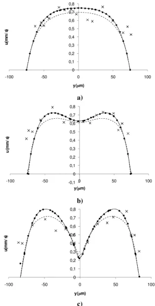
Figures 6 and 7 show the velocity profiles for both computational and experimental results before and after the bifurcation, respectively.
a)
b)
c)
Fig. 6 Velocity profiles for both computational and experimen-tal results before the bifurcation: a) region 1; b) region 2; c) re-gion 3. ( ) Newtonian; (- - -) Power Law; (•) Carreau Model; (×) Confocal micro-PIV.
0 0,1 0,2 0,3 0,4 0,5 0,6 0,7 0,8
-100 -50 0 50 100
u
(m
m
/s
)
y ( m)
-0,1 0 0,1 0,2 0,3 0,4 0,5 0,6 0,7 0,8
-100 -50 0 50 100
u
(m
m
/s
)
y ( m)
0 0,1 0,2 0,3 0,4 0,5 0,6 0,7 0,8
-100 -50 0 50 100
u
(m
m
/s
)
a)
b)
c)
d)
Fig. 7 Velocity profiles for both computational and experimen-tal results after the bifurcation: a) region 4; b) region 5; c) re-gion 6; d) rere-gion 7. ( ) Newtonian; (- - -) Power Law; (•) Car-reau Model; (×) Confocal micro-PIV.
A qualitative examination of Figures 6a) and 6b) does not show a clear correspondence of the experi-mental results with the numerical models used in this study. However Figure 6c) reveals a tendency of the in vitro blood to behave more closely as a Power law model. This is further confirmed by the results obtained in Figure 7, in particular Figures 7 a) and b). An on going study to obtain more detailed
quan-titative measurements of the blood flow behavior through a bifurcation is currently under way.
ACKNOWLEDGMENTS
This study was supported in part by the Portuguese Government by a grant of the Science and Technol-ogy Foundation (BII/UNI/0532/EME/2008).
REFERENCES
Abramoff, M., Magalhaes, P., et al., “Image Processing with ImageJ”. Biophotonics International, 7, 11, 36-42, 2004. Fernandes, C. S., Dias, R. P., et al, “Laminar flow in
chevron-type plate heat exchangers: CFD analysis of tortuosity, shape factor and friction factor”, Chemical Engineering and Processing: Process Intensification, 46, 825-833, 2007. Fernandes, C. S., Dias, R. P., et al, “Friction factors of
power-law fluids in plate heat exchangers”, Journal of Food Engi-neering, 89, 441-447, 2008
Lima, R., Wada, S., et al., “In vitro blood flow in a rectangular PDMS microchannel: experimental observations using a confocal micro-PIV system”. Biomedical Microdevices, 2, 10, 153-67, 2008.
Lima, R., Ishikawa, T., et al., “Measurement of individual red blood cell motions under high hematocrit conditions using a confocal micro-PTV system,”, Annals of Biomedical Engi-neering, 37, 8, 1546-1559, 2009.
Lima, R.., Wada, S., et al., “Confocal micro-PIV measurements of three dimensional profiles of cell suspension flow in a square microchannel”. Meas. Sci. Tech.,17, 797-808, 2006. Lima, R., Wada, S., et al., “In vitro blood flow in a rectangular PDMS microchannel: experimental observations using a confocal micro-PIV system”. Biomedical Microdevices, 2, 10, 153-67, 2008.
Maeda, N., “Erythrocyte rheology in microcirculation”. Japa-nese Journal of Physiology 46, 1-14, 1996.
Meijering, E., Smal, I., and Danuser, G., “Tracking in Molecu-lar Bioimaging”. IEEE Signal Processing Magazine, 3, 23, 46-53, 2006.
Oliveira, B., Lagoela, M., et al., Analyses of the blood flow in a microchannel with a bifurcation, Actas do 3º Congresso Nacional de Biomecânica, Bragança, Portugal, 2009. Pries, A., Secomb, T., et al, “Resistance to blood flow in
mi-crovessels in vivo”. Circulation Research 75, 904-915, 1994.
Pries, A., Secomb, T., “Rheology of the microcirculation”. Clinical Hemorheology and Microcirculation 29, 143-148, 2003.
Suzuki, Y., Tateishi, N., Soutani M. and N., Maeda, “Deforma-tion of erythrocytes in microvessels and glass capillaries: ef-fects of erythrocyte deformability”. Microcirculation 3, 49-57, 1996. 0 0,1 0,2 0,3 0,4 0,5 0,6 0,7 0,8 0,9
0 20 40 60 80 100
u
(m
m
/s
)
y ( m)
0 0,1 0,2 0,3 0,4 0,5 0,6 0,7 0,8 0,9
-100 -80 -60 -40 -20 0
u
(m
m
/s
)
y ( m)
0 0,1 0,2 0,3 0,4 0,5 0,6 0,7 0,8 0,9
0 20 40 60 80 100 120
u
(m
m
/s
)
y ( m)
0 0,1 0,2 0,3 0,4 0,5 0,6 0,7 0,8 0,9
-100 -80 -60 -40 -20 0
u
(m
m
/s
)