RAPID COMMUNICATION
Trunk stabilization among women with chronic
lower back pain: a randomized, controlled, and
blinded pilot study
Silvia Ferreira Andrusaitis, Guilherme Carlos Brech, Gabriela Faller Vitale, Ju´lia Maria D9Andre´a Greve Laborato´rio de Estudos do Movimento do Instituto de Ortopedia e Traumatologia do Hospital das Clı´nicas - Faculdade de Medicina da Universidade de Sa˜o Paulo, Sa˜o Paulo/SP, Brazil.
Email: [email protected] / [email protected] Tel.: 55 11 3069-6000
INTRODUCTION
Treatment for mechanical lower back pain is a challenge in Western society, in which its occurrence can now be considered to have reached epidemic proportions. The origin of such pain and the factors that cause it to become chronic and recurrent remain poorly understood. Abnor-malities in motor control and trunk muscle function have been found in individuals with chronic lumbar pain.1-4 Many studies have demonstrated that the deep muscles of the lumbar column and abdomen, especially the multifidus and transversus abdominis, present late activation, weak-ness, and diminished resistance during episodes of lower back pain.5 These changes persist even when the painful condition goes into remission, and they contribute to episodes of lower back pain recurrence.1,5,6 However, it is still difficult to determine whether the neuromuscular imbalance occurs because of the pain or whether the imbalance causes the pain.1,6 Although the mechanisms that lead to these abnormalities are incompletely under-stood, rehabilitation programs aiming to stabilize the lumbar spine and improve its musculature and propriocep-tive action have been used with posipropriocep-tive effects on pain and functional capacity in individuals with mechanical lower back pain.7
This pilot study compared stabilization exercises with strengthening exercises for the trunk and hips in women with chronic lower back pain in terms of their effects on pain, functional capacity, and postural balance.
MATERIALS AND METHODS
Fifteen female volunteers were prospectively studied in a randomized, controlled and blinded manner between April 2008 and April 2010 with approval from the ethics committee (no. 1248/07).
This study included ten women between the ages of 30 and 55 years who had been referred for physical therapy because of nonspecific, chronic lower back pain. These women had sedentary habits, had no significant radiological abnormalities, and had no neurological impairments. To be
included in the study, patients needed to be free from vestibular abnormalities and musculoskeletal disorders of the hips and lower limbs. In addition, the study had a control group composed of five women between the ages of 30 and 55 who did not present with lower back pain but who fulfilled the same inclusion criteria.
The exclusion criteria were the following: abandonment of the physical therapy, more than three consecutive absences from the treatment sessions, worsening of the symptoms, and a patient’s desire, for any reason, to have her data excluded from the study without this harming the continuity of her treatment.
After the study had been explained to the patients and they had signed the consent form, they were assessed in accordance with the evaluation protocol. Randomization was performed by means of a draw using opaque envelopes containing folded papers that allocated patients to one of two treatment groups: group A (strengthening) or B (stabilization).
All the volunteers were assessed in relation to balance (questionnaire and balance tests at a force plate) and pain scales at the time that they were selected for the study.
Three groups were created, consisting of groups A and B, each with five patients with lower back pain, and a control group with five volunteers. The patients in groups A and B were evaluated with respect to the balance and pain scales before and after the treatment, whereas the patients in the control group were only evaluated before the treatment.
Evaluation protocol
The evaluation protocol was administered one week before the treatment was started and one week after it was terminated by two experienced evaluators who had been trained to handle the assessment instruments and were blinded regarding the treatment groups.
All the patients in the treatment groups (both A and B) gave responses to the Oswestry Disability Questionnaire using the version translated and validated for the Portuguese language,8and they completed a visual analog pain scale (VAS)9regarding the frequency and intensity of their lower back pain.
In addition to the questionnaire and pain scale, all the volunteers (groups A and B and the control group) underwent four balance tests on the Balance MasterH
System (Neurocom International, Inc., Clackamas, Oregon, USA).
Copyrightß2011CLINICS– This is an Open Access article distributed under
The tests undertaken were the following.
Modified clinical test of sensory interaction and balance (mCTSIB). Static balance was assessed using the modified clinical test of sensory interaction and balance, which consists of an assessment of body sway under four sensory conditions while the individual remains on a force platform: eyes open and closed on a stable surface and eyes open and closed on an unstable surface.
Each condition was repeated three times for ten seconds, and the average of the attempts was used. This test measures the displacement velocity at the individual’s center of pressure in degrees per second. A force platform with four coupled sensors is used for the test. Decreases in the displacement velocity are considered to be a positive out-come.
The variables studied were the mean sway velocities with eyes open and with eyes closed on a stable surface (firm surface) and an unstable surface (foam surface) and the mean sway velocities in the anteroposterior (mean-Y) and side-to-side (mean-X) directions with eyes open and with eyes closed on a stable surface (firm surface) and an unstable surface (foam surface).
The following three tests were undertaken to assess the subjects’ functional limitations affecting the activities of daily life.
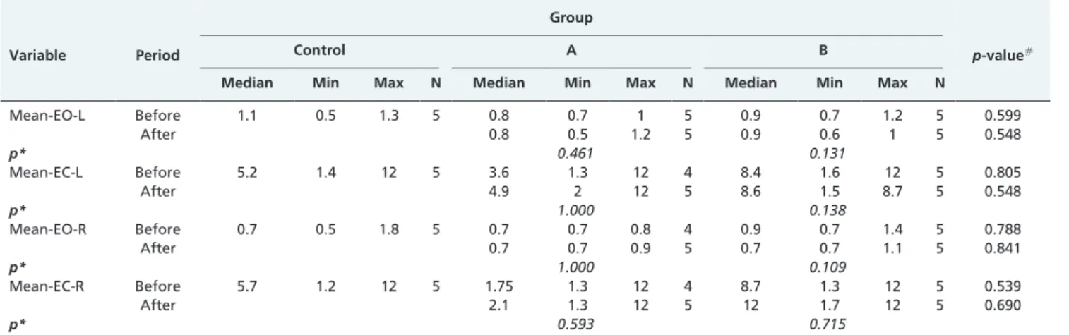
Single-leg stance test. This test was conducted with the subject standing on one leg on a force platform under four conditions: with eyes open or closed and on the left or right leg. Similar to the mCTSIB, each condition was repeated three times for ten seconds, and the mean from the attempts was used. The variables studied were the mean sway velocity with eyes open and closed on the left and right legs.
Get-up-and-go test. The get-up-and-go test was conducted on a platform with the individual initially in a seated position on a bench 30 cm in height without a seat back. The patient’s knees were flexed at 90
˚
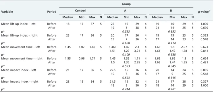
, and her feet were separated by 10 cm at the heels for base support. Her arms were kept along the sides of her body. The patient was instructed to stand up quickly but safely. There were three repetitions of the movement separated by 30-second intervals. The parameters measured were the mean weight transfer time, the mean rising index and the mean sway velocity while rising.Step-up test. In this test, the patients were instructed to climb a step 10 cm in height while putting only one foot on the step. They were told that the other foot should go directly to the platform without contacting the step. When both feet had reached the platform after crossing the step, the patients were then supposed to remain in as static a position as possible. In this protocol, three attempts were made for each leg, beginning with the left leg. The variables evaluated were the mean weight transfer index (mean lift-up index), the mean movement time, and the mean impact index.
In all the balance tests conducted in this study, the volunteers were only allowed a maximum of three attempts to perform each test.
Treatment protocol
For both groups A and B, the treatment consisted of a 40-minute physical therapy session three times a week for a total of 20 sessions. All the sessions began with a ten-minute warm-up on an ergometric bicycle. Following the warm-up,
the patients performed exercises in accordance with their treatment group.
In group A, the exercises had the goal of strengthening the abdominal, back, and hip muscles. The patients performed an average of three series of ten repetitions of each exercise. Increases in the number of exercises performed in each session (or load progression) occurred according to individual tolerance.
In group B, stabilization exercises were taught, starting with the dorsal decubitus and progressing to the ventral decubitus, in seated, four-support, and standing positions. Increases in the number of exercises performed in each session (or load progression) occurred according to indivi-dual tolerance.
All the sessions were conducted individually with the same physical therapist, who was blinded to the results of the initial assessment.
Statistical analysis
Fifteen women participated in this study: ten with lower back pain and five without any history of pain. A descriptive analysis was performed on the following sample parameters: number of cases (N), median, minimum, and maximum.
To meet the aims of the study, the 10 patients were evaluated using the scale values at the time that they were selected, and tests (Kolmogorov-Smirnov) were performed to determine whether the results presented a normal distribution.10
To compare the scales between the groups, the Kruskal-Wallis test was used before the treatment, and the Mann-Whitney test was used after the treatment.10To compare the scales in each group between the evaluations before and after the treatment, the Wilcoxon paired signed-rank test was used.10
The data were analyzed using a significance level of 5%.
RESULTS
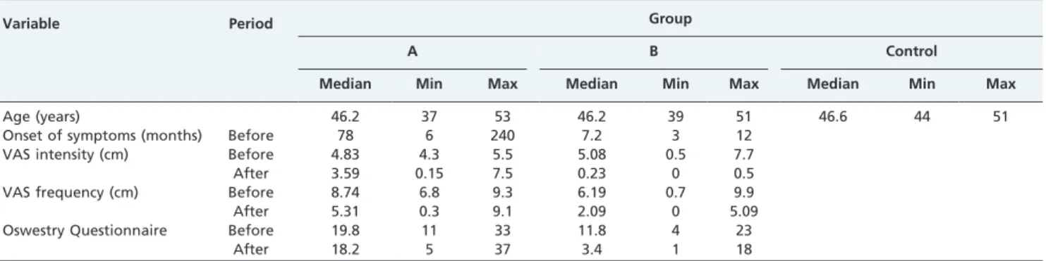
This study was conducted with five patients in each of three groups: group A (strengthening), group B (stabiliza-tion), and a control group. Descriptive information for the groups (median, minimum, and maximum values), includ-ing age, start of symptoms, pain scale (VAS), and Oswestry questionnaire data before and after treatment, is shown in Table 1.
A Pearson correlation analysis between the pain scale and the Oswestry questionnaire responses revealed that higher pain intensity (as assessed using the VAS) correlated with higher Oswestry scores (r = 0.754 andp= 0.007) (Table 2).
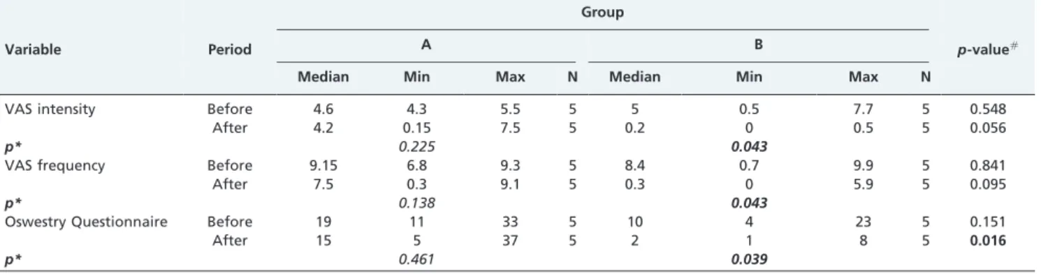
Group B presented significant reductions in both pain (intensity and frequency) (p,0.043) and disability (mea-sured by the Oswestry questionnaire) (p,0.05) after the treatment. However, group A did not show any significant changes (p.0.05). Comparisons between the two groups at each assessment time showed that after the treatment, the Oswestry values of group B were lower than those of group A (p= 0.016) (Table 3).
DISCUSSION
Physical exercise is one of the most widely used methods for the rehabilitation of individuals with chronic low back pain. The primary goals of treating lumbar pain with physical exercise are to improve muscle strength, to maintain or improve flexibility, to heal tissue lesions and to promote spinal segment stability.7,11,12
There are many exercise programs for lower back pain, and they differ in terms of the duration, frequency, and intensity of the training and especially regarding the type of exercise and the way it is performed.11,13 The term ‘‘stabilization exercise’’ is a generic term for any type of exercise that challenges the stability of the spine while training muscle activity patterns and postures that ensure sufficient stability without unnecessarily over-loading tissue.14,15 Trunk stabilization exercises are based on co-contraction of the abdominal and multifidus muscles, and they are performed in a variety of body positions. It has been well documented that individuals with chronic lower back pain present differences in the activation patterns of these muscles (along with histo-morphological abnormalities) compared with the mus-cles of individuals without a history of lumbar pain.16 Stabilization exercises thus aim to improve these muscle activation patterns, thereby diminishing both incapacity and lumbar pain through improvements in trunk muscle contraction.12,15
The results of studies relating to improvements in pain and functional capacity in lower back pain are contra-dictory. In a literature review, Macedo et al.17 found that stabilization exercise programs effectively diminished pain and functional incapacity, but they did not outperform other treatments. Another point of contention is whether stabili-zation training actually interferes with motor control.
In this study, women with chronic lower back pain were divided into two exercise groups (A and B) and were compared in terms of improvements in pain and functional capacity. To ascertain the effects of the postural control
exercises, the patients underwent balance assessments before and after the treatment. They were compared with each other and with a group of women without any history of lumbar pain. The principal result was that the group B (stabilization) patients presented significant reductions after the treatment in both pain (measured using the VAS) and functional capacity (measured using the Oswestry ques-tionnaire). These two evaluations correlated positively with one another. In Table 2, it can be seen that higher pain intensity scores (as assessed using the VAS) correlated with higher Oswestry scores (r = 0.754 and p= 0.007). Similar results were found by Goldby et al.12 in a study in which trunk stabilization exercises were compared with manual therapy and an educational program for patients with chronic lumbar pain. Those authors observed that stabiliza-tion exercises were significantly superior to other interven-tions with regard to pain and function. However, Cairns et al.13 did not find any significant difference between programs using stabilization exercises, general exercises and manual therapy and programs using only general exercises, and manual therapy. Both of their groups presented notable reductions in pain and improvements in functional capacity. They offered two hypotheses to explain these results: the stabilization exercises may not have been as effective as expected, or the general exercises may have actually provided the positive results that were observed.
In the present study, for group A (strengthening), there was no significant change from before to after the treatment. Comparisons between the groups at the two assessment times showed that after the treatment, group B9s values were lower than group A9s, although group B9s values had also been lower before the treatment. Furthermore, the patients in group A presented symptoms of longer duration (a mean of 78 months) than group B9s symptoms (a mean of 7.2 months), which may have influenced group A negatively with regard to the response to treatment.
In relation to the balance measurements, in the get-up-and-go test (Table 6), the mean weight transfer time variable, which measures the time required to transfer the body from a seated position to a standing position, increased significantly from before to after the treatment in group B. The higher the score on this test, the slower this process, thereby signifying a reduction in the ability to move the center of gravity forward and an increased need to prolong the muscular contraction. Changes in the strength or flexibility of the hip or trunk or difficulty in moving the pelvis forward might delay this response. Despite the poor result from this particular test, the difficulty group B Table 1 -Descriptions of group A, group B and the control group (median, minimum and maximum) in relation to age, onset of symptoms, pain scale (VAS) and Oswestry questionnaire before and after treatment.
Variable Period Group
A B Control
Median Min Max Median Min Max Median Min Max
Age (years) 46.2 37 53 46.2 39 51 46.6 44 51
Onset of symptoms (months) Before 78 6 240 7.2 3 12
VAS intensity (cm) Before 4.83 4.3 5.5 5.08 0.5 7.7
After 3.59 0.15 7.5 0.23 0 0.5
VAS frequency (cm) Before 8.74 6.8 9.3 6.19 0.7 9.9
After 5.31 0.3 9.1 2.09 0 5.09
Oswestry Questionnaire Before 19.8 11 33 11.8 4 23
After 18.2 5 37 3.4 1 18
Table 2 -Pearson correlations between the pain scale results and the Oswestry questionnaire responses.
Correlation VAS intensity VAS frequency
VAS frequency r 0.535
p 0.090
Oswestry Questionnaire r 0.754 0.409
patients had in moving the pelvis may have reflected the process of learning to stabilize the trunk and consequently the pelvis. Excessive muscle co-contraction increases joint
stability, but it also increases joint overload and reduces the efficiency of balance strategies, with a negative repercussion on hip joint mobility.15,18,19
Table 3 -Pain scale and Oswestry questionnaire data for the treatment groups and the results of the comparative tests.
Group
Variable Period A B p-value#
Median Min Max N Median Min Max N
VAS intensity Before 4.6 4.3 5.5 5 5 0.5 7.7 5 0.548
After 4.2 0.15 7.5 5 0.2 0 0.5 5 0.056
p* 0.225 0.043
VAS frequency Before 9.15 6.8 9.3 5 8.4 0.7 9.9 5 0.841
After 7.5 0.3 9.1 5 0.3 0 5.9 5 0.095
p* 0.138 0.043
Oswestry Questionnaire Before 19 11 33 5 10 4 23 5 0.151
After 15 5 37 5 2 1 8 5 0.016
p* 0.461 0.039
#Result of the Mann-Whitney test.
*Result of the Wilcoxon paired test.
All significant values are in bold.
Table 4 -Descriptions of the clinical tests of sensory interaction and balance measurements in the treatment groups and the results of the comparative tests.
Group
Variable Period Control A B p-value#
Median Min Max N Median Min Max N Median Min Max N
Mean-Firm-EO Before 0.2 0.1 0.2 5 0.4 0 0.7 5 0.3 0.2 0.4 5 0.515
After 0.2 0.1 0.5 5 0.2 0.2 0.4 5 0.421
p* 0.680 0.317
Mean-Firm-EC Before 0.2 0.1 0.3 5 0.3 0.1 0.8 5 0.3 0.2 0.4 5 0.519
After 0.3 0.2 1.6 5 0.3 0.2 0.4 5 1.000
p* 0.461 0.157
Mean-Foam-EO Before 0.6 0.5 0.9 5 0.7 0.4 1 5 0.8 0.5 1.1 5 0.335
After 0.6 0.3 1.2 5 0.6 0.5 1.2 5 0.548
p* 0.408 0.683
Mean-Foam-EC Before 1.1 0.7 1.5 5 1.4 1 2.1 5 1.4 1.1 1.8 5 0.344
After 1.2 1 2.3 5 1.1 0.8 1.7 5 0.421
p* 0.705 0.279
Firm-EO-Mean-X Before 21 22.2 0.8 5 20.5 20.6 1.2 5 20.7 22.1 0.4 5 0.249
After 20.1 20.6 1.8 5 21 22.5 0.5 5 0.310
p* 0.269 0.893
Firm-EO-Mean-Y Before 20.2 21.9 0.8 5 22.1 23 1.1 5 21 21.7 0.1 5 0.346
After 21.6 22.4 20.2 5 20.8 21.9 0.5 5 0.548
p* 0.893 0.498
Firm-EC-Mean-X Before 20.8 22.1 0.5 5 20.1 20.7 1.1 5 21.5 22.1 0.5 5 0.171
After 0.1 20.5 1.4 5 20.8 22.9 0 5 0.056
p* 0.343 0.893
Firm-EC-Mean-Y Before 20.2 22.2 1.6 5 21.6 22.5 0.7 5 21.2 21.4 0.5 5 0.465
After 21.3 21.9 20.3 5 21 21.8 1.5 5 0.548
p* 0.893 0.715
Foam-EO-Mean-X Before 21 21.5 0.1 5 21.1 22.3 0.9 5 20.8 22.1 20.2 5 0.599
After 20.7 21.2 0 5 21.4 22.5 20.9 5 0.056
p* 0.498 0.176
Foam-EO-Mean-Y Before 2.9 1.8 5.1 5 2.8 23.1 3 5 3.2 1.5 4.6 5 0.295
After 2.6 1.3 3.4 5 2.4 0.9 3.1 5 0.690
p* 0.345 0.345
Foam-EC-Mean-X Before 20.5 21.5 20.4 5 21.1 22.1 0.5 5 20.8 21.1 20.2 5 0.293
After 21.2 21.3 0.2 5 21.2 21.9 0.3 5 0.690
p* 0.588 0.498
Foam-EC-Mean-Y Before 3.5 2 4 5 2.9 22 3.1 5 2.6 1.6 3.9 5 0.175
After 2.5 1.1 4.3 5 2.6 0 2.9 5 0.841
p* 0.465 0.588
#Result of the Kruskal-Wallis test (before) or the Mann-Whitney test (after).
The other measurements from the other tests did not change and did not differ between the groups. Studies have shown that individuals with chronic lumbar pain show postural balance abnormalities relative to individuals with-out any history of pain, especially under conditions that make greater postural demands. Although Mientjes et al.20 and Della Volpe et al.21 did not find any significant difference in static balance in individuals with lumbar pain, when the patients were subjected to more challenging postures, such as closing the eyes and remaining upright on an unstable surface, the subjects with lower back pain presented greater sway than did the control group. In the present study, such abnormalities were not observed. The three groups (A, B, and control) behaved similarly in relation to the four balance tests. After the treatment, groups A and B presented no changes in their results relative to before the intervention. Similar data were obtained by Kuukkanen et al.,22who did not observe any differences in
balance among patients with lower back pain who under-went an exercise program.
There are several hypotheses that might help explain this result. In principle, the step-up test and the get-up-and-go test are sensitive to changes in balance and mobility that may have arisen through many conditions, not only lumbar pain but also conditions associated with aging. The postural balance abnormalities in individuals with lower back pain may be subtle, and the equipment used in this study might not have been sensitive enough to pick up differences between the groups. Such hypotheses were also raised by Kuukkanen et al.,22who came to the conclusion that balance abnormalities could be observed both in patients with significant balance abnormalities and in patients with severe lower back pain.
There are some limitations that must be highlighted in this study, including the small number of subjects in the sample and the differences in the duration of symptoms between the groups.
Table 5 -Step-up balance measurements in the treatment groups and the results of comparative tests.
Group
Variable Period Control A B p-value#
Median Min Max N Median Min Max N Median Min Max N
Mean lift-up index - left Before 18 17 37 5 22 16 29 4 19 16 29 5 1.000
After 19 8 38 5 21 14 25 5 0.690
p* 0.593 0.892
Mean lift-up index - right Before 23 17 36 5 20 17 26 4 19 15 23 5 0.323
After 19 7 36 5 17 14 23 5 0.548
p* 0.180 0.414
Mean movement time - left Before 1.45 1.07 1.82 5 1.465 1.42 2.4 4 1.63 1.5 2.07 5 0.623
After 1.51 1.29 3.21 5 1.61 1.49 1.78 5 0.841
p* 0.109 0.588
Mean movement time - right Before 1.55 0.96 1.74 5 1.45 1.36 1.71 4 1.69 1.66 1.8 5 0.624
After 1.5 1.35 2.95 5 1.63 1.44 1.85 5 0.421
p* 0.593 0.345
Mean impact index - left Before 21 17 36 5 25.5 15 36 4 20 14 24 5 0.805
After 19 6 36 5 17 9 25 5 0.548
p* 0.593 0.345
Mean impact index - right Before 28 19 34 5 21.5 15 32 4 21 17 28 5 0.327
After 18 9 50 5 18 14 29 5 1.000
p* 0.414 0.461
#Result of the Kruskal-Wallis test (before) and the Mann-Whitney test (after).
*
Result of the Wilcoxon paired test.
Table 6 -Get-up-and-go test results for the treatment groups and the results of the comparative tests.
Group
Variable Period Control A B p-value#
Median Min Max N Median Min Max N Median Min Max N
Mean weight transfer time Before 0.24 0.17 0.68 5 0.655 0.58 0.71 4 0.21 0.16 0.59 5 0.050
After 0.55 0.29 0.89 5 0.26 0.23 0.71 5 0.310
p* 1.000 0.043
Mean rising index Before 20 12 26 5 16.5 8 20 4 20 10 23 5 0.319
After 8 7 29 5 16 11 21 5 0.548
p* 0.655 0.223
Mean sway velocity while rising Before 4.3 2 5 5 2.65 1.4 2.8 4 4.4 2.6 4.9 5 0.086
After 2.8 2.3 3.1 5 4.6 1.5 5.4 5 0.690
p* 0.285 0.715
#Result of the Kruskal-Wallis test (before) and the Mann-Whitney test (after).
CONCLUSION
The trunk stabilization exercises were more effective at relieving pain and improving functional capacity than the strengthening exercise program in the patient sample we studied. With regard to the balance measurements, only the mean weight transfer time presented a sig-nificant increase, which was seen in group B after the intervention. The other balance measurements did not change and did not differ between the three groups evaluated.
Stabilization exercises appear to be an important tool for improving lower back pain. This pilot study has helped us to improve the assessment methodology, both in relation to balance and in relation to lower back pain itself. The duration of symptoms needs to be investigated as an important prognostic factor for lumbar pain. Further studies should be conducted taking these factors into account and incorporating adequate sample sizes.
REFERENCES
1. Tsao H, Galea MP, Hodges PW. Reorganization of the motor cortex is associated with postural control deficits in recurrent low back pain. Brain. 2008;131:2161-71, doi: 10.1093/brain/awn154.
2. Suni J, Rinne M, Natri A, Statistisian MP, Parkkari J, Alaranta H. Control of the lumbar neutral zone decreases low back pain and improves self-evaluated work ability – A 12-month randomized controlled study. Spine. 2006;31:E611-E20, doi: 10.1097/01.brs.0000231701.76452.05. 3. Panjabi MM. Clinical spinal instability and low back pain. Journal of
Electromyography and Kinesiology. 2003;13:371-9, doi: 10.1016/S1050-6411(03)00044-0.
4. Ebenbichler GR, Odsson LIE, Kollmitzer J, Erim Z. Sensory motor-control of the lower back: implications for rehabilitation. Med Sci Sports Exerc. 2001;33:1889-98, doi: 10.1097/00005768-200111000-00014. 5. Barr KP, Griggs M, Cadby T. Lumbar stabilization – core concepts and
current literature, part 1. Am J Phys Med Rehabil. 2005;84:473-80, doi: 10. 1097/01.phm.0000163709.70471.42.
6. McGill SM, Karpowicz A. Exercises for spine stabilization: motion/ motor patterns, stability progressions, and clinical technique. Arch Phys Med Rehabil. 2009;90:118-26, doi: 10.1016/j.apmr.2008.06.026. 7. Barr KP, Griggs M, Cadby T. Lumbar stabilization – a review of core
concepts and current literature Part 2. Am J Phys Med Rehabil. 2007;86:72-80, doi: 10.1097/01.phm.0000250566.44629.a0.
8. Vigatto R, Alexandre NM, Correa Filho HR. Development of a Brazilian Portuguese version of the Oswestry Disability Index: cross-cultural adaptation, reliability, and validity. Spine (Phila Pa 1976). 2007;32:481-6, doi: 10.1097/01.brs.0000255075.11496.47.
9. Dolan P, Greenfield K, Nelson R, Nelson I. Can exercise therapy improve the outcome of microdiscectomy? Spine (Phila Pa 1976). 2000;25:1523-32, doi: 10.1097/00007632-200006150-00011.
10. Kirkwood B, Sterne J. Essentials of medical statistics. 2nd ed. Oxford: Blackwell Science. 2003.
11. Kofotolis N, Kellis E. Effects of two 4-week proprioceptive neuromus-cular facilitation programs on muscle endurance, flexibility, and functional performance in women with chronic low back pain. Phys Ther. 2006;86:1001-10.
12. Goldby LJ, Moore AP, Doust J, Trew ME. A randomized controlled trial investigating the efficiency of musculoskeletal physiotherapy on chronic low back disorder. Spine (Phila Pa 1976). 2006;31:1083-93, doi: 10.1097/ 01.brs.0000216464.37504.64.
13. Cairns MC, Foster NE, Wright C. Randomized controlled trial of specific spinal stabilization exercises and conventional physiotherapy for recurrent low back pain. Spine (Phila Pa 1976). 2006;31:E670-81, doi: 10.1097/01.brs.0000232787.71938.5d.
14. Akuthota V, Nadler SF. Core strengthening. Arch Phys Med Rehabil. 2004;85(Suppl 1):S86-S92, doi: 10.1053/j.apmr.2003.12.005.
15. Kavcic N, Grenier S, McGill SM. Quantifying tissue loads and spine stability while performing commonly prescribed low back stabilization exercises. Spine (Phila Pa 1976). 2004;29:2319-29, doi: 10.1097/01.brs. 0000142222.62203.67.
16. Mayer J, Mooney V, Dagenais S. Evidence-informed management of chronic low back pain with lumbar extensor strengthening exercises. Spine J. 2008;8:96-113, doi: 10.1016/j.spinee.2007.09.008.
17. Macedo LG, Maher CG, Latimer J, McAuley JH. Motor control exercise for persistent, nonspecific low back pain: A systematic review. Phys Ther. 2009;89:9-25, doi: 10.2522/ptj.20080103.
18. Henry SM, Hitt JR, Jones SL, Bunn JY. Decreased limits of sta-bility in response to postural perturbations in subjects with low back pain. Clin Biomech. 2006;21:881-92, doi: 10.1016/j.clinbiomech.2006.04.016. 19. Brumagne S, Janssens L, Knapen S, Clayes K, Suuden-Johanson E.
Persons with recurrent low back pain exhibit a rigid postural control strategy. Eur Spine J. 2008;17:1177-84, doi: 10.1007/s00586-008-0709-7. 20. Mientjes MIV, Frank SJ. Balance in chronic low back pain patients
compared to healthy people under various conditions in upright standing. Clin Biomech (Bristol, Avon). 1999;14:710-6, doi: 10.1016/ S0268-0033(99)00025-X.
21. Della Volpe R, Popa T, Ginanneschi F, Spidalieri R, Mazzochio R, Rossi A. Changes in coordination of postural control during dynamic stance in chronic low back pain patients. Gait Posture. 2006;24:349-55, doi: 10. 1016/j.gaitpost.2005.10.009.
22. Kuukkanen TM, Malkia EA. An experimental controlled study on postural sway and therapeutic exercise in subjects with low back pain. Clin Rehabil. 2000;14:192-202, doi: 10.1191/026921500667300454. Table 7 -Single-leg stance test results for the treatment groups and the results of the comparative tests.
Group
Variable Period Control A B p-value#
Median Min Max N Median Min Max N Median Min Max N
Mean-EO-L Before 1.1 0.5 1.3 5 0.8 0.7 1 5 0.9 0.7 1.2 5 0.599
After 0.8 0.5 1.2 5 0.9 0.6 1 5 0.548
p* 0.461 0.131
Mean-EC-L Before 5.2 1.4 12 5 3.6 1.3 12 4 8.4 1.6 12 5 0.805
After 4.9 2 12 5 8.6 1.5 8.7 5 0.548
p* 1.000 0.138
Mean-EO-R Before 0.7 0.5 1.8 5 0.7 0.7 0.8 4 0.9 0.7 1.4 5 0.788
After 0.7 0.7 0.9 5 0.7 0.7 1.1 5 0.841
p* 1.000 0.109
Mean-EC-R Before 5.7 1.2 12 5 1.75 1.3 12 4 8.7 1.3 12 5 0.539
After 2.1 1.3 12 5 12 1.7 12 5 0.690
p* 0.593 0.715
#Result of the Kruskal-Wallis test (before) and the Mann-Whitney test (after).