Rev Saúde Pública 2005;39(5) www.fsp.usp.br/rsp
Prognosis for patients with unilateral Wilms’
tumor in Rio de Janeiro, Brazil, 1990-2000
M arilia Fornaciari Graboisa and Gulnar Azevedo and Silva M endonçab
aServiço de Oncologia Pediátrica. Hospital do Câncer I. Instituto Nacional do Câncer. Rio de Janeiro,
RJ, Brasil. bDepartamento de Epidemiologia. Instituto de Medicina Social. Universidade do Estado do Rio de Janeiro. Rio de Janeiro, RJ, Brasil
Based on master’s dissertation presented to Instituto Medicina Social da Universidade Estadual do Rio de Janeiro, in 2003. Received on 15/9/2003. Reviewed on 2/5/2005. Approved on 5/6/2005.
Correspondence:
Marilia Fornaciari Grabois Serviço de Oncologia Pediátrica Hospital do Câncer I - INCA Praça da Cruz Vermelha, 23 Centro 20230-130 Rio de Janeiro, RJ, Brasil E-mail: [email protected]
Keywords
Wilms tumor. Nephroblastoma, diagnosis. Nephroblastoma, pathology. Prognosis. Anaplasia. Survival analysis.
Abstract
Objective
To analyze the survival and the main prognostic factors among patients with unilateral Wilms’ tumor patients.
Methods
The study cohort included 132 patients with unilateral Wilms’ tumor aged under 15 years, who were enrolled in a pediatric oncology service. Survival curves were calculated using the Kaplan-Meier method and the prognostic factors were analyzed using the Cox proportional hazards model.
Results
The overall survival rate for five years was 84.6%. The survival probabilities for disease in stages I, II, III and IV stages were: 100%, 94.2%, 83.2% and 31.3%, respectively. The survival rate was 89.4% for patients with favorable histology, 66.7% for focal anaplasia and 40% for diffuse anaplasia. All patients with stage IV disease and diffuse anaplasia died (n=4). All patients with stage I disease, regardless of histology, remained alive at the end of the follow-up period.
Conclusions
Among the variables selected for the final model, only the staging and the histology remained associated with high risk of death risk, while patients aged 24 - 47 months presented better prognosis than the other patients. These results showed the importance of establishing the diagnosis at an early stage, and that the histology is fundamental for guiding the appropriate therapy.
INTRODUCTION
Wilms’ tumor is the most frequent primary malig-nant neoplasm of the kidney in childhood worldwide. In Europe it accounts for 97% of the malignant neo-plasms of the kidney.14 The highest incidence rate is observed in the black population of the United States and the lowest in the Asian populations of Shanghai, Philippines and Japan. The incidence may be up to three times greater in high-rate regions than in low-rate regions.6
According to data from the Survival
Epidemiol-ogy and End Results Program (SEER), which includes records of several types of cancer in cities in the United States, this is the childhood cancer with the most fa-vorable prognosis.6
Rev Saúde Pública 2005;39(5) www.fsp.usp.br/rsp Prognosis for Wilms’ Tumor
Grabois MF & Mendonça GAS
ning of any form of treatment, staging, histology, possibility of surgical resection when the tumor was diagnosed, rupture of the tumor (prior to surgery or during the surgical procedure), microscopic involve-ment of the surgical margins and presence of abdomi-nal lymph nodes compromised by the neoplasm. Age at the time of diagnosis was divided into three levels: zero to 23 months, 24 to 47 months and 48 or more months. The cutoff point for the variable “time elapsed between diagnosis and the beginning of any form of treatment” was 15 days, since this was the median time for the study cohort. Staging was assessed ac-cording to the system used by the NWTSG.11 For the final analysis, considering that no patient with stage I disease died, the staging of the disease was strati-fied into three categories. Category 1 included stages I (EI) and II (EII), category 2 included stage III (EIII) and category 3 included stage IV (EIV). The vari-ables of time elapsed between diagnosis and the be-ginning of any form of treatment, possibility of sur-gical resection when the tumor was diagnosed, rup-ture of the tumor (prior to surgery or during the surgi-cal procedure), and microscopic involvement of the surgical margins, were considered to be dichotomous variables. Despite the small number of cases of tumors with anaplasia, the histological variation was classi-fied into three levels: favorable histology (FH), focal anaplasia (FA) and diffuse anaplasia (DA). The com-promising of abdominal lymph nodes by neoplastic cells was also classified into three levels: negative, positive and not found, when there were no lymph nodes present in the surgical specimen.
The assessment of survival took into account deaths that occurrence as a result of the Wilms’ tumor or as a direct consequence of the chosen treatment (date of death). Patients who did not present this event were classified as “censored”. For censored cases, the sur-vival was counted up to the last date recorded in the medical files, or up to the end date of the study. The start time for the observation of each individual (T0) was defined as the date of the medical meeting that decided upon the initial type of treatment for the pa-tient (surgery or chemotherapy).
The survival functions were estimated by means of the Kaplan-Meier method. The log-rank test was ap-plied to verify whether the curves differed between the categories of a given variable.12
The evaluation of the factors associated with the prognosis for Wilms’ tumor was based on the calcula-tion of hazard ratios(HR), with 95% confidence in-tervals (95% CI), using the Cox proportional risks model.12 In the bivariate analysis, the raw HR values were calculated for each variable. Variables consid-Wilms’ tumor (GCBTTW)5 since 1986. Because of
the therapeutic advances achieved by these coopera-tive groups, the prognosis for patients with tumors presenting favorable histology has been improving considerably over the recent decades, reaching four-year survival rates of 90%.13
A variety of factors associated with the prognosis have been identified. The most prominent and per-sistently relevant of these over the course of time have been the stage (lymph node involvement and rupture of the tumor) and the histology.4,11,16
The presence of diffuse anaplasia has been associ-ated with lower survival rates in an statistically sig-nificant manner. This finding is thus characterized as a marker of “unfavorable histology” and one of the most relevant indicators of poor prognosis.1,11 A study by the NWTSG9 showed that tumors presenting diffuse anaplasia had worse prognosis than those with focal anaplasia. Furthermore, some studies have reported that local recurrence of the disease is more frequent in pa-tients with local or diffuse rupture of the tumor during surgery, or in cases where there was microscopic in-volvement of the surgical resection margins.11,16
Thus, the present study aimed to assess the sur-vival and the principal prognostic factors among pa-tients with unilateral Wilms’ tumor.
M ETH O D S
The study cohort included all children with unilat-eral Wilms’ tumor who were enrolled in the pediatric oncology service of a specialized hospital, during the period from January 1990 to December 2000. The criteria for eligibility were that they should be under 15 years of age at the time of the diagnosis and should have unilateral kidney disease, Wilms’ tumor con-firmed by histology and disease that was not so ad-vanced that treatment via surgery, chemotherapy and/ or radiotherapy would be impossible. Patients were not considered eligible for the study if they presented Wilms’ tumors located outside the kidney, or bilat-eral synchronic or metachronic tumors.
All data utilized were obtained from information in medical records up to December 31, 2004. The information was recorded on a clinical form that was designed specifically for this study and was based on clinical forms from the GCBTTW and NWTSG.
begin-!
Rev Saúde Pública 2005;39(5) www.fsp.usp.br/rsp
Prognosis for Wilms’ Tumor Grabois MF & Mendonça GAS
ered to be of clinical importance, as reported in the literature, and those that showed statistical signifi-cance, were selected for the multivariate model.
The graphical methods of log minus log (log-log) and observed versus expected, and also the Schoenfeld residuals test,7 were used to verify the proportionality of risks in the Cox model. The vari-ables analyzed were shown not to violate this princi-ple, as proven by the overall result from the Schoenfeld residuals test(p-value of 0.82), and this test result was not statistically significant for any of the vari-ables included in this final model. All the analyses were performed via the Stata 7.0 statistical software.
RESU LTS
Out of all the patients registered at the hospital with a diagnosis of unilateral Wilms’ tumor during the period of this study, five were not included in the analyses: three of them because they refused ment, and two others because they opted for treat-ment in other States within Brazil.
Table 1 summarizes the characteristics of the sam-ple population. The female to male ratio was 1.4. The median age was 49.11 months, ranging from five to 154 months. The distribution of the staging in rela-tion to age groups showed that EI and EII were the most frequent stages in children under 48 months old, while EIII and EIV were predominant in patients
aged 48 months and over. No child under 24 months old presented stage EIV of the disease.
Fifty patients received chemotherapy prior to ne-phrectomy, because they presented tumors that were considered by the surgeon to be too large and there-fore inoperable, at the time of diagnosis. Among these, 14 presented metastatic disease, of which two were to the liver, ten to the lungs, and two to the liver and lungs. The other 36 patients with non-re-sectable tumors had their staging established after nephrectomy.
Among the 132 patients studied, only 16 presented tumors with anaplasia, of which six were AF and 10 were AD. All other patients were classified as having favorable histology. Twenty children died: 19 of them as a direct consequence of progressive disease and one due to treatment toxicity. Eleven of these 20 pa-tients presented metastatic disease.
The overall survival for the cohort of 132 patients was 84.6%. This rate was achieved after three years and four months, and was maintained up to the end of the follow-up period (Figure 1). The median length of follow up was six years and five months (mean of six years and four months), ranging from 40 days to 14 years.
The five-year survival rates for patients with EI, EII, EIII and EIV, regardless of histology, were 100%,
Table 1 - General characteristics of the patients studied. Rio de Janeiro, 1999-2000.
Variable N % D eath (N ) %
Gender
Femal e 77 58.3 12 15.6
M al e 55 41.7 8 14.5
Age group
0-23 months 30 22.7 3 10.0
24-47 months 43 32.6 3 6.9
≥48 months 59 44.7 14 23.7
Time elapsed between diagnosis and the beginning of any formof treatment
≤15 days 82 62.1 18 22.0
>15 days 50 37.9 2 4.0
Resectability at the time of diagnosis
Yes 82 62.1 5 6.0
N o 50 37.9 15 30.0
Rupture of tumor during the surgical procedure
N o 112 84.9 12 10.7
Local 6 4.5 1 16.7
D iffuse 14 10.6 7 50.0
Stagi ng
I 26 19.7 0 —
II 54 40.9 3 5.5
III 36 27.3 6 16.6
IV 16 12.1 11 68.7
H istopathology
Favorable histology 116 87.9 12 10.3
Focal anaplasia 6 4.5 2 33.3
Diffuse anaplasia 10 7.6 6 60.0
Abdominal lymph nodes
N egati ve 77 58.3 9 11.6
Positive 9 6.8 3 33.3
Not found 46 34.9 8 17.4
M icroscopically compromised margins
N egati ve 122 92.4 14 11.5
" Rev Saúde Pública 2005;39(5) www.fsp.usp.br/rsp Prognosis for Wilms’ Tumor
Grabois MF & Mendonça GAS
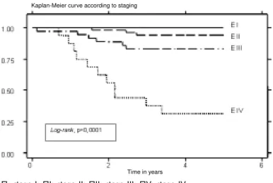
94.2%, 83.2% and 31.3%, respectively. The log-rank test showed highly significant statistical differences between these curves (p<0.0001) (Figure 2). Clearly, the patients with EIV had much lower survival rates than those in other stages.
The five-year survival rate stratified according to the histology, regardless of staging, was 89.4% for patients with FH, 66.7% for patients with FA and 40% for DA. The log-rank test showed highly significant statistical differences between the curves (p<0.0001) (Figure 3), and that the patients with DA had the worst survival rate.
In the bivariate analysis, considering lymph nodes that were positive for neoplastic cells versus those that were negative or not found, the risk of death was not associated with compromised lymph nodes (HR=3.23; 95% CI: 0.87-11.94). This was probably due to the small number of patients with positive lymph nodes (n=9). Comparing the “lymph nodes not found” cases with the “lymph nodes
negative” cases, a tendency towards in-creased risk of death was seen, although not reaching significance (HR=1.51; 95% CI: 0.58-3.91) (Table 2).
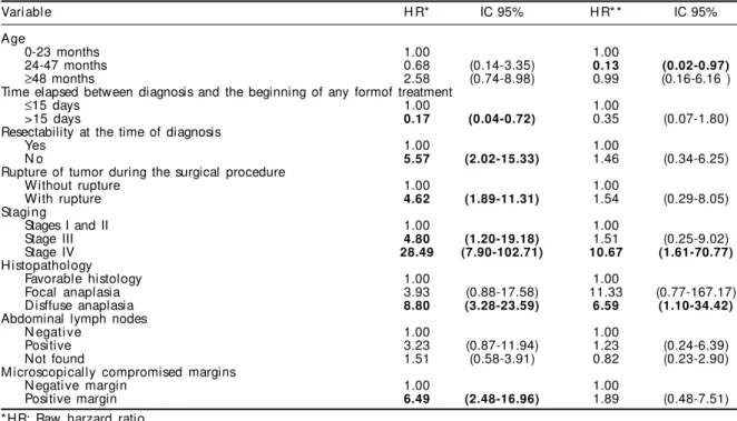
Table 2 also presents the results from the Cox proportional risks model. It was ob-served that the raw HR values pointed to-wards greater risk of death for patients pre-senting disease in advanced stages, espe-cially for patients with EIV and EIII (EIV: HR=28.49; 95% CI: 7.90-102.71; and EIII: HR=4.80; 95% CI: 1.20-19.18), and also for patients presenting AD (HR=8.80; 95% CI: 3.28-23.59), non-resectable tumors at the time of diagnosis (HR=5.57; 95% CI: 2.02-15.33), surgical margins involved in the
dis-ease (HR=6.49; 95% CI: 2.48-16.96) and tumor rupture during the surgical procedure (HR=4.62; 95% CI: 1.89-11.31).
In the multivariate analysis, only the stag-ing and histology maintained statistical sig-nificance. The risk of death for patients with disease in EIV remained high (HR=10.67; 95% CI: 1.61-70.77), while for EIII the sta-tistical significance was lost (HR=1.51; 95% CI: 0.25-9.02). Patients with DA presented six times greater risk of death than those with FH (HR=6.59; 95% CI: 1.10-34.42). The age group of 24 to 47 months now presented de-creased risk of death (HR=0.13; 95% CI: 0.02-0.97). On the other hand, the variables of resectability at the time of diagnosis, sur-gical margins compromised by the neoplasm and rup-ture of the tumor during the surgical procedure lost their statistical significance (Table 2).
D ISCU SSIO N
In the cohort of the present study, the overall five-year survival rate at five five-years was 84.6%. This rate is similar to what was found in the European Cancer Registries Study on Cancer Patients’ Survival and Care (EUROCARE), which presented an overall five-year survival rate of 83%,14 and close to the results reported by SEER6(88%). When only the cases with FH are considered, the survival estimate reaches 89.4%, thus becoming similar to the rates observed in more developed countries, for patient populations with the same characteristics.13
The SIOP17 and NWTS10 studies found survival of 92% and 96% over five and two years, respectively. However, these results refer to selected patients, since
Figure 1 - Overall survival curve via the Kaplan-Meier method.
Time in years Overall survival - Kaplan-Meier curve
Figure 2 - Overall survival according to staging. EI: stage I; EII: stage II; EIII: stage III; EIV: stage IV
#
Rev Saúde Pública 2005;39(5) www.fsp.usp.br/rsp
Prognosis for Wilms’ Tumor Grabois MF & Mendonça GAS
the first group analyzed only tumors with FH, and the second group excluded patients with EIV.
The Brazilian study carried out by GCBTTW showed that for 602 assessable patients with EI to IV and FH, the overall survival was 80% and 73% over two and four years, respectively.*
In the present study, there was no selection of patients, i.e. even those with EIV and ana-plasia (both the focal and diffuse types) were included in the analysis. For these patients, the five-year survival rates, according to staging were: 100% for EI, 94.2% for EII, 83.2% for EIII and 31.2% for EIV. In relation to histology, the survival rates were: 89.4%
for FH, 66.7% for FA, and 40% for AD. The children who presented EIV associated with FA (n=1) or DA (n=4) died. On the other hand, all the patients with EI, regardless of histology, remained alive up to the end of the follow-up period. These findings are in accord-ance with several publications that have reported that there is still no efficient treatment for tumors with diffuse anaplasia, in particular for those that are dis-seminated (EIV), for which the prognosis remains poor.8 Faria et al9 (1996), in a review of 165 cases of Wilms’ tumor with anaplasia that came from the NWTSG, observed 59 cases of death among 126
pa-tients with DA, and 22 of 23 children who presented EIV were among these deaths. On the other hand, only one out of the 39 patients with FA died. In a more recent study carried out by SIOP,3 the patients with EIV treated with chemotherapy prior to nephrectomy and who presented Wilms’ tumor found to be com-pletely necrotic via histology, achieved excellent survival. This again confirms that DA is a predictor for worse prognosis.
In the various clinical trials carried out by NWTSG and SIOP, factors associated with the prognosis for
Figure 3 - Overall survival according to histology.
FH: Favorable histology; FA: Focal anaplasia; DA: Diffuse anaplasia
Kaplan-Meier curve according to histology
Time in years
FH
FA
DA
Table 2 - Results from the bivariate and multivariate analyses. Rio de Janeiro, 1999-2000.
Variable H R* IC 95% H R* * IC 95%
Age
0-23 months 1.00 1.00
24-47 months 0.68 (0.14-3.35) 0.13 (0.02-0.97)
≥48 months 2.58 (0.74-8.98) 0.99 (0.16-6.16 )
Time elapsed between diagnosis and the beginning of any formof treatment
≤15 days 1.00 1.00
>15 days 0.17 (0.04-0.72) 0.35 (0.07-1.80)
Resectability at the time of diagnosis
Yes 1.00 1.00
N o 5.57 (2.02-15.33) 1.46 (0.34-6.25)
Rupture of tumor during the surgical procedure
Without rupture 1.00 1.00
With rupture 4.62 (1.89-11.31) 1.54 (0.29-8.05)
Stagi ng
Stages I and II 1.00 1.00
Stage III 4.80 (1.20-19.18) 1.51 (0.25-9.02)
Stage IV 28.49 (7.90-102.71) 10.67 (1.61-70.77)
H istopathology
Favorable histology 1.00 1.00
Focal anaplasia 3.93 (0.88-17.58) 11.33 (0.77-167.17)
Disffuse anaplasia 8.80 (3.28-23.59) 6.59 (1.10-34.42)
Abdominal lymph nodes
N egati ve 1.00 1.00
Positive 3.23 (0.87-11.94) 1.23 (0.24-6.39)
Not found 1.51 (0.58-3.91) 0.82 (0.23-2.90)
M icroscopically compromised margins
N egati ve margi n 1.00 1.00
Positive margin 6.49 (2.48-16.96) 1.89 (0.48-7.51)
*HR: Raw harzard ratio
**HR: Hazard ratio adjusted for all variables in the table
$ Rev Saúde Pública 2005;39(5) www.fsp.usp.br/rsp Prognosis for Wilms’ Tumor
Grabois MF & Mendonça GAS
Wilms’ tumor cases were identified. Among these, the most prominent were the staging (particularly the compromising of lymph nodes), rupture of the tumor and histology.4,8,16
The results from the bivariate analysis relating to the compromising of lymph nodes, although not sta-tistically significant, point in the same direction as the report by Shamberger et al16 (1999). In this, the patients with EI whose lymph nodes were not repre-sented in the surgical specimen (lymph nodes not found) had six times greater risk of relapse than those with negative lymph nodes.
The NWTSG and SIOP groups have been adopting somewhat different therapeutic approaches for pa-tients with Wilms’ tumor. NWTS recommends ne-phrectomy prior to any other form of treatment, while SIOP recommends the inverse, i.e. chemotherapy prior to nephrectomy. However, both approaches have achieved excellent results that are similar for the majority of patients.2,3,13
In relation to the resectability of the tumor at the time of diagnosis, it was observed among the cases in which the tumor was non-resectable at the time of diagnosis (and in which chemotherapy was received prior to nephrectomy) that there was a larger number of patients with EIV. By excluding these patients, the survival of the group went up from 69.9% to 86.1%, thus showing that the worse survival ob-served in the “non-resectable at the time of diagno-sis” group was due to the higher frequency of cases with EIV (14 cases out of 50 patients). This was proven when this statistically significant variable in the bivariate analysis lost its significance when adjusted for other factors (HR=1.46; 95% CI: 0.34-6.25). Rupture of the tumor and compromised sur-gical margins also lost their statistical significance when adjusted for other variables.
The age at the time of diagnosis deserves attention. Patients aged over 48 months presented greater fre-quency of EIII and IV tumors. This greater frefre-quency of disease at more advanced stages in these patients may be explainable, at least in part, by the delay in diagnos-ing it. A study from the NWTS4 showed that children older than 48 months at the time of diagnosis had a higher probability of relapse and death. This associa-tion was attributed to the delay in the diagnosis, which is more frequently found among older children.
The results presented allow the conclusion that only the staging and histology remained as prognostic fac-tors associated with higher risk of death. These should therefore guide the therapeutic approach. However, other data that might provide prognostic indicators, such as molecular markers, need to be added to the factors that have now been established.
Interdisciplinary studies integrating epidemiology, clinical studies, pathology, surgery and molecular bi-ology, including patients from several treatment cent-ers, should be encouraged with the aim of furnishing further evidence concerning the pathological mecha-nisms implicated in the prognosis. This would thus contribute more towards guiding and planning the treat-ment protocols for patients with Wilms’ tumor.
ACKN O W LED GEM EN TS
To the medical team from the pediatric oncology and pediatric surgery services of Hospital do Câncer (INCA), represented by Dr. Sima Esther Ferman and Dr. Alberto R. Gonçalves, for their collaboration in retrieving the medical records; to Dr. Paulo Antônio Faria and Dr. Teresinha C. F. Sá of the anatomical pa-thology service of INCA for their assistance with the anatomopathological reports; and to Mr Idálio Espinheira of the medical filing service for his col-laboration in data collection.
REFEREN CES
1. Beckwith JB. Renal neoplasms of children. In: Sternberg SS, editor. Diagnostic surgical pathology. 2nd ed. New York: Raven Press; 1994. p. 1741-66.
2. Beckwith JB. Revised SIOP working classification of renal tumors of childhood. Med Pediatr Oncol 2002;38:77-8.
3. Boccon-Gibod L, Rey A, Sandstedt B, Delemarre J, Harms D, Vujanic G et al. Complete necrosis induced by preoperative chemotherapy in Wilms tumor as an indicator of low risk: report of the International Society of Paediatric Oncology (SIOP)
%
Rev Saúde Pública 2005;39(5) www.fsp.usp.br/rsp
Prognosis for Wilms’ Tumor Grabois MF & Mendonça GAS
4. Breslow N, Sharples K, Beckwith JB, Takashima J, Kelalis PP, Green DM et al. Prognostic factors in nonmetastatic, Favorable Histology Wilms´Tumor: Results of the Third National Wilms´Tumor Study. Cancer 1991;68:2345-53.
5. Camargo B de, Franco EL. A randomized clinical trial of single-dose versus fractionated-dose dactinomycin in the treatment of Wilms’ tumor. Results after extended follow-up. Brazilian Wilms’ Tumor Study Group. Cancer 1994;73:3081-6.
6. Chow W-H, Linet MS, Liff JM, Greenberg RS. Cancers in children. In: Schottenfeld D, Fraumeni Jr JF, editors. Cancer epidemiology and prevention. 2nd ed. New
York: Oxford University Press; 1996. p. 1331-69.
7. Cleves MA, Gould WW, Gutierrez RG. An introduction to survival analysis using stata. Texas: Stata Press Corporation; 2002.
8. D´Angio GJ, Breslow N, Beckwith JB, Evans A, Baum E, de Lorimier A et al. Treatment of Wilms´tumor: results of the third national Wilms´tumor study. Cancer 1989;64:349-60.
9. Faria P, Beckwith JB, Mishra K, Zuppan C, Weeks DA, Breslow N et al. Focal versus diffuse anaplasia in Wilms tumor – new definitions with prognostic significance: a report from the National Wilms Tumor Study Group. Am J Surg Pathol 1996;20:909-20.
10. Green DM, Breslow NE, Beckwith JB, Finklestein JZ, Grundy PE, Thomas PR et al. Comparison between single-dose and divided-dose administration of dactinomycin and doxorubicin for patients with Wilms’ tumor: a report from the National Wilms’ Tumor Study Group. J Clin Oncol 1998;16:237-45.
11. Grundy PE, Green DM, Coppes MJ, Breslow NE, Ritchet ML, Perlman EJ et al. Renal tumors. In: Pizzo PA, Poplack DG, editors. Principles and practice of pediatric oncology. 4th ed. Philadelphia: Lippincott
Williams &Wilkins, 2002. p. 865-93.
12. Kleinbaum DG. Survival analysis: a self-learning text. In: Dietz K, Gail M, Krieckeberg K, Singer B, series editors. Statistics in the health sciences. New York: Springer; 1996.
13. Neville HL, Ritchey ML. Wilms’ tumor. Overview of National Wilms’ Tumor Study Group Results. Urol Clin North Am 2000;27:435-42.
14. Plesko I, Kramárová E, Stiller CA, Coebergh J-W, Santaquilani M and the EUROCARE Working Group. Survival of children with Wilms´ tumour in Europe. Eur J Cancer 2001;37:736-43.
15. Pritchard J, Imeson J, Barnes J, Cotterill S, Gough D, Marsden HB et al. Results of United Kingdom Children´s cancer study Group First Wilms´ Tumor Study. J Clin Oncol 1995;13:124-33.
16. Shamberger RC, Guthrie KA, Ritchey ML, Haase GM, Takashima J, Beckwith JB et al. Surgery-related factors and local recurrence of Wilms tumor in National Wilms Tumor Study 4. Ann Surg 1999;229:292-7.