Rev Odontol UNESP. 2016 July-Aug; 45(4): 207-213 © 2016 - ISSN 1807-2577
ORIGINAL ARTICLE
Doi: http://dx.doi.org/10.1590/1807-2577.28515
Counseling and oral splint for conservative treatment of
temporomandibular dysfunction: preliminary study
Aconselhamento e dispositivo interoclusal para tratamento conservador da disfunção temporomandibular:
estudo preliminar
Ana Paula Varela Brown MARTINS
a*, Luana Maria Martins de AQUINO
b, Carolina Beraldo
MELOTO
c, Célia Marisa Rizzatti BARBOSA
daUFJF – Universidade Federal de Juiz de Fora, Governador Valadares, MG, Brasil bUFRN – Universidade Federal do Rio Grande do Norte, Natal, RN, Brasil
cThe Alan Edwards Centre for Research on Pain, McGill University, Montreal, QC, Canada
dFaculdade de Odontologia de Piracicaba, UNICAMP – Universidade Estadual de Campinas, Piracicaba, SP, Brasil
Resumo
Introdução: As desordens temporomandibulares (DTM) envolvem a musculatura mastigatória, as articulações temporomandibulares (ATM) ou ambas. O sintoma mais frequente é a dor, geralmente localizada nos músculos da mastigação, na região pré-auricular, e/ou na ATM, principalmente durante as funções mandibulares. O tratamento principal para pacientes com DTM consiste no alívio da dor. Objetivo: O objetivo deste estudo de relato de casos foi avaliar a redução da sintomatologia dolorosa, utilizando Escala Visual Analógica (EVA), de pacientes com DTM por meio do aconselhamento e utilização de dispositivos interoclusais (DI). Material e método: Um total de 16 indivíduos participaram deste estudo, composto de 4 consultas com intervalo de 7 dias entre cada uma delas (CEP FOP/Unicamp – 137/2009). Na primeira consulta, foi realizado o diagnóstico através do questionário para diagnóstico de DTM, o Research Diagnostic Criteria for Temporomandibular Disorders (RDC/TMD) e entregue a EVA para registrar a intensidade de dor diária. Na segunda consulta, foi realizado o aconselhamento, moldagem para confecção dos DI e a entrega de nova EVA. Na terceira, houve a instalação e ajuste dos DI e a entrega de outra escala, e na última, realização de possíveis ajustes dos DI. Os dados foram analisados por meio do ANOVA a 2 critérios e utilizado o pós-teste de Tukey, com nível de significância de 5%. Resultado: Observou-se diferença significante quando se comparou a intensidade de dor dos indivíduos após instalação dos DI com as informações inicias da pesquisa e após o aconselhamento (p=0,05). Conclusão: A partir dos dados obtidos, pode-se concluir que o tratamento de DTM por meio do DI, seguido do aconselhamento, é eficaz na redução da intensidade da dor.
Descritores: Disfunção temporomandibular; aconselhamento; medição da dor; dor orofacial; placas
oclusais.
Abstract
Introduction: Temporoamndiular Disorders (TMD) involve the masticatory muscles, temporomandibular joint (TMJ) or both. The most common symptom is pain, which is usually located in the muscles of mastication, pre-auricular region, and / or ATM, especially during mandibular function. The main treatment for TMD is related to pain relief.
Objective: The purpose of this case report was to evaluate the reduction of pain symptoms using Visual Analogue Scale (VAS) of patients with TMD treated with counseling and use of occlusal splint (OS). Material and method: 16 subjects had participated in this study, that was composed by 4 appointment with 7-day interval between each (CEP FOP / Unicamp – 137/2009). In the first, an examiner used the Research Diagnostic Criteria for Temporomandibular Disorders (RDC / TMD) to diagnose each patient and delivered a VAS to register the intensity of daily pain. In the second, counseling, molding of both dental arcs to fabricate the OS and the delivery of new VAS were performed. In the third, there was the installation and adjustment of the OS and the delivery of another scale, and in the last, possible adjustments on the OS were done. Data were analyzed by ANOVA two way and Tukey post-test at 5% significance level. Result: There was significant difference when comparing the intensity of pain of individuals after installation of splint with the baseline data and after counseling (p = 0.05). Conclusion: According to the result of this study, the treatment of TMD associating counseling occlusal splint is effective in reducing pain intensity.
Descriptors: Temporomandibular joint disorders; counseling; pain measurement; myofascial pain; occlusal
INTRODUCTION
Orofacial pain is the main symptom of Temporomandibular Disorder (TMD), which motivates the patient to seek treatment. he painful symptomology, which ranges from sensitivity or discomfort to the worst pain possible, may afect the quality of life of the patient on diferent levels. It may interfere with activities of daily living, work performance, and reduce the quality of sleep, among others.
TMD is a collective expression, which involves a series of clinical signs and symptoms that afect the masticatory musculature, temporomandibular joint and associated structures1. here are
several treatment possibilities for patients with this disorder, which may be conservative or invasive, found in the literature. Treatment for TMD is not standardized due to the diferent etiologies and the distinct structures that might be afected.
Treatment for TMD aims for the elimination or remission of the pain and the reestablishment of normal functions. Occlusal Splints (OS) are widely used to treat patients with TMD, with the aim of stabilizing the occlusion, reorganizing the function of the TMJ and masticatory muscles and reducing or eliminating the painful symptoms2. hey are also indicated for patients with
bruxism, especially sleep bruxism, to prevent wear or fractures to the teeth3. In addition to the OS, counseling therapy (CT), consisting
of explaining the etiology, the patient’s current condition and new habits, have been used in the treatment of TMD. Whether combined with other therapies or isolated, CT has obtained favorable results in the control of pain4-6.
Based on the information above, the objective of the present study was to evaluate the reduction of painful symptomology using the Visual Analog Scale (VAS) in patients with TMD, through counseling and using the OS.
Case Report
In the present study, patients with orofacial pain who sought dental services at the outpatient center of the College of Dentistry of Piracicaba, Unicamp, were treated. Individuals were excluded from this report who required treatment for caries and/or periodontal disease, whose pain could interfere with the data analysis; minors under 18 years of age; patients with previous experience or currently using OS and those who did not show up regularly for
treatment, did not use the OS regularly or who required associated pharmacological therapy during the period of the study. he inal sample totaled 16 patients.
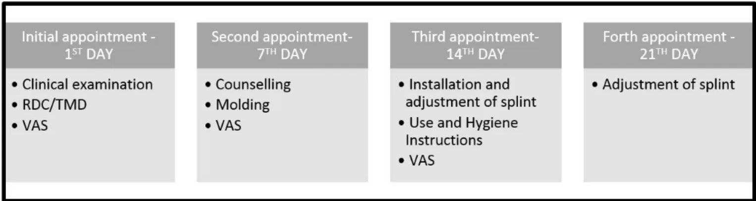
he present study was approved by the Committee for Ethics in Research of the Piracicaba Dental School/Unicamp (protocol no. 137/2009). All patients signed the Free and Informed Consent form. he treatment chosen was based on the conservative treatment protocol, consisting of: diagnostic appointment, counseling and molding appointment, installation of the intraoral splint and occlusal adjustment appointment, and follow up appointment, as illustrated in Figure 1.
At the irst appointment, the Research Diagnostic Criteria for Temporomandibular Disorder (RDC/TMD) were applied so that a inal diagnosis could be made Group I – Muscular Disorders; Group II – Dislocated Disks; Group III - arthralgia, osteoarthritis, osteoarthrosis. A single, calibrated examiner was responsible for implementing and verifying the RDC/TMD. At the end of this appointment, the patient received a VAS to record the pain intensity daily. he VAS was a 100-millimeter straight line, on which there were 2 reference points: at the beginning, identiied as “No pain” and at the end, identiied as “Worst pain possible”. he patient was instructed to draw a vertical line on this line between these reference points, to correspond to the quantitative assessment of the pain sensation. Using a millimetered ruler, the initial point of the scale (absence of pain) and the mark made by the patient were measured. he patients were also instructed not to take any pain medication during the entire study and, if they did, they should register the day and the name of the drug on the VAS.
Ater 7 days, the patient returned to deliver the VAS and begun counseling therapy. his procedure consisted of informing the patient of the diagnosis obtained with the RDC, including possible etiological factors; the prevalence of the disease; explaining and showing visually the standard range of the TMJ as well as the alteration found; and, instructing the patient on new habits which could improve the symptomology. Some of these new habits were: maintaining, whenever possible, the relaxed postural position of the mandible (teeth separated, lips lightly touching, tongue touching the anterior part of the hard palate, not pushing against the teeth); positioning the pillow supporting the head and neck during sleep; avoiding placing objects on the chin and/or biting objects; avoiding foods of thick consistency; limiting the maximum range of the
oral opening, especially when yawning and laughing; performing bilateral chewing, simultaneously, in order not to overtax the muscles and/or joint individually; placing warm compresses, alternating every 20 minutes. Subsequently, the patient would be molded for the construction of the plane lat OS. At the end of this session, the patient received a new VAS to be illed out during the week in which that patient would incorporate these habits, newly-acquired from the counseling.
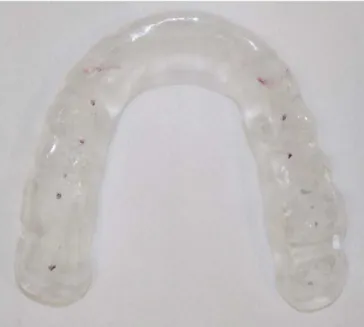
At the third appointment, 7 days ater counseling, the patient returned to deliver the VAS and for installation of the OS (Figure 2), adjustment of the occlusal contacts (Figure 3), coniguration with simultaneous bilateral contacts during closing (Figure 4) and lateral disclusion by canine guide (Figure 5), and protrusion performed by the anterior teeth and disclusion on the posterior teeth. Instructions for the use (24 hours, in the irst week) and hygiene of the device were provided, besides receiving another VAS. At the fourth appointment, one week ater installation of the OS, the patient delivered the VAS and the occlusal contacts were reassessed. For the other appointments, the follow up intervals became broader, at 15, 30 and 60 days from this fourth session.
Having all the VAS, measurements of pain intensities were made. hese data were analyzed using the 2-way Analysis of Variance and the Tukey parametric test (p<0.05).
RESULT
he values obtained from the VAS represent, quantitatively, the intensity of pain that the patients recorded during all stages of the study: ater the initial exam, ater counseling and subsequent installation of the OS. hus, the values obtained were divided by 7 days, which represents the interval between appointments. All data are in Table 1.
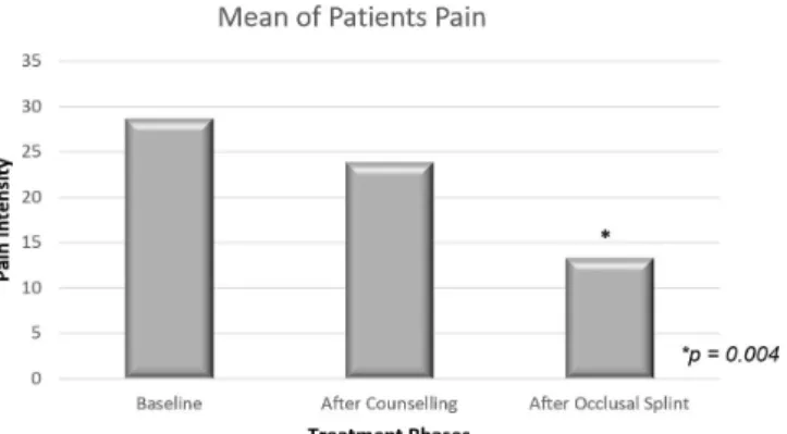
Analyzing the means of the intensity of pain of all patients in the 3 evaluation periods, initial, post-counseling and post-installation of the OS, a reduction in symptomology was found. A reduction in pain intensity was noted in 10 patients ater counseling and in 14 patients ater the installation of the OS, when compared to the initial data (Figure 6).
When the mean values of the baseline were compared with those of the post-counseling, there was no statistically signiicant diference, even noting a reduction in pain intensity values. When comparing the means of the patients between the post-counseling periods and the post-installation of the OS, no statistically signiicant diference was found. However, a reduction in the intensity of the pain reported by the patients was noted. he comparison of the data taken in the moments following the initial exam and ater the inal analysis showed a statistically signiicant diference in the reduction of pain symptomology (Figure 7).
DISCUSSION
he objective of the present study was to evaluate the symptomology of patients treated using the association between counseling and an occlusal splint. he patients treated show the trend for TMD among females (100%) with a mean age of 36.6 years, in the age
Figure 2. Installation of Occlusal Splint.
Figure 3. Adjustment of occlusal contacts.
Figure 4. Coniguration of simultaneous bilateral oclusal contacts during the mandibular closure and canine disclusion (in red).
range characteristic for the prevalence of TMD as shown in the literature7,8.
All patients who participated in the present study presented with a diagnosis of myofascial pain (Group Ia or Ib – RDC/TMD). Myofascial pain is a condition of regional myogenic pain characterized
Table 1. Measurement, in mm, of pain intensity gotten by VAS ater initial exam, ater counseling and ater OS installation periods
Baseline (mm) Ater Counselling (mm) Ater Occlusal Splint (mm)
P1 16.28 27.14 6.14
P2 62.57 48.71 1.14
P3 67.16 57.71 19
P4 53.85 2.71 1.57
P5 39 52.85 33.85
P6 34.71 20.14 37.57
P7 16.71 20.71 14.57
P8 22.71 19.42 3.14
P9 73.28 79.14 32.57
P10 10.71 8.14 0.28
P11 1.85 0 0
P12 7.71 8 41.71
P13 9.85 0 0
P14 15.85 5.57 3.71
P15 4.14 3.42 3
P16 23.28 29.28 15.42
Figure 6. Bar graph of mean pain intensity for each patient throughout the study.
Figure 7. Bar graph of the mean of pain intensity of all patients during the study.
by hypersensitive localized areas of irm muscle tissues, known as trigger points that represent the most common clinical characteristic of myofascial pain9. Palpation produces pain, but the most common
symptom is usually associated with excitatory efects of the trigger points, which induce referred pain9.
he treatment for myofascial pain involves several modalities ranging from conservative to more invasive measures10. Among the
options, the occlusal splint, either in isolation or in combination with other modes of treatment, is the most common form of pain control in patients with TMD6,11-17. A systematic review by Freitas et al.10
revealed the efectiveness of counseling and self-control therapies for the treatment of myofascial TMD, compared with the use of the occlusal splint. hey found similar results between the therapies for spontaneous pain, muscular rigidity upon palpation and maximum opening with or without pain.
he present study revealed an improvement in the symptomology following the installation of the splint, when compared with the initial data and ater counseling. he action mechanisms of the OS are not completely clear, and several hypotheses are proposed to explain its efectiveness18-23. A few assumptions may be cited:
alteration of the positions of the condyles and/or the articulator disks in the TMJ; reduction of electromyographic activity of the masticatory muscles; modiications in parafunctional activities; changes in the occlusal condition of the patients and in the vertical dimension; change in the peripheral impulses (motor or aferent) to the central nervous system; greater awareness of the patient; cognitive placebo efect9,19,21,24-26.
sleep and/or teeth clenching27. Widmalm25 adds that the OS is
efective for testing the efects that changes in occlusion could have on the function of the TMJ and the masticatory muscles prior to restorative treatment, when inding the inluence of the occlusion on the disorder.
It is estimated that the efectiveness of the OS in reducing TMD symptoms is between 70 and 90%12, when used in association
with other methods of treatment. Based on the data recorded by the patients of this case report, a reduction was found in the mean pain intensity in patients among the moments evaluated. his reduction was signiicant only when the post-installation data for the OS were compared to the initial and post-counseling data. he objective of any treatment modality for myofascial pain is to relieve or eliminate the symptom as well as to reestablish muscular function5. he myorelaxant occlusal splints may improve the pain
and the amplitude of the mandibular movements signiicantly. hus, it is considered an efective treatment for patients with myofascial pain (Subgroup I) and Dislocated Disks (Subgroup II)28.
Alencar, Becker5 claimed that the treatment of patients with
myofascial pain should involve low-cost, conservative therapies (counseling and self-care). hey also claimed that the type of format or material of the OS did not afect the results between the groups using the rigid, sot and uncovered interocclusal splints, when only the reduction of pain intensity ater treatment was analyzed.
According to the data from the present study, it was found that some patients had an increase in the intensity of the pain, as seen in patients numbers 1, 5, 7, 9 and 16 ater the installation of the OS. It is currently known that biomedical factors and psychosocial impacts are involved in the development of TMD29.
his may explain the increase in pain among these patients, and highlights the need for a biopsychosocial approach that takes into account the multifactorial nature of this disease in addition to an interdisciplinary treatment involving physiological, psychological
and social aspects which may trigger or perpetuate the chronic pain of TMD30. hus, the treatment for TMD is characterized as
reversible and conservative, and involves self-care strategies and behavioral therapies31,32.
he counseling includes extensive education about the disease and guarantees its benign progress associated with the use of heat or cold therapies, mandible exercises and guidance on reducing parafunctional mandibular activities, progressive muscular relaxation, diaphragmatic breathing training and improved sleep and posture4-6,33-35. his approach does not require profound knowledge
of psychology and treatment strategies, such as cognitive-behavioral treatment and psychotherapies10. So this therapy may be applied
to the treatment of TMD pain by any professional with experience in the prevention and elimination of possible etiological factors responsible of triggering and perpetuating the signs and symptoms of TMD health10.
Regarding the atteendance of patients to other appointments, ater the irst follow up appointment of the OS, a considerable reduction was noticed. his suggests that there was a reduction in pain, and conirms that this symptom is primarily responsible for seeking treatment. Additionally, the acquisition of knowledge about the disease and new habits may promote greater longevity in treatment eicacy.
CONCLUSION
According to the data obtained in the present study, the association between counseling and the OS can be found to be an excellent option for the treatment of patients with myofascial pain. In addition to being conservative, non-invasive and low-cost, this therapeutic modality suggests greater longevity in pain reduction because the patient acquires new habits or behaviors which avoid muscular and joint overtaxing.
REFERENCES
1. Sipilä K, Suominen AL, Alanen P, Heliövaara M, Tiittanen P, Könönen M. Association of clinical findings of temporomandibular disorders (TMD) with self-reported musculoskeletal pains. Eur J Pain. 2011 Nov;15(10):1061-7. http://dx.doi.org/10.1016/j.ejpain.2011.05.001. PMid:21664847.
2. Nelson SJ. Principles of stabilization bite splint therapy. Dent Clin North Am. 1995 Apr;39(2):403-21. PMid:7781834.
3. Boero RP. The physiology of splint therapy: a literature review. Angle Orthod. 1989;59(3):165-80. PMid:2672904.
4. Michelotti A, Iodice G, Vollaro S, Steenks MH, Farella M. Evaluation of the short-term effectiveness of education versus an occlusal splint for the treatment of myofascial pain of the jaw muscles. J Am Dent Assoc. 2012 Jan;143(1):47-53. http://dx.doi.org/10.14219/jada. archive.2012.0018. PMid:22207667.
5. Alencar F Jr, Becker A. Evaluation of different occlusal splints and counselling in the management of myofascial pain dysfunction. J Oral Rehabil. 2009 Feb;36(2):79-85. http://dx.doi.org/10.1111/j.1365-2842.2008.01913.x. PMid:18976268.
6. Truelove E, Huggins KH, Mancl L, Dworkin SF. The efficacy of traditional, low-cost and nonsplint therapies for temporomandibular disorder: a randomized controlled trial. J Am Dent Assoc. 2006 Aug;137(8):1099-107, quiz 1169. http://dx.doi.org/10.14219/jada.archive.2006.0348. PMid:16873325.
7. Macfarlane TV, Blinkhorn AS, Davies RM, Kincey J, Worthington HV. Oro-facial pain in the community: prevalence and associated impact. Community Dent Oral Epidemiol. 2002 Feb;30(1):52-60. http://dx.doi.org/10.1034/j.1600-0528.2002.300108.x. PMid:11918576.
8. Carlsson GE. Epidemiology and treatment need for temporomandibular disorders. J Orofac Pain. 1999;13(4):232-7. PMid:10823035.
10. Freitas RF, Ferreira MÂ, Barbosa GA, Calderon PS. Counselling and self-management therapies for temporomandibular disorders: a systematic review. J Oral Rehabil. 2013 Nov;40(11):864-74. http://dx.doi.org/10.1111/joor.12098. PMid:24102692.
11. Fricton J. Current evidence providing clarity in management of temporomandibular disorders: summary of a systematic review of randomized clinical trials for intra-oral appliances and occlusal therapies. J Evid Based Dent Pract. 2006 Mar;6(1):48-52. http://dx.doi.org/10.1016/j. jebdp.2005.12.020. PMid:17138397.
12. Wassell RW, Adams N, Kelly PJ. The treatment of temporomandibular disorders with stabilizing splints in general dental practice: one-year follow-up. J Am Dent Assoc. 2006 Aug;137(8):1089-98, quiz 1168-9. http://dx.doi.org/10.14219/jada.archive.2006.0347. PMid:16873324.
13. Ekberg E, Nilner M. Treatment outcome of appliance therapy in temporomandibular disorder patients with myofascial pain after 6 and 12 months. Acta Odontol Scand. 2004 Dec;62(6):343-9. http://dx.doi.org/10.1080/00016350410010063. PMid:15848979.
14. Wassell RW, Adams N, Kelly PJ. Treatment of temporomandibular disorders by stabilising splints in general dental practice: results after initial treatment. Br Dent J. 2004 Jul;197(1):35-41, discussion 31, quiz 50-1. http://dx.doi.org/10.1038/sj.bdj.4811420. PMid:15243608. 15. Ekberg E, Vallon D, Nilner M. The efficacy of appliance in patients with temporomandibular disorders of mainly myogenous origin. A
randomized, controlled, short-term trial. J Orofac Pain. 2003;17(2):133-9. PMid:12836501.
16. Ekberg EC, Vallon D, Nilner M. Occlusal appliance therapy in patients with temporomandibular disorders a double-blind controlled study in a short-term perspective. Acta Odontol Scand. 1998 Apr;56(2):122-8. http://dx.doi.org/10.1080/00016359850136102. PMid:9669465.
17. Rubinoff MS, Gross A, McCall WP Jr. Conventional and nonoccluding splint therapy compared for patient with myofascial pain dysfunction syndrome. Gen Dent. 1987 Nov-Dec;35(6):502-6. PMid:3481734.
18. Scopel V, Alves da Costa GS, Urias D. An electromyographic study of masseter and anterior temporalis muscles in extra-articular myogenous TMJ pain patients compared to an asymptomatic and normal population. Cranio. 2005 Jul;23(3):194-203. http://dx.doi.org/10.1179/ crn.2005.028. PMid:16128354.
19. Dubé C, Rompré PH, Manzini C, Guitard F, de Grandmont P, Lavigne GJ. Quantitative polygraphic controlled study on efficacy and safety of oral splint devices in tooth-grinding subjects. J Dent Res. 2004 May;83(5):398-403. http://dx.doi.org/10.1177/154405910408300509. PMid:15111632.
20. Turp JC, Komine F, Hugger A. Efficacy of stabilization splints for the management of patients with masticatory muscle pain: a qualitative systematic review. Clin Oral Investig. 2004 Dec;8(4):179-95. http://dx.doi.org/10.1007/s00784-004-0265-4. PMid:15179561.
21. Kreiner M, Betancor E, Clark GT. Occlusal stabilization appliances: evidence of their efficacy. J Am Dent Assoc. 2001 Jun;132(6):770-7. http://dx.doi.org/10.14219/jada.archive.2001.0274. PMid:11433856.
22. Yap AU. Effects of stabilization appliance on nocturnal parafunctional activities in patients with and without signs of temporomandibular disorders. J Oral Rehabil. 1998 Jan;25(1):64-8. http://dx.doi.org/10.1046/j.1365-2842.1998.00194.x. PMid:9502129.
23. Dao TTT, Lavigne GJ, Charbonneau A, Feine JS, Lund JP. The efficacy of oral splints in the treatment of myofascial pain of the jaw muscles: a controlled clinical trial. Pain. 1994 Jan;56(1):85-94. http://dx.doi.org/10.1016/0304-3959(94)90153-8. PMid:8159444.
24. Ekberg E, Nilner M. A 6-and 12 month follow-up of appliance therapy in TMD patients: a follow-up of a controlled trial. Int J Prosthodont. 2002 Nov-Dec;15(6):564-70. PMid:12475163.
25. Widmalm SE. Use and abuse of bite splints. Compend Contin Educ Dent. 1999 Mar;20(3):249-54, 256, 258-9, quiz 260. PMid:11692335.
26. Okeson JP. The effects of hard and soft occlusal splints on nocturnal bruxism. J Am Dent Assoc. 1987 Jun;114(6):788-91. http://dx.doi. org/10.14219/jada.archive.1987.0165. PMid:3475357.
27. Magdaleno F, Ginestal E. Side effects of stabilization occlusal splints: a report of three cases and literature review. Cranio. 2010 Apr;28(2):128-35. http://dx.doi.org/10.1179/crn.2010.018. PMid:204912Apr;28(2):128-35.
28. Botelho LC, Messora MR, Pereira CV, Pereira SM, Marques LS, Pereira LJ. Estudo longitudinal dos sinais e sintomas de disfunção temporomandibular frente a tratamento conservador com placa estabilizadora em clínica de graduação. Arq Odontol. 2012 Abr-Jun;48(2):76-81. http://dx.doi.org/10.7308/aodontol/2012.48.2.03.
29. Suvinen TI, Reade PC, Kemppainen P, Könönen M, Dworkin SF. Review of aetiological concepts of temporomandibular pain disorders: towards a biopsychosocial model for integration of physical disorder factors with psychological and psychosocial illness impact factors. Eur J Pain. 2005 Dec;9(6):613-33. http://dx.doi.org/10.1016/j.ejpain.2005.01.012. PMid:15978854.
30. Sherman JJ, Turk DC. Nonpharmacologic approaches to the management of myofascial temporomandibular disorders. Curr Pain Headache Rep. 2001 Oct;5(5):421-31. http://dx.doi.org/10.1007/s11916-001-0053-7. PMid:11560807.
31. Dionne RA. Pharmacologic treatments for temporomandibular disorders. Oral Surg Oral Med Oral Pathol Oral Radiol Endod. 1997 Jan;83(1):134-42. http://dx.doi.org/10.1016/S1079-2104(97)90104-9. PMid:9007937.
32. Feine JS, Widmer CG, Lund JP. Physical therapy: a critique. Oral Surg Oral Med Oral Pathol Oral Radiol Endod. 1997 Jan;83(1):123-7. http:// dx.doi.org/10.1016/S1079-2104(97)90102-5. PMid:9007935.
33. Michelotti A, Steenks MH, Farella M, Parisini F, Cimino R, Martina R. The additional value of a home physical therapy regimen versus patient education only for the treatment of myofascial pain of the jaw muscles: short-term results of a randomized clinical trial. J Orofac Pain. 2004;18(2):114-25. PMid:15250431.
35. Wright EF, Domenech MA, Fischer JR Jr. Usefulness of posture training for patients with temporomandibular disorders. J Am Dent Assoc. 2000 Feb;131(2):202-10. http://dx.doi.org/10.14219/jada.archive.2000.0148. PMid:10680388.
CONFLICTS OF INTERESTS
he authors declare no conlicts of interest.
*CORRESPONDING AUTHOR
Ana Paula Varela Brown Martins, UFJF – Universidade Federal de Juiz de Fora, Rua Afonso Pena, 2130, apt. 401, Centro, 35010-000 Governador Valadares - MG, Brasil, e-mail: [email protected]