UDC: 636.7.09:[616.98:579.881.3.083.3 636.7.09:616.993.161]:616-097
Case Report
ABSTRACT
INTRODUCTION
Leishmaniasis in dogs is a serious protozoan zoonosis that infects millions worldwide. Dogs and human get infected by leishmania promastigotes induced by bite of infected sand fl y Phlebotomus spp. Promastigotes multiply into the macrophages as intracellular amastigotes. Clinical signs may appear months or years after the infection (10). Visceral leishmaniasis or Kala-azar is a life-threatening disease that has medical, social and economic importance in the endemic areas. Dogs are the main reservoir for Leishmania infantum. The clinical fi ndings are very different which impedes the diagnosis. Some dogs show clinical
CASE REPORT OF CANINE CO-INFECTION WITH
LEISHMANIA INFANTUM
AND
EHRLICHIA CANIS
Atanaskova Elena
1, Kocevski Zoran
2, Nikolovski Goran
1, Stefanovska Jovana
21Department of internal diseases of companion animals and horses at the Faculty of Veterinary Medicine in Skopje;
2 Department of Parasitology and parasitic diseases at the
Faculty of Veterinary Medicine in Skopje;
Canine leishmaniasis (CanL) due to Leishmania infantum and canine monocytic ehrilichiosis (CME) due to Ehrlichia canis are common diseases with zoonotic potential in the Mediterranean area. Their prevalence in R. Macedonia as a neighboring Mediterranean county is expected. In both diseases similar clinical symptoms can be manifested in dogs such as: lethargy, anorexia, weight loss, epistaxis, fever, pale mucous membranes, enlarged lymph nodes, splenomegaly, ocular signs. This case report present an atypical case of 11 year old female Samoyed with starting single clinical symptom epistaxys. Initial diagnostic procedures revealed the presence only of CanL, which was diagnosed using indirect immunofl uorescence method and ELISA. First laboratory fi ndings showed normal hematological and renal profi les. Dog was put on a treatment with Allopurinol (20mg/kg, p/o) for at least 9 months. Termination of the therapy after 6 months brought a numerous clinical symptoms involving weakness, dehydration, pale mucous membranes lost pupilar refl ex, uremic breath and biochemical parameters revealed a renal failure. Using a commercial ELISA kit Ehrlichia canis as a co-infection was diagnosed. Most probably the second infectious agent was induced in the past 6 months, causing more severe pathological effects than CanL infection alone.
Key words: L. infantum, Ehrichia canis, dog, IFAT, ELISA,
to chronic renal failure or nephritic syndrome. Renal insuffi ciency is one of the classic general symptoms that follow leishmaniasis, but are rarely the only manifestation of the disease (1; 4). Due to the capacity of the kidneys it is estimated that over 75% of the tissue should be destroyed before the appearance of serum creatinine and urea elevations (12).
Canine monocytic ehrlichiosis (CME) is a diseases in dogs caused by rickettsia Ehrlichia canis. Rhipicephalus sanguineus, known as a brown dog tick, traditionally found in southern Europe represent a main vector of the canine ehrlichiosis. (17). When the infected tick ingests a blood meal, the infective agent is inoculated on the feeding site through the salivary secretions. The course of ehrlichiosis in infected dog can be divided into three phases: acute, subclinical and chronic (18). In all three phases a wide variety of clinical signs can be manifested in dogs (6 ). It is proven by Schouls, L.M. et al. (1999) that the same tick species can be a vector for several pathogens and co-infection with different infectious agent by individual ticks can occur. The common clinical signs between leishmaniasis and ehrlichiosis that can mislead to a correct diagnosis are : weakness, lethargy, anorexia, chronic weight loss, epistaxis, fever, pale mucous membranes, enlarged lymph nodes, splenomegaly, ocular signs. Furtheremore L. infantum kDNA in salivary glands of R. sanguineus ticks has been reported (3). Hence, it is very crucial to run several diagnostic test for detection of different pathogens and perform detailed laboratory analyses before defi ning the fi nal diagnosis, especially in cases where ticks were registered on dogs.
MATERIALS AND METHODS
Case history
Our patient was a dog, 11 year old Samoyed, weighing 25kg.The origin of the dog was from town
Clinical observation and diagnosis
First physical examination revealed normal temperature, respiration and pulse and good quality of the coat. Submandibular and popliteal lymph nodes were enlarged with normal temperature and no pain. The clinical signs and medical history didn’t give enough information for diagnosis, so additional examinations were made, such as hematological and biochemical analyses. Based on a single clinical symptom epystaxis a doubt was placed for L. infantum and Ehrlichia canis, hence a few diagnostic methods were performed. Serum sample was tested for Leishmania-specifi c antibodies by an indirect fl uorescent-antibody test (IFAT) according to recommendation from OIE (5) (Figure 1). Samples showing fl uorescence with serum dilutions equal or higher than 1:80 were regarded as positive. Antibody detectionagainst CanL was also performed using anindirect ELISA (INGENASA). For the detection of E. canis antibodies a commercial ELISA kit (SNAP® 4Dx®; IDEXX Laboratories, Inc.
U.S.A.), was performed twice, after the fi rst clinical examination and 6 months after. Both ELISA test were performed according to the manufacturer’s instruction listed in the product package insert.
metabolized by the parasites of leishmania and produces inactive analog inosine that incorporates into parasite’s RNA and produces false protein synthesis. It is used in dogs independently or in combination with pentavalent antimonials, because of its limited toxicity, effi ciency and low price (2). The most common doses are 10-30 mg/kg daily,
divided in two doses every 12 hours, in a long period of time. Side effects are rare. Some of the combined therapeuticprotocols are given in Table 1. Due to the availability of medications, Allopurinol (Zyloprim; GSK, 100mg) therapy (20 mg/kg, p/o 9-12 months) was chosen, with recommendation for controls every 3 months.
Table 1. Recommended therapy protocols
Medicine Doses and applications Duration of therapy
Alopurinol 10-30 mg 2x24 p/o 9-12 months
Amfotericin B 0,5-0,8 mg/kg
2 x per week
Total dose 6-12 mg/kg
creatinin controle once a week.
Meglumin antimonat +
Alopurinol
50 mg/kg;
10-30 mg 2x24 p/o
20-40 days
9-12months
Amfotericin B+
Alopurinol
0,5-0,8 mg/kg; 10-30 mg 2x24 p/o
9-12 months creatinin control
RESULTS
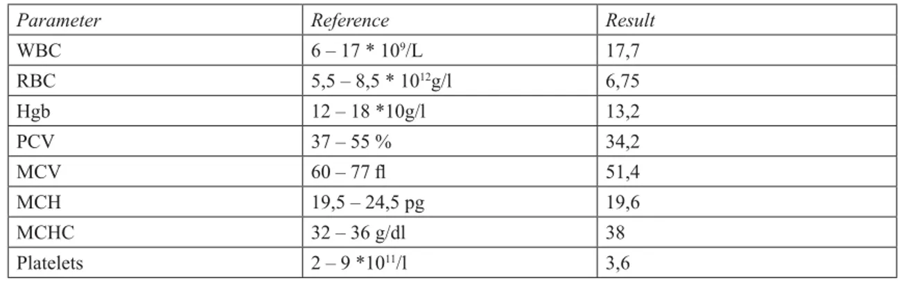
The haematological and biochemical results after the fi rst clinical examination given in the Table
2 and 3 revealed only light leucocitosis. All other results were normal.
Table 2. Hematology analysis
Parameter Reference Result
WBC 6 – 17 * 109/L 17,7
RBC 5,5 – 8,5 * 1012g/l 6,75
Hgb 12 – 18 *10g/l 13,2
PCV 37 – 55 % 34,2
MCV 60 – 77 fl 51,4
MCH 19,5 – 24,5 pg 19,6
MCHC 32 – 36 g/dl 38
Table 3. Biochemical analysis
Biochemical analysis
Parameter Unit Reference Result
ALT U/l 8,02-57,3 15
GGT U/l 1,0-9,7 3
AST U/l 8,9 – 48,5 29,1
Creatinine μmol/L 44,3-138,4 106,7
Urea mmol /L 3,1-9,2 7,9
Glucose mmol /L 3,4-6,0 3.96
Total protein g/L 55,0-75,0 62,3
Albumins g/L 26,0-40,0 27,4
BUN
(blood urea nitrogen)
mmol/L 0-0.12 0.02
Ca mmol /L 2,2 - 3 1,3
Phosphates mmol /L 1 - 2 1,19
Case monitoring
Because of the distance, the owner wasn’t able to bring the dog for controls. After 6 months, therapy was ended by the owners, without prior consultation. Week after the therapy was ended weakness and reduced appetite were noticed. The symptoms after short period of time elevated to low coordination in space, probably blindness, incontinent urination and complete loss of appetite. By our recommendation, the patient was delivered for further examinations. During the clinical examination weakness, dehydration, pale mucous membranes and uremic breath were noticed, pupilar refl ex was lost, eye balls were drawn into the orbit. Palpation of the lymph nodes revealed enlargement of the submandibular
lymph nodes. Body temperature was 36, 6°C, the pulse was barely sensed, shallow breathing was registered. The patient was kept in the clinic’s stationary. Biochemical serum analyses were made and enormously elevated serum creatinin and urea, elevation of the total serum proteins and decreased albumins were noticed. These parameters, especially the levels of creatinin and urea indicated that the glomerular fi ltration was disturbed. Depression of serum albumins and increased activity of serum GGT, gave us indication of early stages of hepatic damage. Elevation of the total serum proteins indicates nephrotoxic condition in the organism. The results are given in table 4.
Table 4. Biochemical analysis 6 month after the initial examination
Biochemical analysis
Indirect immunofluorescent-antibody test (IFAT) was regarded as positive since the titter of the examined serum was 1:320. The other serological method performed with ELISA test confi rm the positivity for L.infantum, because it resulted with an optical density (0,525) higher than the cut off value.
The commercial ELISA test for detection of the presence of E. canis antibodies was negative after the fi rst clinical examination and positive after six months, respectively after the second examination.
Due to the poor general condition of the patient and owner’s approval human euthanasia with Dorminal 20% (200mg pentobarbital sodium, Alfasan Holland) was undertaken.
DISCUSSION
Generally visceral leishmaniasis in dogs has similar symptoms to that in people including: irregular fever, pale mucous membranes and progressive weight loss to cachexia. Hypertrophy of the mononuclear-fagocitic systemis which is often present leads to splenomegalia, hepatomegalia and generalized adenopathy. The most common signs are related to skin lesions as local or diffuse lost of hair, isolate ulcerations of the snout and legs, dermatitis, as well as purulent conjunctivitis and keratitis, apathy, diarrhea and vomiting (15). According to the same authors no-regenerative anemia is an often fi nding. A.Blavier et al. (2001) found that membrane-proliferative glomerulo-nephritis appears as consequence of the circulating immune-complex deposits, which later results in proteinuria. These lesions can progress to chronic renal insuffi ciency or nephritic syndrome. Renal insuffi ciency is one of the classic general symptoms that fallow leishmaniasis, bur rarely are the only manifestation of the disease (1). According to the research done by Soares, M.J.V. et al. (2005) it was noticed that the concentrations of serum creatinin and urea are elevated in 5, 88% from the examined dogs with leishmaniasis. In 61, 76% from the group and 17, 65% from the control group membrane-proliferative glomerulo-nephritis was present. They concluded that kidneys are one of the fi rst organs impacted by the parasite, but due to absence of clinical signs these changes are revealed much later. This case of leishmaniasis was very atypical, since at the beginning of the appearance of the disease only one clinical symptom such as epistaxis was present. This clinical symptom is not present only in leishmaniosis, since platelet-related bleeding, such
REFERENCES
1. Blavier, S. Keroack, Ph. Denerolle, I. Goy-Thollot, L. Chabanne, J.L. Cadore and G. Bourdoiseau. (2001). Atypical forms of canine leishmaniosis; The Veterinary Journal, 162, 108-120
2. Beneth G., Shaw, S.E., (2002). Chemotherapy of canine leishmaniosis. Veterinary Parasitology 106, 315-324.
3. Dantas-Torres F, Lorusso V, Testini G, de Paiva-Cavalcanti M, Figueredo LA, Stanneck D, Mencke N, Brandão-Filho SP, Alves LC, Otranto D. (2010). Detection of Leishmania infantum in Rhipicephalus sanguineus ticks from Brazil and Italy. Parasitol Res. Mar;106(4):857-60.
4. Gad Baneth and Itmar Aroch (2008). Canine leishmaniasis: A diagnostic and clinical challenge, The Veterinary Journal 175(2008) 14-15
5. Gomes, Y.M., Pavia Cavalcanti, M., Lira, R.A., Abath, F.G.C., Alves, L.C., (2008). Diagnosis of canine visceral leishmaniasis: biotechnological advances. The Veterinary Journal 175, 45-52
6. Harrus, S., Waner, T. and Bark, H.: Canine monocytic ehrlichiosis: an update.(1997). Compend. Contin. Educ. Prac. Vet. 19: 431-444
7. Langoni H.; Lucheis S. B., Da Silva R. C.; Castro A. P. B.; Paes A. C. (2005). American visceral Leishmaniasis: a case report. J. Venom. Anim. Toxins incl. Trop. Dis [online], vol.11, n.3, pp. 361-372. ISSN 1678-9199.
8. Laia Solano-Gallego, Guadalupe Miró, Alek Koutinas, Luis Cardoso, Maria Grazia Pennisi, Luis Ferrer, Patrick Bourdeau, Gaetano Oliva, Gad Baneth. (2011). LeishVet guidelines for the practical management of canine leishmaniosis. Parasites & Vectors , 4:86
9. Murray, H.W., (2001). Clinical and
techniques in a cohort of naïve dogs exposed to three consecutive transmission seasons. Journal of Clinical Microbiology 44, 1318-1322;
11. OIE-(Office International des Epizooties) (2008): Manual of Standards Diagnostic tests and Vaccines, Part 2, Section 2.2, Chapter 2.2.11.: LEISHMANIOSIS
12. S.A. Brown, W.A. Crowell, C.A. Brown, J.A. Barsanti and D.R.Finco (1997). Pathophysiology and management of progressive renal disease, The Vererinary jurnal , 154, 93-109
13. Scalone, A., De Luna, R., Oliva, G., Balde, L., Satta, G., Vesco, G., Mignone, W., Turilli, C., Mondesire, R.R., Simpson, D., Donoghue, A.R., Frank, G.R., Gradoni, L.(2002). Evaluation of the Leishmania recombinant K39 antigen as a diagnostic marker for canine leishmaniasis and validation of a standardized enzyme-linked immunosorbent assay. Veterinary Parasitology 104, 275-285
14. Schouls, L.M. et al. (1999). Detection and identifi cation of Ehrlichia, Borrelia burgdorferi sensu lato and Bartonella species in Dutch Ixodes ricinus ticks. J. Clin. Microbiol. 37, 2215–2222
15. Soares M. J. V.; Moraes J. R. E.; Palmeira Borges V.; Miyazato L. G.; Moraes F. R. Renal involvement in visceral leishmaniasis dogs. J. Venom. Anim. Toxins incl. Trop. Dis [online]. 2005, vol.11, n.4, pp. 579-593. ISSN 1678-9199.
16. Smith, R.D., Ristic, M., Huxsoll, D.L. and Baylor, R.A. (1975). Platelet kinetics in canine ehrlichiosis: Evidence for increased platelet destruction as the cause of thrombocytopenia. Infect. Immun. 11: 1216-1221.
17. Shaw SE, Day MJ, Birtles RJ, Breitschwerdt EB. (2001). Tick-borne infectious diseases of dogs. Trends Parasitol , 17:74-80.