5 artigo 497
ORIGINAL ARTICLE
1 – Orthopedist in the Hand Group, Santa Casa de Porto Alegre, Porto Alegre, RS, Brazil. 2 – Resident Physician in Orthopedics, Santa Casa de Porto Alegre, Porto Alegre, RS, Brazil. 3 – Orthopedist and Head of the Hand Group, Santa Casa de Porto Alegre, Porto Alegre, RS, Brazil. 4 – Orthopedist at Hospital Fremap, Madrid, Spain.
Work performed at the Santa Casa Hospital Complex and Hospital Mãe de Deus, Porto Alegre, RS.
Correspondence: Rua Leopoldo Bier 825, sala 301, Santana, 90620-100 Porto Alegre, RS. E-mail: [email protected]
Work received for publication: February 23, 2011; accepted for publication: August 8, 2011.
OSTEOTOMY OF THE DISTAL RADIUS USING A
FIXED-ANGLE VOLAR PLATE
Ricardo Kaempf de Oliveira1, Mário Arthur Rockenbach Binz2, Marco Tonding Ferreira2, Paulo Henrique Ruschel3,
Pedro Delgado Serrano4, Rafael Pêgas Praetzel1
AbSTRACT
Objective: Skewed consolidation of the distal radius, due to sequelae of fractures, may cause functional incapacity, thus leading such patients to present pain, loss of strength and diminished mobility. Based on the excellent results obtained from surgical treatment of unstable fractures of the distal ra-dius through a volar approach and use of rigid fixation with a fixed-angle volar plate, we started to use the same method for osteotomy of the distal radius. Methods: A retrospective review was conducted, and 20 patients treated between Fe-bruary 2002 and October 2009 were found. The mean length of follow-up was 43.9 months (range: 12 to 96 months). The surgical indications were persistent pain, deformity and func-tional limitation subsequent to a dorsally displaced fracture.
Results: The mean preoperative deformity was 27º of dorsal tilt of the distal radius, 87º of ulnar tilt, and 7.3 mm of shor-tening of the radius. All the osteotomies consolidated and the final mean volar tilt was 6.2°, with ulnar tilt of 69.3° and shortening of 1 mm. The mean mobility of the wrist increa-sed by 19.9° (flexion) and by 24° (extension). Mean forearm supination increased by 23.5° and pronation by 21.7°. Grip strength increased from 13.4 to 34.5 pounds. Conclusion: Use of a fixed-angle volar plate for a volar approach towards osteotomy of the distal radius enables satisfactory correction of the deformities and eliminates the need for removal of the synthesis material caused by tendon complications
Keywords – Colles’ Fracture/complications; Osteotomy/uti-lization; Fracture Fixation, Internal
INTRODUCTION
Fractures of the distal radius were originally des-cribed by Abraham Colles in the nineteenth century as a benign pathological condition that did not cause functional limitations, but only esthetic limitations(1). Critical analyses on the results from conservative tre-atment of these fractures, conducted by DePalma in the 1950s, Frykman in the 1960s and Cooney in the 1980s, demonstrated that there was a high rate of un-satisfactory results. Poor results were found in the cases of more than 30% of the patients in these series, and most of these were related to skewed consolida-tion(2). Several authors have now demonstrated the possible sequelae that may occur when such fractures are not treated correctly(3).
Conservative treatment is still widely used, parti-cularly in cases of non-displaced extra-articular frac-tures and in stable displaced facfrac-tures after reduction that do not require immobilization in uncomfortable positions of flexion and ulnar deviation of the wrist. Nonetheless, these fractures may displace secon-darily, thus gradually losing the reduction that was obtained in the emergency treatment(4,5). Skewed consolidation is the most frequent complication following fractures of the distal radius, presenting variable prevalence that ranges from five to 70% of the cases(3,6).
Skewed consolidation of the distal radius can be divided into extra-articular, intra-articular and com-bined (intra and extra-articular)(6). The definition of
The authors declare that there was no conflict of interest in conducting this work
It is also contraindicated for patients with irreducible mediocarpal instability, mediocarpal arthrosis and signs of sympathetic reflex dystrophy(6).
There are several studies on the clinical and radio-logical results from surgical treatment of this deformi-ty. However, these are very heterogenous series, with results that do not focus much on functional evolution.
In the present study, we analyze the clinical and radiological results from surgical treatment of skewed consolidation of the distal radius by means of extra-articular osteotomy in the form of an addition wedge, using a volar approach and fixation with a fixed-angle plate.
MATERIAL AND METHOD
All the patients who underwent corrective surgery for skewed consolidation of the distal radius at the Orthopedics and Traumatology Service of Santa Casa Hospital and Hospital Mãe de Deus, Porto Alegre, RS, between February 2002 and October 2009, were eva-luated retrospectively. This study was analyzed and authorized by the ethics committee of our hospital.
The criteria for indicating surgery were persistent pain, loss of mobility, unacceptable radiographic parameters, functional limitation and incapacity to return to previous professional activities. The exclu-sion criteria were taken to be presence of mild radio-logical deformities, degenerative joint abnormalities and clinical signs of sympathetic-reflex dystrophy. Osteopenia and osteoporosis were not considered to be contraindications, since diminished bone quality is common among patients with sequelae from distal radius fractures. Patients with incomplete follow-up and follow-up of less than 12 months were also ex-cluded from the study.
Out of a total of 32 patients who underwent opera-tions to treat skewed consolidation of the distal radius, 20 patients (14 women and six men) were included in this study because they underwent extra-articular osteotomy with a volar approach. The patients are presented in Tables 1 and 2.
The patients’ mean age was 57.9 years (ranging from 39 to 72 years). The left side was more affec-ted (11 patients), and the other nine patients were affected on the right side. Most of the patients (17) were treated for an extra-articular fracture of the distal radius orthopedically (i.e. conservatively). The other three patients initially underwent closed reduction and intra-articular skewed consolidation was determined by
Knirk and Jupiter as a joint step of more than 2 mm, which is a situation that is considered to present a risk of development of arthrosis(7). In cases of extra-articular skewed consolidation, the definition is based on the an-gle and shortening of the distal radius. The wide range of prevalence of skewed consolidation (5-70%) is due to the disparity of the criteria for defining it. Small changes in the orientation of joint surfaces and length and/or rotational discrepancies should not strictly be considered to be skewed consolidation, since these do not give rise to significant deformities or functional repercussions. They should therefore be excluded from this definition(7,8).
Approximately 25% of distal radius fractures that are treated orthopedically present secondary misa-lignment that evolves into skewed consolidation(9,10). With the development of fixation systems, this rate was reduced to 10%, although 80% of corrective osteotomy procedures are carried out on fractures that were initially treated orthopedically (i.e. con-servatively)(9,11,12).
Skewed consolidation of distal radius fractures alters the normal function of the radiocarpal distal radioulnar joints(13). Shortening and loss of volar tilt of the radius give rise to loss of strength, diminished mobility, deformity and pain(14). Loss of radial tilt greater than 20º in the sagittal plane, 10º in the coronal plane, rotation greater than 10º or shortening greater than 4 mm may produce symptomatic alterations, with indication of surgical correction(15).
The objective of osteotomy is to modify the orientation of the wrist, thus enabling, through bone realignment, homogenous distribution of forces and normal kinematics of the radiocarpal, mediocarpal and distal radioulnar joints.
The results from distal osteotomy of the radius performed because of skewed consolidation are better in patients with extra-articular deformity (80% of the results are good and excellent), with good preoperative mobility (70% mobility compared with the contralateral side), without degenerative joint abnormalities, with good mediocarpal alignment and with radial shortening less than 10 mm(8).
percutaneous fixation using Kirschner wires. In all the cases, the fractures were Fernandez type I(16) and extra-articular(17).
With regard to occupation, retired individuals predo-minated in our sample, forming 55% of the population. All the patients were treated for their initial frac-ture in other clinics or hospitals, and were referred to our service because of their deformities and residual symptoms (Figures 1 and 2).
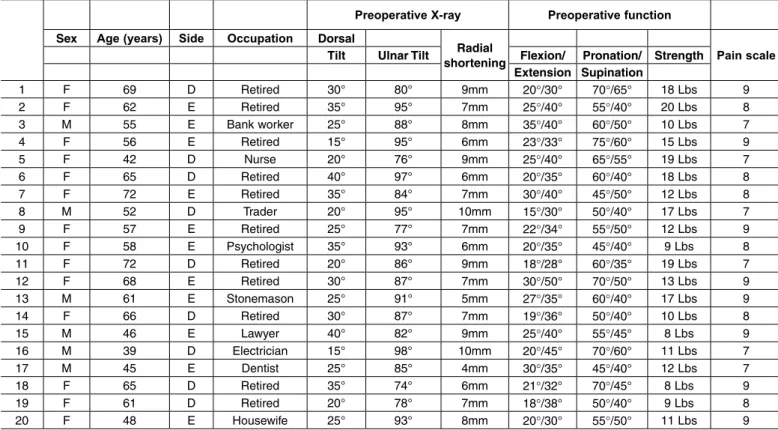
The most frequent complaints among the patients were pain and loss of mobility, followed by esthetic deformity, loss of strength and numbness in the areas innervated by the median nerve. All the patients presented diminished mobility at the preoperative assessment (Figure 3 – A, B, C and D). The mean extension was 36.3° (range: 28-50°) and the mean flexion was 23.1° (range: 15-35°). The mean supination was 46.7° (range: 35-65°) and the mean pronation was 58.2° (range: 45-75°).
Grip strength was measured using a Jamar dyna-mometer placed at setting 3. The mean before surgery was 13.4 pounds (lbs) (range: 8-20 lbs). Among the 20 patients, four underwent carpal tunnel release during the same surgical procedure as the osteotomy. The mean time that elapsed between the fracture and the osteotomy was nine months (range: 3-38 months). The
mean duration of the follow-up after the osteotomy was 43.9 months (range: 12-96 months).
Our retrospective review analyzed the clinical and radiological results and the possible compli-cations consequent to the surgery. The clinical analysis was performed using a visual analogue scale (0-10; in which 0 signified absence of pain and 10 was the worst pain that could be felt), wrist and forearm mobility assessment, grip strength and the DASH questionnaire, applied before and after the operation.
By means of wrist radiographs in posteroante-rior and lateral view, the following radiographic parameters of the distal radius were analyzed: ra-dial tilt, volar angle and shortening of the radius (ulnar variance). The time taken for consolidation of the osteotomy to be achieved was also determi-ned from radiographs.
The synthesis material used varied according to the surgeon’s choice and the health insurance funding that was released. All the fixation systems used the same principle of locked plates, with distal pins at fixed angles. The Synthes volar fixation system was used for 35% of the patients, the GMReis PBA system for 35%, the Engiplan volar plate for 20%, and the Johnson and Johnson DVR plate for 10%.
Table 1 – Epidemiology and preoperative data.
Preoperative X-ray Preoperative function
Sex Age (years) Side Occupation Dorsal
Radial
shortening Pain scale
Tilt Ulnar Tilt Flexion/ Pronation/ Strength
Extension Supination
1 F 69 D Retired 30° 80° 9mm 20°/30° 70°/65° 18 Lbs 9
2 F 62 E Retired 35° 95° 7mm 25°/40° 55°/40° 20 Lbs 8
3 M 55 E Bank worker 25° 88° 8mm 35°/40° 60°/50° 10 Lbs 7
4 F 56 E Retired 15° 95° 6mm 23°/33° 75°/60° 15 Lbs 9
5 F 42 D Nurse 20° 76° 9mm 25°/40° 65°/55° 19 Lbs 7
6 F 65 D Retired 40° 97° 6mm 20°/35° 60°/40° 18 Lbs 8
7 F 72 E Retired 35° 84° 7mm 30°/40° 45°/50° 12 Lbs 8
8 M 52 D Trader 20° 95° 10mm 15°/30° 50°/40° 17 Lbs 7
9 F 57 E Retired 25° 77° 7mm 22°/34° 55°/50° 12 Lbs 9
10 F 58 E Psychologist 35° 93° 6mm 20°/35° 45°/40° 9 Lbs 8
11 F 72 D Retired 20° 86° 9mm 18°/28° 60°/35° 19 Lbs 7
12 F 68 E Retired 30° 87° 7mm 30°/50° 70°/50° 13 Lbs 9
13 M 61 E Stonemason 25° 91° 5mm 27°/35° 60°/40° 17 Lbs 9
14 F 66 D Retired 30° 87° 7mm 19°/36° 50°/40° 10 Lbs 8
15 M 46 E Lawyer 40° 82° 9mm 25°/40° 55°/45° 8 Lbs 9
16 M 39 D Electrician 15° 98° 10mm 20°/45° 70°/60° 11 Lbs 7
17 M 45 E Dentist 25° 85° 4mm 30°/35° 45°/40° 12 Lbs 7
18 F 65 D Retired 35° 74° 6mm 21°/32° 70°/45° 8 Lbs 9
19 F 61 D Retired 20° 78° 7mm 18°/38° 50°/40° 9 Lbs 8
Table 2 - Postoperative data.
Postoperative X-ray Postoperative function
Plate Follow-up (months)
Consolidation
(weeks) Radial
shortening
Pain scale
Volar angle Ulnar angle Flexion/ Pronation/ Grip Extension Supination
1 Synthes 96 8 5° 65° 2mm 40°/60° 85°/80° 26 Lbs 3
2 G M ReisPBA 60 7 2° 75° 2mm 40°/50° 85°/75° 30 Lbs 1
3 Synthes 84 9 1° 68° 3mm 45°/55° 80°/70° 45 Lbs 4
4 G M ReisPBA 90 6 5° 75° 0mm 50°/65° 90°/80° 25 Lbs 1
5 G M ReisPBA 72 16 1° 65° 0mm 40°/55° 80°/70° 30 Lbs 2
6 Hand InnovationsDVR 18 8 2° 67° 1mm 45°/55° 80°/65° 35 Lbs 3
7 Engiplan 36 9 5° 70° 2mm 50°/70° 75°/65° 28 Lbs 1
8 G M Reis PBA 40 7 9° 75° 3mm 40°/60° 80°/75° 37 Lbs 2
9 Engiplan 70 8 3° 67° 0mm 43°/57° 80°/80° 30 Lbs 3
10 Hand InnovationsDVR 10 10 10° 65° 1mm 40°/60° 75°/70° 40 Lbs 4
11 Engiplan 30 6 7° 70° 0mm 30°/45° 80°/60° 28 Lbs 2
12 Synthes 60 9 4° 65° 1mm 50°/65° 85°/70° 47 Lbs 2
13 Synthes 24 10 12° 75° 2mm 45°/65° 80°/70° 32 Lbs 3
14 G M Reis PBA 42 7 5° 73° 2mm 40°/50° 85°/70° 30 Lbs 2
15 G M Reis PBA 36 7 13° 68° 0mm 45°/65° 70°/65° 53 Lbs 1
16 G M ReisPBA 24 8 6° 75° 1mm 40°/65° 90°/85° 48 Lbs 3
17 Synthes 18 10 4° 71° 2mm 55°/70° 85°/70° 33 Lbs 2
18 Synthes 36 8 10° 69° 3mm 40°/60° 80°/70° 27 Lbs 4
19 G M Reis PBA 12 11 2° 67° 0mm 45°/65° 65°/55° 32 Lbs 1
20 Synthes 20 7 9° 62° 0mm 50°/70° 70°/60° 34 Lbs 2
Figure 1 – 62-year-old female patient presenting fracture of the distal third of the radius: anteroposterior (A) and lateral (B) ra-diographs with dorsal angle, shortening and signs of instability. Initially treated with reduction and plaster cast immobilization.
Post-traumatic wrist arthrosis seen on pre and pos-toperative radiographs was graded according to the criteria of Knirk and Jupiter(7). Mild arthrosis was defined as a small decrease in the joint space; mode-rate arthrosis as an evident decrease in the joint space and presence of osteophytes; and severe arthrosis as alteration and complete loss of the joint space and presence of osteophytes and subchondral cysts. None of the patients presented signs of arthrosis on the pre-operative radiographs.
The statistical analysis was performed by compa-ring the preoperative parameters of pain, mobility, radiographic data, strength and DASH with the same parameters after the operation to correct the skewed consolidation of the distal radius by means of extra--articular osteotomy with a volar approach, using the SPSS statistical test. The quantitative variables were investigated regarding whether they were of parame-tric nature using the Kolmogorov-Smirnov test. To
evaluate the means from the pre and postoperative results, the paired Student t test was used.
SURGICAL TECHNIQUE
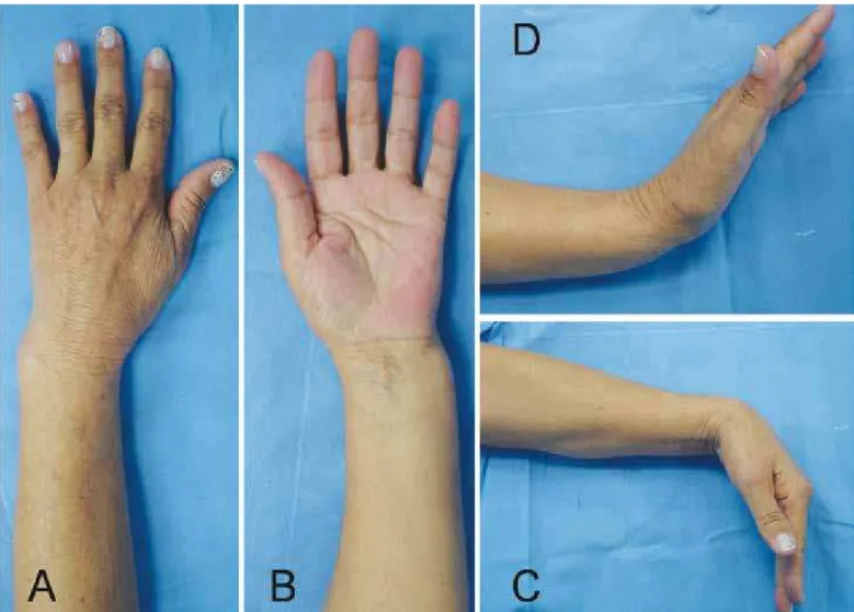
In first place, planning for osteotomy should be done outside of the time of the surgery. Posteroanterior and lateral radiographs of both wrists are produced, and drawings are made to observe the correct site for the osteotomy and the degree of correction necessary, which will determine the positioning of the plate and the quantity of bone graft (Figure 4 – A and B).
Once in the surgical theater, the patient is firstly positioned in dorsal decubitus and a pneumatic tour-niquet is set up on the affected upper limb. The wing of the contralateral iliac is prepared for bone graft harvesting. This facilitates the procedure, because a second surgical team can harvest the bone graft during the procedure on the wrist.
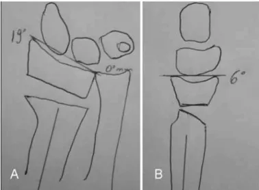
The approach taken for the wrist is from the exten-ded flexor carpi radialis (FCR)(18). A longitudinal volar cutaneous incision is made above the distal portion of the FCR tendon. A Z-shaped break in the incision is made when crossing the flexion crease of the wrist. After making the incision in the skin and subcutaneous cellular tissue, the superficial and deep portions of the FCR tendon sheath are then opened and moved away to the medial side, thereby protecting the median nerve. Going in deeper, the Parona space is encountered, and an incision is made in the pronator quadratus muscle, to elevate it in an L-shape. In this manner, the distal and radial portions of the muscle are deinserted and the muscle belly is pushed out medially, thus preserving its vascularization coming from interosseous arteries. Once this has been done, the volar osseous portion of the distal radius is reached (Figure 5 – A, B and C).
After this stage, the radial septum is released. This is a structure formed by the combined insertion of the tendon of the brachioradial muscle and the first ex-tensor compartment(19). This facilitates the release and future positioning of the metaphysis and epiphysis of the radius. Tenotomy and stretching of the brachiora-dial muscle also makes it easier to maintain the bone reduction, since this removes one of the deforming factors relating to flexion and radial deviation of the distal portion of the radius.
Through this approach, the stage of osteotomy itself is reached. To correctly plan and position the osteotomy site, Kirschner wires are put in place, and these serve as guides for the osteotomy. It is done 1.5 cm proximally to the joint, and following the joint surface, both in
lateral and in anteroposterior projection. This provides enough space to position the plate. The osteotomy is generally performed at the site of the initial fracture.
Once the osteotomy has been done, the cut in the bone makes it possible to pronate the diaphysis (pro-ximal fragment) of the radius, thereby allowing access to the posterior portion of the wrist. At this time, the portion of the dorsal periosteum of the radius that became shortened and thickened due to the skewed consolidation of the fracture should be resected. If the periosteum is not released, correct positioning of the distal fragment of the radius becomes impossible (Figure 6 – A and B).
Correct positioning of the epiphysis of the radius is achieved with the aid of a lamina spreader, which is placed on the most radial and dorsal portion of the bone. This helps to stretch and angle the distal frag-ment of the radius, while the osteotomy is fixed using the plate (Figure 7 – A, B and C). In some cases, the technique described by Prommersberger is used. In this, the distal extremity of the radius is firstly fixed by means of a fixed-angle implant that already is set at the desired corrected angle, and then the plate is used as a fulcrum for stretching and angling the distal epiphysis of the radius(20).
Fixation of the osteotomy is done using a fixed--angle volar plate. This type of plate was initially designed for fixation of distal fractures of the radius and its use was expanded to include fixation of distal osteotomy of the radius. Application of the volar plate to the distal radius automatically corrects the pronated deformity of the distal fragment.
This type of plate fixes the distal bone fragment by means of locking pins that provide subchondral sup-port. The distal pins are inserted individually with the aid of a guide that is fixed to the plate. When fixed the plate, they are incorporated into its structure. Becau-se the distal pins are located juxta-proximally to the subchondral bone, they have the function of providing support and neutralizing the forces exerted on the joint fragment, thereby impeding displacement after the cor-rect alignment of the distal radius has been achieved(4,9). Each pin is angled differently, to follow the anatomical angulation of the distal radius. After the first pin has been inserted, radiographic confirmation of its correct positioning is required in order to avoid placement of pins inside the joint. Proximal bone fixation is achieved by means of 3.5 mm screws (Figure 8 – A and B).
After placement of all of the pins and screws, the cortical-spongy bone graft that was harvested from the iliac wing is implanted. This does not have a me-chanical or structural function but, rather, the function of stimulating bone consolidation in a rapid manner (osteoinduction).
After this, the plate and bone graft are covered by reinserting the pronator quadratus and thus separating them from the flexor tendons. Complementary osteosyn-thesis methods with Kirschner wires or external fixators do not need to be used (Figure 9 – A and B).
Osteotomy to shorten the ulna(21) can be performed in cases of severe shortening of the radius (more than 4 mm). This facilitates reduction of the radioulnar joint and diminished the quantity of bone graft needed. Des-pite this criterion, no additional procedure was perfor-med on the radioulnar joint in our series of patients, or any shortening of the ulna.
The postoperative management for our patients consisted of immediate stimulation of active mo-bilization of the fingers and elevation of the ope-rated limb. The radiocarpal joint was immobilized for three to four weeks, using a plaster-cast splint going from the forearm to the palm, while leaving the metacarpal-phalangeal joints free. Twenty days after the operation, the patients were released to perform tasks of daily living, but without applying force using the operated upper limb. The patients were released to use the upper limb without protec-tion only after radiographic bone consolidaprotec-tion had been achieved.
For all the procedures performed, informed consent was obtained from the patient. During the immediate postoperative period, the patients received prophylaxis against infection using cefazolin (2 g every 8 hours) for a total of 24 hours.
Figure 6 – Osteotomy performed one centimeter proximally to the joint surface (A). After pronation of the radial diaphysis, the dorsal periosteum can be accessed, which is thickened because of consolidation of the fracture (*). In Figure B, after resection of the dorsal periosteum (#), thereby enabling correct positioning and stretching of the distal radius, the extensor tendons of the fingers can be seen (Ω). In the same photo, the brachioradial tendon after Z-shaped stretching can be seen (@).
Figure 7 – Marking of the site for the osteotomy with placement of two Kirschner wires, 1 cm from the joint, in both radiographic views (anteroposterior (A) and lateral (B)). After the osteotomy had been performed, tweezers (lamina spreaders) were placed on the dorsal-radial portion of the bone. This enables stretching and correct positioning of the joint surface.
RESULTS
From a clinical point of view, the patients presented a great improvement in symptoms. The mean on the analogue pain scale went from 8.1 to 2.3 during the postoperative period (with a range from 1 to 4 during the postoperative period), which was a statistically significant improvement (p < 0.0001).
The patients’ mean wrist extension increased sig-nificantly, from 36.3° (range: 28-50°) before the opera-tion to 60.3° after the operaopera-tion (range: 45-70°) (p < 0.001). The mean wrist flexion increased from 23.1° (range: 15-35°) before the operation to 43° after the operation (range: 40-55°) (p < 0.0001). The mean su-pination after the operation was 70.2° (range: 55-85°), which was a significant increase in relation to before the operation, which was a mean of 46.7° (range: 35-65°) (p < 0.0001). The mean pronation increased from 58.2° (range: 45-75°) before the operation to 80° after
the operation (range: 65-90°) (p < 0.001).
Grip strength was measured using a Lamar dy-namometer placed at setting 3. After the operation, grip strength was significantly greater, going from a mean of 13.4 lbs (range: 8-20 lbs) to a mean of 34.5 lbs (range: 26-53 lbs) (p < 0,0001).
With regard to complaints of numbness and tin-gling in the area innervated by the median nerve, car-pal tunnel release was performed only in the patients who had presented symptoms prior to the fracture. All the patients who were operated achieved improve-ments regarding their nerve compression complaints. The DASH questionnaire was not applied to all of the patients after the operation, and complete evolu-tion was only recorded in the cases of 11 of the 20 patients. Among these patients, the mean score was 62 before the operation (range: 24-83) to 11 after the operation (range: 2-28).
From a radiographic point of view, the mean dorsal angle of the radius was 27.2° (range: 15-40°) before the operation to a volar angle of 6.2° after the ope-ration (range: 1-13°) (p < 0.0001). The mean ulnar angle of the radius was 87° (range: 74-98°) before the operation to 69.3° after the operation (range: 62-75°) (p < 0.0001). The mean shortening of the radius was 7.3 mm (range: 4-10) before the operation to 1.1 mm after the operation (range: 0-3) (p < 0.0001).
There was no significant difference in the radio-graphic indices between assessments in the immediate postoperative period and after complete evolution.
At the time of the final assessment, with a mean follow-up of 43.9 months (range: 12-96 months), all the patients presented bone consolidation at the frac-ture sites. The mean time taken for consolidation to be achieved, as seen in radiological examination, was 8.5 weeks (range: 6-16 weeks) (Figure 10 – A and B).
There were no complications relating to absence or breakage of synthesis material or loss of the bone cor-rection that had been obtained (Figure 11 – A and B). There were also no severe complications such as deep infection or neurotendinous lesions. There was no need for a second procedure relating to complications con-sequent to the fracture or to tendon irritation caused by the synthesis material.
Although five patients demonstrated moderate dege-nerative alterations on postoperative radiographs, particu-larly with long-term follow-up, there were no clinical re-percussions like pain, loss of strength or loss of mobility.
Postoperative physiotherapy was indicated starting four weeks after the osteotomy, for all the 20 patients. Of these, five did not do the rehabilitation with a trai-ned professional, for personal reasons. All of the pa-tients achieved full mobility of their fingers; i.e. they remained able to touch the distal flexor crease of the palm with the pulp of the fingers. There was no statis-tically significant difference between the patients who did and those who did not do physiotherapy, when assessed after long evolution (more than 12 months).
At the time of the final assessment, the functional evaluation was done in accordance with the Gartland and Werley scale(17), from which 16 cases were con-sidered to be excellent and four, good.
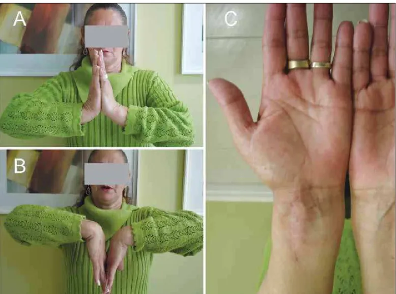
To assess the esthetics, the Frykman scale was used, and 100% of the cases were considered to be excellent(22) (Figure 12 – A, B and C).
DISCUSSION
Deformity of the distal radius may give rise to bio-mechanical changes to the carpus and radiocarpal joint. Restoration of the length and angulation is often a chal-lenge(23). When there is a dorsal angle in the sagittal pla-ne greater than 20°, the contact surfaces and load axes undergo dorsal translation, thus giving rise to dorsal subluxation of the first row of the carpus, which incre-ases the load per surface unit at the level of this joint by 50%, with a dorsal tilt of 20%, and reaches 67% with dorsal deformity of 45 degrees(12). Thus, a change in
Figure 10 – Three months after the operation. Anteroposterior ra-diograph (A) showing consolidation with 2 mm of radial shortening; and lateral radiograph (B) showing complete recovery of the volar angle of the distal radius (15 degrees).
the center of rotation of the wrist is generated, which influences the normal translation of the tendons and re-duces their lever arm, thereby causing a loss of strength of 50-60% in relation to the contralateral limb(10,24).
Skewed consolidation in association with shorte-ning of the radius gives rise to a discrepancy in the distal radioulnar joint. Under normal conditions, 17% of the axial load acting on the wrist is located on the ulnocarpal joint(25). Stretching of the ulna in relation to the radius gives rise to an additional, reaching 42% of the axial load in deformities with positive variance of 2.5 mm(26). The incongruence generated negatively influences the range of motion of the distal radioulnar joint and may cause reduction of forearm rotation by 47% in pronation and 29% in supination(27).
The commonest symptom of extra-articular skewed consolidation is pain. This may occur in the radiocarpal, distal radioulnar or mediocarpal joint. Loss of movement
and weakness of the wrist are often noted. Other compli-cations from skewed consolidation of the radius include tendon tearing – most frequently of the long extensor of the fingers – and compression of the median nerve(2).
The need for correction of deformities in cases of fractures of the distal radius that present symptomatic skewed consolidation is not a new concept. In 1932, Ghormley and Mroz(28) described surgical correction of a deformity of the radius and, in 1946, Speed and Knight(23) presented treatment for skewed consoli-dation of the distal radius by means of a graft from the iliac crest and internal fixation. Nonetheless, the reference technique over the last few years has been the one described by Fernandez in the 1980s(29).
Because it is known that unstable fractures may become displaced, and because reliable new fixation systems for the distal radius have appeared, surgical treatment for distal radius fractures has become refined,
thus diminishing the incidence of skewed consolidation. Different authors have used different approached for correcting the deformities, using dorsal and volar osteotomy to correct the deformity. Anatomical and imaging studies have demonstrated that the volar re-gion of the distal portion of the radius has greater space, which makes it possible to emplace synthesis material. Furthermore, the distal and volar portions of the radius are anatomically planar, which facilitates bone fixation. This location is mechanically favorable for this fixation, as well as avoiding complications with flexor and extensor tendons, and removal of the synthesis material is unnecessary. Using an extended approach from the FCR makes it possible to fully view the operative field and facilitates carrying out the osteotomy that had previously been planned(18,30-32).
It is clear that the best time for treating distal radius fractures is in the acute phase. There is less morbidity and the results are better, especially compared with the results obtained from treating sequelae (skewed consolidation), which requires a longer immobiliza-tion period and a second associated procedure, such as bone graft harvesting from the iliac crest, thereby increasing the morbidity. In agreement with the lite-rature, our series also confirms that most patients who evolve to skewed consolidation of the distal radius are initially treated conservatively (orthopedically).
Misalignment of the fragments of distal radius fractures and their subsequent skewed consolidation may occur in all three spatial axes: sagittal, origi-nating dorsal or volar angulation (less frequently); coronal or frontal, through collapse of the radial column; and axial, through rotation of a distal frag-ment in supination, in fractures with dorsal angula-tion, or in pronation if the angulation is volar(5). Just as joint steps greater than 2 mm are considered to be precursors of arthrosis, sagittal angulation of the distal radius greater than 20° or shortening greater than 4 mm also give rise to degenerative proces-ses in the joint and should be treated surgically in young patients.
According to Fernandez(29), for patients treated by means of osteotomy of the distal radius, there should be an associated procedure to save the distal radioul-nar joint. This author took the view that correct align-ment of the distal radius during osteotomy would not produce sequelae in the distal radioulnar joint. For this reason, and because of the absence of symptoms
of instability in our series, we did not perform any associated procedures on the distal radioulnar joint during the osteotomy on the radius.
By using a volar approach for performing osteo-tomy, it become possible to release the pronator qua-dratus muscle. This is often shortened due to tearing and subsequent fibrosis, caused by the fracturing of the distal radius(18). For this reason, almost complete recovery of supination was justifiable in our group of patients. Moreover, the volar approach enabled re-lease and stretching of the volar portion of the distal radioulnar fragments that were found to be shortened.
Differing from other series that we reviewed, which showed heterogenous groups of patients (both young and old adults) and different deformity charac-teristics (intra or extra-articular), our group of patients was as homogenous as possible, formed by patients with similar deformities and fractures. Like the pu-blished results from previous series, our results show that volar fixation systems for the radius present lower complication rates than do those with dorsal fixation. This incidence diminishes the need for a second sur-gical procedure for removing the synthesis material or performing neurotendinous repairs(33).
A new technique for performing corrections on dorsal deformities of the radius by means of a con-ventional volar approach expanded to the FCR was recently described(20). The distal extremity of the ra-dius was fixed by means of a fixed-angle implant and proximal osteotomy was performed on the distal ra-dius. Through the same approach, a compacted spon-gy bone graft was introduced to fill the bone defect that had been created. In this manner, the need for a second approach to place the bone graft and fix it with dorsal implants was avoided.
Internal fixation systems for osteotomy procedures using plates with locking screws, in which the distal screws are fixed with a plate, reduces the risk of losing the bone alignment after the operation and does not re-quire such a long immobilization time. In many cases, conventional fixation systems do not have the capacity to bear the loads and other associated fixation methods, such as an external fixator, are required(34).
methods, which require preoperative planning and very precise graft fragments in order to maintain the corrective osteotomy in the right position.
Jupiter and Ring(30) demonstrated that performing early osteotomy facilitates the surgical procedure (osteotomy by means of immature bone tissue, with less soft-tissue retraction and less instability of the distal radioulnar joint) and diminishes the time off work. For this reason, young patients with dorsal angulation greater than 20° should be considered to be in a precursor situation of the degenerative process (arthrosis), and a surgical procedure should be indicated.
CONCLUSION
We can affirm that treatment for skewed consolida-tion of the distal radius by means of extra-articular cor-rective osteotomy using a fixed-angle volar plate
produ-ces satisfactory clinical and radiological results among symptomatic patients, like in the series presented.
Our results obtained using a fixed-angle volar plate are good compared with other series of similar lesions that were treated with other types of fixation. We be-lieve that these positive results were due to the lower damage to tendons and the release of the pronator qua-dratus muscle, in comparison with the dorsal approach. This could be seen through the rapid postoperative re-turn to functioning and the low need for physiotherapy. For these reasons, we believe that it is possible, through using fixed-angle volar plates, to routinely manage severe deformities due to sequelae from distal radius fractures, by means of stable fixation and early mobilization of the wrist. Because of the rapid functio-nal recovery, this form of treatment can be considered to be beneficial for all adult patients, including those with lower demands and elderly patients.
REFERENCES
1. Colles A. On the fracture of the carpal extremity of the radius. Edinb Med Surg J. 1814;10:1813-5.
2. Sharpe F, Stevanovic M. Extra-articular distal radial fracture malunion. Hand Clin. 2005;21(3):469-87.
3. Cooney WP 3rd, Dobyns JH, Linscheid RL. Complications of Colles’ fractures. J Bone Joint Surg Am. 1980;62(4):613-9.
4. De Pedro JA, Blanco J, De Cabo A, Garcia de Lucas F, Martin AP, Persson I, et al. Resultados del tratamiento quirúrgico de las fracturas del radio distal. Rev Ortop Traumatol. 2004;48(Supl 1):83-7.
5. Albertoni WM, Faloppa F, Belotti JC. Tratamento das fraturas da extremidade distal do rádio. Rev Bras Ortop. 2002; 37(1):1-4.
6. Jupiter J, Fernandez D. Complications following distal radial fractures J Bone Joint Surg Am. 2001;83(1):1244-65.
7. Knirk JL, Jupiter JB. Intra-articular fractures of the distal end of the radius in young adults. J Bone Joint Surg Am. 1986;68(5):647-59.
8. Pino J, Bartolomé del Valle E, López Graña G, Ferreira Villanova J. Consoli-daciones viciosas tras fracturas del extremo distal del radio: patogenia, indi-caciones y técnicas quirúrgicas. Rev Ortop Traumatol. 2003;47(Supl 1):55-69. 9. Earnshaw SA, Aladin A, Surendran S, Moran CG. Closed reduction of colles
fractures: comparison of manual manipulation and finger-trap traction: a prospective, randomized study. J Bone Joint Surg Am. 2002;84(3):354-8. 10. Prommersberger KJ, Van Schoonhoven J, Lanz UB. Outcome after corrective
osteotomy for malunited fractures of the distal end of the radius. J Hand Surg Br. 2002;27(1):55-60.
11. Fernandez DL. Radial osteotomy and Bowers arthroplasty for malunited frac-tures of the distal end of the radius. J Bone Joint Surg Am. 1988;70(10):1538-51. 12. França Bisneto EN, Paula EJL, Resende MR, Mattar Júnior R, Zumiotti AV.
Fratura distal do rádio em pacientes com mais de 60 anos: placas ortogonais versus placa volar. Rev Bras Ortop. 2010;45(6):590-5.
13. Prommersberger KJ, Lanz U. Corrective osteotomy for malunited Colles frac-tures. Orthop Traumatol. 1998;6:75-87.
14. Fernandez DL, Jupiter JB. Fractures of the distal radius. A practical approach to management. New York: Springer-Verlag; 1995.
15. Castaing J. Les fractures récentes de l´extremité inférieure du radius chez l´adulte. Rev Chir Orthop. 1964;50:581-696.
16. Fernandez DL. Fractures of the distal radius: operative treatment. Instr Course Lect. 1993;42:73-88.
17. Gartland JJ Jr, Werley CW. Evaluation of healed Colles’ fractures. J Bone Joint Surg Am. 1951;33(4):895-907.
18. Orbay J, Badia A, Khoury RK, Gonzalez E, Indriago I. Volar fixed-angle fixa-tion of distal radius fractures: the DVR plate. Tech Hand Up Extrem Surg. 2004;8(3):142-8.
19. Orbay JL, Fernandez DL. Volar fixation for dorsally displaced fractures of the
distal radius: a preliminary report. J Hand Surg Am. 2002;27(2):205-15. 20. Prommersberger KJ, Lanz UB. Corrective osteotomy of the distal radius through
volar approach. Tech Hand Up Extrem Surg. 2004;8(2):70-7.
21. Milch H. Cuff resection of the ulna for malunited Colles fractures. J Bone Joint Surg Am. 1941;23(2):311-3.
22. Frykman G. Fracture of the distal radius including sequelae - shoulder hand- finger syndrome, disturbance in the distal radio-ulnar joint and impairment of nerve function. A clinical and experimental study. Acta Orthop Scand. 1967;(Su-ppl 108): 3+.
23. Speed JS, Knight RA. The treatment of malunited Colles´s fractures. J Bone Joint Surg. 1945;27(3):361-7.
24. Shea K, Fernandez DL, Jupiter JB, Martin C Jr. Corrective osteotomy for mal-united, volarly displaced fractures of the distal end of the radius. J Bone Joint Surg Am. 1997;79(12):1816-26.
25. Iwasaki N, Minami A, Miyazawa T, Kaneda K. Force distribution through the wrist joint in patients with different stages of Kienböck’s disease: using com-puted tomography osteoabsorptiometry. J Hand Surg Am. 2000;25(5):870-6. 26. Werner FW, Palmer AK, Fortino MD, Short WH. Force transmission through
the distal ulna: effect of ulnar variance, lunate fossa angulation, and radial and palmar tilt of the distal radius. J Hand Surg Am. 1992;17(3):423-8.
27. Bronstein AJ, Trumble TE, Tencer AF. The effects of distal radius fracture malalignment on forearm rotation: a cadaveric study. J Hand Surg Am. 1997;22(2):258-62.
28. Ghormley RK, Mroz RJ. Fractures of the wrist. A review of one hundred seventy-six cases. Surg Gynec Obstet. 1932;57:377-81.
29. Fernandez DL. Correction of post-traumatic wrist deformity in adults by osteotomy, bone-grafting, and internal fixation. J Bone Joint Surg Am. 1982;64(8):1164-78. 30. Jupiter JB, Ring D. A comparison of early and late reconstruction of malunited fractures of the distal end of the radius. J Bone Joint Surg Am. 1996;78(5):739-48. 31. Linder L, Stattin J. Malunited fractures of the distal radius with volar angulation: corrective osteotomy in 6 cases using the volar approach. Acta Orthop Scand. 1996;67(2):179-81.
32. Kamano M, Honda Y, Kazuki K, Yasuda M. Palmar plating for dorsally displaced fractures of the distal radius. Clin Orthop Relat Res. 2002;(397):403-8. 33. Kambouroglou GK, Axelrod TS. Complications of the AO/ASIF titanium distal
radius plate system (pi plate) in internal fixation of the distal radius: a brief report. J Hand Surg Am. 1998;23(4):737-41.
34. Ring D, Roberge C, Morgan T, Jupiter JB. Osteotomy for malunited fractures of the distal radius: a comparison of structural and nonstructural autogenous bone grafts. J Hand Surg Am. 2002;27(2):216-22.