04 artigo 486
ORIGINAL ARTICLE
1 – Head of the Pediatric Orthopedics Clinic of Hospital de Acidentados – Goiânia and of the Pediatric Orthopedics Clinic of Hospital das Clínicas da Universidade Federal de Goiás (UFG) – Goiânia, GO, Brazil.
2 – Resident of the Orthopedics and Traumatology Department of Hospital das Clínicas da Universidade Federal de Goiás (UFG) – Goiânia, GO, Brazil.
3 – Master’s Degree, Assistant Professor of the Department of Orthopedics and Traumatology of Hospital das Clínicas da Universidade Federal de Goiás (UFG) – Goiânia, GO, Brazil.
Study conducted at the Pediatric Orthopedics Clinic of the Department of Orthopedics and Traumatology of Hospital das Clínicas da Universidade Federal de Goiás (UFG) – Goiânia, GO. Mailing address: Dr. Frederico Barra de Moraes, Departamento de Ortopedia e Traumatologia e Cirurgia Plástica do HC-FMUFG, Primeira Avenida, Sem Número, 3° andar, Setor Universitário – 74605-085 – Goiânia, GO. Email: [email protected]
Study received for publication: 5/31/2010, accepted for publication: 6/22/2011.
CLINICAL AND RADIOLOGICAL EVALUATION ON
DEVELOPMENTAL HIP DYSPLASIA AFTER SALTER
AND OMBRÉDANNE PROCEDURE
Válney Luiz da Rocha1, André Luiz Coelho Thomé2, Daniel Labres da Silva Castro2, Leandro Zica de Oliveira2, Frederico Barra de Moraes3
ABSTRACT
Objective: To evaluate the clinical and radiological medium-term results from surgical treatment of developmental hip dysplasia through Salter innominate bone osteotomy and Ombrédanne femoral shortening. Methods: Fourteen patients were evaluated, with surgical treatment on 18 hips (seven right-side hips and eleven left-side hips) using the proposal technique, performed between 1998 and 2008. The Dutoit and Severin criteria were used respectively for clinical and radiographic evaluations. Results: The average preoperative index for the seven right-side hips was 43.3º (40º to 50º), and this was corrected through surgery to an average of 31.57º (24º to 42º). The average preoperative index for the eleven left-side hips was 42.1º (36º to 56º), and this was corrected through surgery to an average of 30.36º (20º to 44º). There was a statistically significant difference between the preope-rative and postopepreope-rative acetabular indexes, with P < 0.05.
INTRODUCTION
Developmental hip dysplasia (DHD) involves se-veral abnormalities, which range from hip ligament laxity, leading to instability, to dislocation with loss of the anatomical relationship between the femoral head and the acetabulum. The femoral head can remain spherical or appear posteromedially flattened, and the acetabulum progressively becomes thick, shallow and oblique. It can be classified as teratologic and typical, and the latter is subdivided into dislocatable, subluxated and dislocated hip.
7KHDXWKRUVGHFODUHWKDWWKHUHZDVQRFRQIOLFWRILQWHUHVWLQFRQGXFWLQJWKLVZRUN
This article is available online in Portuguese and English at the websites: www.rbo.org.br and www.scielo.br/rbort
The clinical evaluation showed that there were seven ex-cellent hips (38.9%), eight good ones (44.4%), three fair hips (16.7%) and no poor ones (0%). By grouping the hips rated good and excellent as satisfactory and those rated poor and fair as unsatisfactory, 83.3% of the results were seen to be favorable. There were no statistically significant correlations between occurrences of complications and patient age at the time of surgery or between complications and the preopera-tive acetabular index (p > 0.05). The complications observed consisted of one case each of subluxation, osteonecrosis and osteonecrosis together with subluxation. Conclusion: The combined procedure of Salter and Ombrédanne is a viable option for treating developmental hip dysplasia after patients have started to walk.
Keywords - Hip Dislocation, Congenital/surgery; Surgical Procedures, Operative/methods; Bone Diseases, Develop-mental; Hip/growth & development
The etiology of DHD remains unknown. Ethnic and genetic factors are important. The genetic factors can determine acetabular dysplasia, ligament laxity, or both, as reported by Wynne-Davies(1). Mechanical factors, such as the intrauterine position and post-natal habits, are added to the preexisting factors. In several studies in scientific literature, the incidence of DHD has varied from 2 to 17 per 1,000(3). In Brazil, Volpon and Carvalho Filho(2) demonstrated an incidence of 2.31 per 1,000.
651
tive and analytical manner, using the Student’s t, paired Student’s t, chi-square, ANOVA and Pearson methods, with the intention of establishing statistical signifi-cance between the clinical and radiological parameters.
RESULTS
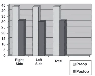
The hips were grouped for analysis according to the affected side. In the seven right-side hips, the preopera-tive index ranged from 40° to 50° (average of 43.3°), and was corrected through surgery to an average of 31.57° (24° to 42°), while the 11 left-sided hips had preoperative mean of 42.1° (36° to 56°), evolving to 30.36° (20° to 44°). The paired Student›s t-test was used to analyze these data, obtaining a statistically sig-nificant result (p < 0.001) (Figure 2).
The clinical evaluation, according to Dutoit
et al(5), showed seven excellent hips (38,9%), eight
good hips (44.4%), three fair ones (16.7%) and no poor ones (0%). By grouping the hips rated good and excellent as satisfactory and those rated poor and fair as unsatisfactory, 83.3% of the results were seen to be favorable (Table 1).
The radiological evaluation showed six excellent hips (33.3%) (Figure 3), 10 good hips (55.6%), no fair ones (0%) and two poor ones (11.2%). By grouping the hips rated good and excellent as satisfactory and those rated poor and fair as unsatisfactory, 88.9% of the results were seen to be favorable (Table 2).
The complications observed consisted of one case each of subluxation, osteonecrosis and osteonecrosis together with subluxation, while the case of isolated subluxation was treated with another surgical proce-dure. There were no cases of infection, fracture, sig-nificant lower limb dysmetria or neurovascular lesion. Both the clinical picture, based on the criteria of Dutoit
et al(5), and the radiological picture, according to Severin(3),
were associated with the preoperative acetabular index. However, statistical relevance was not achieved.
The clinical and radiological pictures were asso-ciated with one another by the chi-square test, yet no results with statistical value were obtained.
After the data analysis, it was observed that the evaluation of pre- and postoperative acetabular indi-ces presented statistical significance with p < 0.05. In applying the Student’s t-test, no statistical signifi-cance was obtained between the occurrence of com-plications and the patient’s age at the time of surgery, and the preoperative acetabular index (p > 0.05). CLINICAL AND RADIOLOGICAL EVALUATION ON DEVELOPMENTAL HIP DYSPLASIA AFTER SALTER
AND OMBRÉDANNE PROCEDURE
degree of acetabular dysplasia and proximal femoral dysplasia. A surgical option for the treatment of DHD, after walking has started, is the association between Salter innominate bone osteotomy and Ombrédanne femoral shortening. This association is geared towards decreasing pressure in the femoral head that will be reduced surgically into the acetabulum, compensating the contracture of soft parts.
The aim of this study is to evaluate the clinical and radiological medium-term result of the surgical treatment of DHD through the Salter procedure and Ombrédanne femoral shortening osteotomy.
MATERIALS AND METHODS
Fourteen patients were evaluated, with surgical treatment on 18 hips between 1998 and 2008 by the Salter and Ombrédanne technique. None of the pa-tients had received previous treatment for DHD, and they did not undergo postoperative physiotherapy or rehabilitation. The study was approved by the Re-search and Ethics Committee of the hospital where the trial was carried out.
The age of the patients, who were all female, ranged from two to eight years, and four presented bilateral DHD. Seven right-side hips and 11 left-side hips were affected. The ideal acetabular index has an acceptable maximum value of 30°(3). The average postoperative immobilization time with pelvic-podalic plaster cast was 2.5 months. In the bilateral cases there was an average interval of six months between the two pro-cedures. All the cases were operated by the same or-thopedic surgeon (Figure 1). No preoperative traction was performed in any case.
The osteosynthesis material used in the procedures was removed, on average, after one year of postopera-tive follow-up. The clinical and radiological evalua-tion occurred with average outpatient follow-up of 56 months (26 to 132 months).
Clinical and radiographic criteria were used to evaluate the results. The radiographs were evaluated by the Severin criteria(3), which take into account the acetabular (AC) and Wiberg’s(4) CE angles, sphericity of the femoral head, dislocation or subluxation of the hip and the presence or absence of arthrosis. The clini-cal profile was analyzed by the criteria of Dutoit et al(5)
based on hip stability and mobility, pain, lameness and the Trendelenburg test.
653
DISCUSSION
The physical examination to identify cases of DHD should be carried out on a routine basis on all new-borns. The Ortolani maneuver, described in 1948 by Marino Ortolani, when positive, allows the diagnosis of DHD; however, a negative result does not rule out the diagnosis, since some hips are unstable, yet not dislocated. The Barlow provocative maneuver allows the diagnosis of hip instability. On the other hand, in children over three months of age, the Ortolani ma-neuver can be negative, since even if the hip remains dislocated, it is no longer possible to place the femoral head in the acetabulum. As regards the Barlow ma-neuver, it should be emphasized that many newborns
Right Side
Left
Side Total 45
40 35 30 25 20 15 10 5 0
Preop
Postop
Figure 2 – 'LVWULEXWLRQRIWKHSUHDQGSRVWRSHUDWLYHDFHWDEXODU
LQGH[DFFRUGLQJWRWKHDIIHFWHGVLGHRIWKHKLS
Table 1 – 5HVXOWRIWKHFOLQLFDOHYDOXDWLRQDFFRUGLQJWR'XWRLW
Dutoit % %
Excellent 7
Good
Fair
Poor 0
Total
Figure 3 – 5DGLRORJLFDOHYROXWLRQRIDIHPDOHSDWLHQWZLWK'+'VXEPLWWHGWR6DOWHUDQG2PEUÑGDQQHRVWHRWRP\1RWHWKHH[FHOOHQW
UDGLRORJLFDOUHVXOWDIWHUPRQWKVRISRVWRSHUDWLYHIROORZXS
Table 2 – 5HVXOW RI WKH UDGLRORJLFDO HYDOXDWLRQ DFFRUGLQJ WR
6HYHULQ
Severin % %
1 – Excellent 6
2 – Good 10
3 – Fair 0
4 – Poor 1
5 – Poor 1
6 – Poor 0
Total
testing positive in the first examination become nega-tive after two or three weeks.
In the dislocated hip, the treatment consists of con-centric and atraumatic reduction of the femoral head inside the acetabulum. Before the walking phase, this treatment can be conservative; however, as the infant starts to walk, there is a tendency for interposition of soft tissues (round ligament, labrum and capsule) in this joint and open reduction is necessary. Once reduction is obtained, this can be maintained through procedures in the acetabulum, in the femur, in the soft parts or in both. Lindstrom et al(6) demonstrated that,
if concentric reduction is obtained and maintained, there will be remodeling of the acetabulum, which is more accentuated up to the age of four years and can occur up to the age of eight years.
Severin(3) developed a system for radiological classification of the results of surgical procedures for the treatment of developmental hip dysplasia, evaluating deformities both of the head and of the neck and acetabulum, using Wiberg’s CE angle and the presence of subluxation/dislocation in the posto-perative period as a reference.
Dutoit et al(5) developed a postsurgical clinical
ra-ting system based on joint mobility and stability and the presence of pain and/or lameness.
Salter(7) described innominate bone osteotomy for the treatment of congenital dislocation and subluxa-tion of the hip, promoting acetabular reposisubluxa-tioning with the formation of a roof to support the femoral head after the reduction.
Salter et al(8) reported that a reduction in which it is
necessary to adopt an extreme position, with hyperfle-xion/abduction of the hip, tended to provoke avascu-lar necrosis of the femoral head due to hyperpressure between the femoral head and acetabulum resulting from the action of the strong abductor muscles of the hip, promoting interruption of the blood supply to the proximal portion of the femur. They also concluded that this alteration evolved with severe complications for the patients affected, with significant worsening of their prognosis. Based on this finding, Klisic proposed the association of innominate bone osteotomy with femoral shortening.
Klisic and Jankovic(9) analyzed, over a minimum period of five years, 60 hips of children aged between five and 15 years, submitted to the Salter procedure associated with femoral shortening, obtaining 3% of
excellent results (clinical and radiological) and 60% of good results.
Klisic et al(10) monitored 225 hips submitted to
in-nominate bone osteotomy (procedures of Salter, Pem-berton or Chiari) associated with femoral shortening in children between seven and 15 years of age, with long follow-up, obtaining satisfactory general results, with good function and absence of pain.
Santili(11), in a study of 42 hips, treated between two years and one month and 10 years and three months of age, with open reduction and Salter’s os-teotomy associated with femoral shortening, referred to 47.6% of excellent results and 40.5% of good re-sults. In this study, the satisfactory results (good and excellent) achieved clinically and radiologically were, respectively, 83.33% and 88.88%.
Of the 18 hips included in this study, satisfacto-ry clinical and radiological results were obtained in 83.33% and 88.9%, respectively; therefore there is concordance with the results of the other series.
Taking into account the frequency, the degree of disability, the duration of the symptoms and morbidi-ty, osteonecrosis is the most formidable complication of the treatment of DHD. Osteonecrosis occurs only in patients who have received some form of closed or open treatment. The positioning of the hip in ab-duction of more than 70° or forced medial rotation is a frequent cause of osteonecrosis. This can occur even in the normal hip, opposite to the one that is being treated. Therefore, hip immobilizations in an appropriate position and a careful open or closed reduction technique, observing the basic principles, can reduce the risk of this serious complication.
655
Some authors such as Galpin et al(13), Browne(14)
and Gibson and Benson(15) prefer femoral osteotomy as a complement of open reduction. Other authors such as Karakas et al(16) and Williamson et al(17)
associate femoral osteotomy with the Salter osteoto-my after open reduction.
Saleh et al(18) demonstrated that pelvic remodeling
after innominate bone osteotomy was not observed in patients with skeletal maturity. In this study, the osteotomy was performed on patients between 2.23 and 7.78 years of age (post-walking age);
neverthe-less, there was no influence on the clinical and radio-graphic results in the medium term, according to the description provided by Volpon and Carvalho Filho(2).
CONCLUSION
It is concluded that the Salter procedure in associa-tion with the femoral shortening osteotomy is a viable option for DHD treatment after the patient has started to walk, with satisfactory results both clinically and radiologically, presenting a low rate of complications.
REFERENCES
1. Wynne-Davies R. Acetabular dysplasia and familial joint laxity: two etiological factors in congenital dislocation of the hip. A review of 589 patients and their families. J Bone Joint Surg Br. 1970;52(4):704-16.
2. Volpon JB, Carvalho Filho G. Luxação congênita do quadril no recém nascido. Rev Bras Ortop. 1985;20(7):317-20.
3. Severin E. Contribution to knowledge of congenital dislocation of hip joint: late results of closed reduction and arthrographic studies of recent cases. Acta Chir Scand. 1941;84(63):1-142.
4. Wiberg G. Studies on dysplastic acetabula and congenital subluxation of the hipjoint: with special reference to the complication of osteoarthritis. Acta Chir
Scand.1939;83:1-135.
5. Dutoit M, Moulin P, Morscher E. [Salter’s innominate osteotomy. 20 years later...]. Chir Pediatr. 1989;30(6):277-83.
6. Lindstrom JR, Ponseti IV, Wenger DR. Acetabular development after reduction in congenital dislocation of the hip. J Bone Joint Surg Am. 1979;61(1):112-8.
7. Salter RB. Innominate osteotomy in treatment of congenital dislocation of the hip. J Bone Joint Surg. 1961;43:72-80.
8. Salter RB, Kostuik J, Dallas S. Avascular necrosis of the femoral head as a complication of treatment for congenital dislocation of the hip in young children: a clinical and experimental investigation. Can J Surg. 1969;12(1):44-61.
9. Klisic P, Jankovic L. Combined procedure of open reduction and shortening of the femur in treatment of congenital dislocation of the hips in older children. Clin Orthop Relat Res. 1976;(119):60-9.
10. .OLVLü3-DQNRYLü/%DVDUD9/RQJWHUPUHVXOWVRIFRPELQHGRSHUDWLYHUHGXF -tion of the hip in older children. J Pediatr Orthop. 1988;8(5):532-4.
11. Santili C. Tratamento da subluxação e luxação congênita do quadril pelo mé-todo associado da operação de Salter com o encurtamento ósseo femoral. Análise dos resultados a longo prazo [tese]. São Paulo: Faculdade de Ciências Médicas, Santa Casa de São Paulo; 1996.
12. Haidar RK, Jones RS, Vergroesen DA, Evans GA. Simultaneous open reduction and Salter innominate osteotomy for developmental dysplasia of the hip. J Bone Joint Surg Br. 1996;78(3):471-6.
13. Galpin RD, Roach JW, Wenger DR, Herring JA, Birch JG. One-stage treatment of congenital dislocation of the hip in older children, including femoral shorte-ning. J Bone Joint Surg Am. 1989;71(5):734-41.
14. Browne RS. The management of late diagnosed congenital dislocation and subluxation of the hip-with special reference to femoral shortening. J Bone Joint Surg Br. 1979;61(1):7-12.
15. Gibson PH, Benson MK. Congenital dislocation of the hip. Review at maturity of 147 hips treated by excision of the limbus and derotation osteotomy. J Bone Joint Surg Br. 1982;64(2):169-75.
16. .DUDNDú(6%DNWLU$$UJQ07UN&<2QHVWDJHWUHDWPHQWRIFRQJHQLWDO dislocation of the hip in older children. J Pediatr Orthop. 1995;15(3):330-6.
17. Williamson DM, Glover SD, Benson MK. Congenital dislocation of the hip presenting after the age of three years. A long-term review. J Bone Joint Surg Br. 1989;71(5):745-51.
18. Saleh JM, O’Sullivan ME, O’Brien TM. Pelvic remodeling after Salter osteotomy. J Pediatr Orthop. 1995;15(3):342-5.