ORIGINAL ARTICLE
1144
Urethral duplication type influences on the complications
rate and number of surgical procedures
_______________________________________________
Roberto Iglesias Lopes
1,2, Amilcar Martins Giron
1, Marcos Figueiredo Mello
1, Cristovao Machado
Barbosa Neto
1, Joana dos Santos
2, Paulo Renato Marcelo Moscardi
1, Victor Srougi
1, Francisco Tibor
Denes
1, Miguel Srougi
11 Unidade de Urologia Pediátrica, Divisão de Urologia, Hospital das Clínicas, Faculdade de Medicina da
Universidade de São Paulo, Brasil; 2 Division of Urology, The Hospital for Sick Children, University of
Toronto, Canada
ABSTRACT
ARTICLE
INFO
______________________________________________________________ ______________________
Introduction: Urethral duplication is rare. Characterized by the presence of two urethral channels. This anomaly presents a great variety of clinical findings that depend on the type of duplication that often is associated with other anomalies.
Material and Methods: We report thirteen boys with urethral duplication managed in our institution between 1988-2015. Clinical findings, associated anomalies, treatment of urethral duplication and our results are described. Patients were classified according to Effmann classification.
Results: Mean patient’s age was 38.3±34.7 months (3-136 months). Mean follow-up was 7.7±3.4 years (3y8m-14y2m). Type II A2 was the most common pattern (8/13 patients, 61.5%), followed by type IA (3/13 patients, 23%) and IIA1 (2/13 patients, 15.3%). The most frequent clinical manifestations were urinary tract infections (UTI) observed in 11/13 patients (84.6%) and anal urinary leakage, found in 7/13 patients (53.8%). Associated anomalies were found in 9/13 patients (69.2%).
Required surgeries were 3.53±2.84 procedures per patient. Considering groups: Type IIA2 4.25±3.28, type IIA1 4±1.41 and type IA 1.33±0.57 needed procedures per patient. Complications rate were 0% for type IA, 50% for type IIA1 and 75% for type IIA2. Conclusions: Patients with incomplete duplication (type I A or I B) can totally be as-ymptomatic, with no need of surgical correction. Type IIA2 is the most complex form of duplication to correct and multiple procedures might be required because of the very hypoplastic orthotopic dorsal urethral tissue. Surgical treatment should be individual-ized and parents should be advised on complications and need of multiple surgeries according to urethral duplication type.
Keywords:
Surgical Procedures, Opera-tive; Urethra; complications [Subheading]
Int Braz J Urol. 2017; 43: 1144-51
_____________________ Submitted for publication: May 21, 2016
_____________________ Accepted after revision: September 14, 2016 _____________________ Published as Ahead of Print: February 03, 2017
INTRODUCTION
Urethral duplication is a rare congenital anomaly characterized by two urethral channels, whose location and extension present variations. It is more common in males, occurring usually in the sagittal plane. In females, this anomaly is rare
and most often associated with bladder duplica-tion (1-4).
Because of the diversity of clinical ma-nifestations, diagnosis is difficult, as well as its classification. Patients can be either asymptomatic or symptomatic, most common clinical findings being incontinence, obstruction, recurrent urinary
Vol. 43 (6): 1144-1151, November - December, 2017
IBJU| TREATEMENT OF URETHRAL DUPLICATION
1145
infection, and occasionally double urinary stre-am (1-4).
The objective of this study was to review our experience in the management of urethral duplication anomalies and to determine whe-ther the type of urethral duplication influences on the number of surgical procedures needed for repair and complication rates.
MATERIALS AND METHODS
Medical records of patients with urethral duplication anomalies were analyzed retrospec-tively. We searched in our hospital database for urethral duplication cases submitted to surgical treatment. Urethral duplication cases without surgical management were not included in this study. Clinical characteristics such as age at pre-sentation, type of urethral duplication, clinical findings, associated anomalies, surgical treat-ment, complications and results were reviewed. Patients were classified according to Effmann et al., as shown in Table-1 (5).
Thirteen male patients with urethral du-plication were managed surgically at our insti-tution between 1988 and 2016. All patients were carefully assessed preoperatively by clinical
history, physical examination, kidney and bla-dder ultrasonography and voiding cystourethro-graphy (VCUG). For surgical planning, an ure-throcystoscopy was performed at the beginning of the operation to aid the surgical decision.
Uroflowmetry and urodynamics studies were only performed in cases of associated dys-functional voiding (irritative or obstructive lower urinary tract symptoms). Follow-up was done by regular clinic visits (every 6 months to 1 year) that included physical examination and ultrasonogra-phy of urinary system. VCUG was carried out only if there was recurrent urinary tract infection or suspicion of urethral obstruction.
After collection of analytical data, ure-thral duplication type was correlated to the number of surgical procedures needed for repair and complications rates.
Statistical analysis
All values were presented as mean ± standard deviation with the ranges. Statistical analysis was done using ANOVA (Bonferroni) for categorical comparisons. Results were con-sidered significant when p value was equal or less than 0.05.
Table 1 - Classification of urethral duplication (Effmann et al., 1976).
Type I Incomplete urethral duplication (accessory urethra)
A - Distal: The meatus is on the dorsal or ventral surface, but it does not have communication with the urethra or the bladder.
B - Proximal: The accessory urethra originates from the normal urethra and ends blindly.
Type II Complete Duplication
A – Two meatus:
1 – two non-communicating urethras originating independently from the bladder
2 – the second urethra originates from the first, with an independent channel until the second meatus
B – One meatus:
1 – two urethras originate from the bladder or of the subsequent urethra joining later in a single channel
IBJU| TREATEMENT OF URETHRAL DUPLICATION
1146
RESULTSMean age at surgical intervention was 38.3±34.7 months (range: 3 to 136 months). Mean age±standard deviation (range) was 72.3±55.1 (38 to 136 months) for group IA, 43±26.8 (24 to 62 months) for group IIA1 and 24.3±19.3 (3 to 61 months) for group IIA2. Mean follow-up was 7.7±3.4 years (3y8m-14y2m).
Type IIA2 was the most common pattern, found in 8/13 patients (61.5%), followed by type IA (3/13 patients, 23%) and IIA1 (2/13 patients, 15.3%). The most frequent clinical manifesta-tions were urinary tract infecmanifesta-tions (UTI) observed in 11/13 patients (84.6%) and anal urinary le-akage, found in 7/13 patients (53.8%). Associated anomalies were found in 9/13 patients (69.2%). The most common associated anomalies were ve-sicoureteral reflux (6/13 patients, 46%) and renal abnormalities (4/13 patients, 30.7%), as demons-trated in Table-2.
Required surgeries were 3.53±2.84 proce-dures per patient (1 to 12 proceproce-dures). Conside-ring groups: type IA needed 1.33±0.57 procedu-res per patient (1 to 2 surgeries), type IIA1 needed 4±1.41 procedures per patient (3 to 5 surgeries) and type IIA2 need 4.25±3.28 procedures per patient (2 to 12 surgeries). Children with types IIA1 and IIA2 of urethral duplication underwent significantly more surgical procedures than type IA (p values 0.05 and 0.05, respectively). Compli-cations rate were 0% for type IA, 50% for type IIA1 (1/2 patients had urethral stenosis) and 75% for type IIA2 (6/8 patients, with 6/8 developing urethral stenosis and 2/8 with a defunctionalized bladder that required augmentation). However, no statistically significant difference was found when number of surgeries for types IIA1 and IIA2 were compared (p=1.0)
DISCUSSION
Urethral duplication is a rare anomaly, with great diversity of clinical presentations. Some explanations of its morphogenesis have been proposed and as a consequence, several hy-potheses were formulated aiming to explain fai-lures in its embryogenesis.
The occurrence of urethral duplication with the accessory urethra epispadic, as in cases 2 and 3 can be associated with the same embryo-logy theory of the exstrophy-epispadias com-plex, in which it might occur failure of the fusion of lateral mesoderm in the midline, between the ectoderm and the endoderm of the cloacal mem-brane (5).
In cases of duplication in which one of the urethral meatus is located in the anal or pe-rineal region (cases 4 to 13), these are probably secondary to failure of the urorectal septum nor-mal development (6).
In the type III urethral duplication, asso-ciated with the syndrome of caudal duplication, that can present duplications of the uterus, va-gina, rectum, colon, among others, the morpho-genetic defect occurs due to the division of the notochord in earlier phase of the embryonic de-velopment (5). In cases of collateral urethral du-plication, that is, when both urethras are side by side, the urethral groove could be divided before forming the urethra in the medium line, origina-ting two urethras (7).
In this study, Effmann et al. (5) classifi-cation was used because it is considered the most complete, as described in Table-1.
Patients’ clinical findings with urethral duplication are variable, depending on the du-plication type. According to Bogaert, (8) an ac-cessory urethra ends blindly (type I A) and can cause few symptoms as elimination of purulent secretion or be asymptomatic. Type IB can also be asymptomatic, many times difficult of being differentiated of urethral diverticulum. In cases of duplication with epispadic accessory urethra, a dorsal curvature of the penis can occur; in the cases of hypospadic accessory urethra, ventral curvature can occur.
Types I B and IIB1 can be asymptoma-tic (8). Probably this is the reason why these are considered rare (difficult diagnosis due to the lack of clinical manifestations).
IBJU
|
TREA
TEMENT OF URETHRAL DUPLICA
TION
1147
Table 2- Clinical features of our cohort.
Pt Age Effmann Clinical finding Associated
anomalies
Treatment # 1 Treatment # 2 Treatment # 3 Treatment # 4 Treatment #5 Complications
Follow-up
1 3y2m IA Hypospadic
subcoronal urethral meatus
No abnormalities
Ventral incision in the accessory
urethra Denis Browne technique (hypospadia repair) No 6y6m
2 11y4m IA UTI +purulent
discharge from epispadic meatus
Left VUR grade III
Accessory urethral resection
No 3y8m
3 3y7m IA UTI + purulent
discharge from epispadic meatus No abnormalities Accesory urethral resection
No 5y1m
4 2y IIA1 Anal urinary
leakage + UTI
No abnormalities
Vesicostomy Perineal
urethrostomy
Second perineal urethrostomy + vesicostomy
closure
Preputial island flap neourethroplasty
2 nd stage urethroplasty
Urethral stenosis (treated
successfully)
7y11m
5 5y2m IIA1 Anal urinary
leakage + UTI
Tetralogy of Fallot/ Left solitary kidney /
Absence of the second right costal arch / Hemivertebra between the fourth and fifth lumbar vertebrae Colostomy performed in another institution Perineal urethrostomy + colostomy closure Preputial island neourethroplasty No 12y
6 1y6m IIA2 Anal urinary
leakage + UTI
Posterior urethral valves
/ Right VUR grade III / Right
solitary kidney Endoscopic transurethral valve resection Resection of the accessory urethra + urethral
stenosis dorsal urethra
T-T anastomosis dorsal urethra
No 6y3m
7 5m IIA2 Anal urinary
leakage + UTI
Left kidney exclusion
/ Right vesicoureteral reflux grade IV
Vesicostomy Resection of
the accessory urethra + urethral
stenosis dorsal urethra
Urethrotomy + Neourethroplasty (distal correction +
meatoplasty)
Urethral stenosis (treated
successfully)
IBJU
|
TREA
TEMENT OF URETHRAL DUPLICA
TION
1148
8 5y1m IIA2 UTI Anal
imperfuration / Left vesicoureteral reflux grade V /
Spina Bifida
Vesicostomy Left nephrectomy
+ cystostomy
Bladder augmentation +
Monti
Urethral stenosis + low capacity bladder
(treatment: augmentation
and Monti)
4y9m
9 2y4m IIA2 Anal urinary
leakage + UTI
Right dysplastic kidney/ Left vesicoureteral
reflux grade III/ Congenital
megacolon
Vesicostomy + sigmoidostomy
Perineal urethrostomy
Preputial island neourethroplasty
Urethrotomy Preputial
neourethroplasty
Urethral stenosis (treated
successfully)
10y3m
10 3y3m IIA2 Anal urinary
leakage + UTI
Right ectopic crossed fused
kidney
8 surgeries to correct the urethra/bladder
in another institution
Vesicostomy Perineal
urethrostomy
Prepuce urethroplasty + vesicostomy closure + cystostomy
Urethral transplantation of
acellular matrix
Urethral stenosis (treated
successfully)
8y5m
11 2y5m IIA2 Anal urinary
leakage
No abnormalities
Perineal urethrostomy
Preputial island neourethroplasty
No 5y4m
12 3m IIA2 UTI (sepsis) Bilateral ureteral
duplication
Cystostomy Vesicostomy Vesicostomy
closure (obstruction)
Mitrofanoff Urethral stenosis
(ended up with Mitrofanoff)
12y5m
13 1y IIA2 Multiple UTI Anal
imperforation / Left vesicoureteral reflux grade IV
Vesicostomy Bladder
augmentation + Mitrofanoff
Urethral stenosis + low capacity bladder
(treatment: augmentation and Mitrofanoff)
IBJU| TREATEMENT OF URETHRAL DUPLICATION
1149
in the anal channel, also called duplication in Y or H, usually presents a more functional ventral urethra. In those children, clinical findings may include urine elimination along with stool. Howe-ver, in this presentation, some patients can present normal dorsal orthotopic urethra also called con-genital urethroperineal fistula. It is considered as a separated entity of the urethral duplications by some authors.
Some patients with complete duplication can present urinary incontinence. Gross and Moo-re Moo-reported this manifestation in seven of 19 exa-mined patients. In our cases, no patient presented
urinary incontinence. In most of the cases, the ventral urethra crosses the sphincter, also contai-ning the accessory glands and the verumontanum (3). These patients can also present stress urinary incontinence. This clinical manifestation is due to the accessory urethra, which is not usually sur-rounded by the urinary sphincter (9).
In cases of collateral duplication, the dou-ble flow is the most common clinical manifesta-tion. These patients usually present several other associated congenital anomalies, such as colon and bladder duplications, hemivertebrae, dyspla-sia and renal agenesis, among others (9).
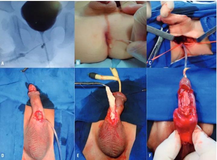
Figure 1- A) Voiding cystourethrogram revealed urethral duplication with a narrow cal-iber dorsal penile orthotopic urethra and the ventral urethra meatus opening in the rec-tum (type IIA urethral duplication); B) Catheterization of the ventral urethra by its opening in the rectum (type IIA urethral duplication); C) Intraoperative picture show-ing dissection of the ventral urethra; D) Intra-operative view depicting catheterization of the accessory urethral meatus located at penoscrotal junction; E) Onlay acellular matrix urethral transplantation; F) Final aspect of onlay acellular matrix urethral transplanta-tion (Patient 10 of Table-2).
A
D
B
E
C
IBJU| TREATEMENT OF URETHRAL DUPLICATION
1150
No study about fertility and ejaculation in patients with urethral duplication was repor-ted in the literature. The association between urethral duplication and posterior urethral val-ves was described by Lorenzo et al., (10) Fern-bach et al. (11) and Ramanujam et al. (12). This association presents more difficulty to provide an accurate diagnosis of urethral duplications, as verified in case 6.
Other anomalies associated with urethral duplication include bifid scrotum, cryptorchi-dism, hypospadias, megalourethra, micropenis, vesicoureteral reflux, agenesis and renal ectopy, dysplastic-multicystic kidney, vertebral anoma-lies (sacral agenesis, thoracic hemivertebra), ano-rectal anomalies, trachea-esophagic fistula and penile, vagina, uterus, bladder and colon dupli-cations (1, 2, 6, 7, 13).
The diagnosis of urethral duplication is performed by clinical history, physical exam and imaging methods, especially voiding cystoure-thrography. Kidney and bladder ultrasonography is recommended to investigate associated ano-malies. Urethrocystoscopy is important for surgi-cal planning. Magnetic resonance urography or excretory urography might be useful to further depict upper tract abnormalities.
Treatment of this anomaly depends on the duplication type and its clinical manifesta-tions. Before any surgical decision, it must be identified which urethra is more functional. Pa-tients with incomplete duplication (type I A or I B) can totally be asymptomatic, with no need of surgical correction. If those patients present pu-rulent secretions or local infection, the accessory urethra should be resected (8). Other option is the opening of the ventral wall of the accessory hy-pospadic urethra and posterior neourethroplasty, as described by Podesta et al. (14) to treat pa-tients with hypospadias and incomplete urethral duplication. In these cases, it was observed fewer surgeries to proper surgical repair and no com-plications in our cohort.
In cases where the patient presents nor-mal dorsal urethra and ventral urethra interfe-ring in the anal canal (anorectal junction) or in the rectum (urethroperineal fistula) – type IIA2, our surgical approach aimed the initial
urethros-tomy of the ventral urethra and preservation of the dorsal orthotopic urethra. After that, a neou-rethroplasty using flaps or grafts was usually performed. In difficult redo cases, even an acellu-lar matrix transplantation was performed for one of our patients (case 10) as shown in Figure 1.
These patients with urethral duplication of hypospadic type, in which the ventral urethra is more functional and located in the perineal or anal area (type IIA2), are challenging to surgi-cal correction and more commonly they present complications such as neourethral dehiscence and stenosis, which are common in these neou-rethroplasty types. In our study, a significant rate of surgical procedures per patient and compli-cations were observed for type IIA duplicompli-cations. Type IIA1 needed 4±1.41 procedures per patient (3 to 5 surgeries) and type IIA2 need 4.25±3.28 procedures per patient (2 to 12 surgeries) and rate of complications were 50% for type IIA1 and 75% for type IIA2 which should be informed for patients and families preoperatively. Type IIA2 is the most complex form of duplication to correct, and in such cases the orthotopic urethra usually has an extensive hypoplastic segment. Hence, it is recommended to mobilize extensively the ventral functional urethra to the perineal-scrotal junction to prevent complications and anticipate that multiple procedures might be required be-cause of the very hypoplastic orthotopic dorsal urethral tissue.
CONCLUSIONS
Urethral duplication is a rare anomaly, with several forms of clinical presentation, often accompanied by other anomalies, and sometimes with difficult diagnosis. The treatment of urethral duplication should be individualized, according to its type. Significantly higher rates of surgical procedures per patient and, possibly complica-tion rates were observed for type IIA duplica-tions, which should be informed for patients and families preoperatively.
CONFLICT OF INTEREST
IBJU| TREATEMENT OF URETHRAL DUPLICATION
1151
Ethical approval
The Institutional Review Board at Hospital das Clínicas da Faculdade de Medicina da Univer-sidade de São Paulo approved this study.
REFERENCES
1. Prasad N, Vivekanandhan KG, Ilangovan G, Prabakaran S. Duplication of the urethra. Pediatr Surg Int. 1999;15:419-21. 2. Salle JL, Sibai H, Rosenstein D, Brzezinski AE, Corcos J.
Urethral duplication in the male: review of 16 cases. J Urol. 2000;163:1936-40.
3. Onofre LS, Gomes AL, Leão JQ, Leão FG, Cruz TM, Carnevale J. Urethral duplication--a wide spectrum of anomalies. J Pediatr Urol. 2013;9:1064-71.
4. Coleman RA, Winkle DC, Borzi PA. Urethral duplication: cases of ventral and dorsal complete duplication and review of the literature. J Pediatr Urol. 2010;6:188-91.
5. Effmann EL, Lebowitz RL, Colodny AH. Duplication of the urethra. Radiology. 1976;119:179-85.
6. deVries PA, Friedland GW. Congenital “H-type” ano-urethral fistula. Radiology. 1974;113:397-407.
7. Kennedy HA, Steidle CP, Mitchell ME, Rink RC. Collateral urethral duplication in the frontal plane: a spectrum of cases. J Urol. 1988;139:332-4.
8. Bogaert GA. Urethral duplication and other urethral anomalies. In: Gearhart JP, Rink RC, Mouriquand PDE, editors. Pediatric urology, 1st ed., Philadelphia: WB Saunders; 2001; pp. 607-19.
9. Atala A. Congenital urethral duplication. In: Marshall FF, editor. Textbook of operative urology. Philadelphia: WB Saunders; 1996; pp. 992-1006.
10. Lorenzo RL, Turner WR, Bradford BF, Upshur J, Sexton FM. Duplication of the male urethra with posterior urethral valves. Pediatr Radiol. 1981;11:39-41.
11. Fernbach SK, Maizels M. Posterior urethral valves causing urinary retention in an infant with duplication of the urethra. J Urol. 1984;132:353-5.
12. Ramanujam TM, Sergius A, Usha V, Ramanathan S. Incomplete hypospadiac urethral duplication with posterior urethral valves. Pediatr Surg Int. 1998;14:134-7.
13. Savanelli A, Schiano A, Esposito C, Russo S, Dolezalova H. Congenital megalourethra associated with urethral duplication and imperforate anus. Pediatr Surg Int. 1998;13:607-9.
14. Podesta ML, Medel R, Castera R, Ruarte AC. Urethral duplication in children: surgical treatment and results. J Urol. 1998;160:1830-3.
_______________________ Correspondence address: