Sao Paulo Med J. 201X; XXX(X):xxx-xxx 1
CASE REPORT
DOI: 10.1590/1516-3180.2017.0083290517Intracranial aneurysm and arachnoid cyst: just a coincidence?
A case report
Guilherme Brasileiro de Aguiar
I, Rafael Gomes dos Santos
II, Aline Lariessy Campos Paiva
III, João Miguel de Almeida Silva
IV,
Rafael Carlos da Silva
V, José Carlos Esteves Veiga
VIFaculdade de Medicina da Santa Casa de São Paulo (FMSCSP), São Paulo (SP), Brazil
ABSTRACT
CONTEXT: Presence of an arachnoid cyst and a non-ruptured intracystic brain aneurysm is extremely rare. The aim of this paper was to describe a case of a patient with an arachnoid cyst and a non-ruptured aneurysm inside it. Clinical, surgical and radiological data were analyzed and the literature was reviewed.
CASE REPORT: A patient complained of chronic headache. She was diagnosed as having a temporal arachnoid cyst and a non-ruptured middle cerebral artery aneurysm inside it. Surgery was performed to clip the aneurysm and fenestrate the cyst.
CONCLUSIONS: This report raises awareness about the importance of intracranial vascular investigation in patients with arachnoid cysts and brain hemorrhage.
IMD, MSc. Attending Neurosurgeon, Faculdade
de Ciências Médicas da Santa Casa de São Paulo (FMSCSP), São Paulo (SP), Brazil.
IIMedical Student, Faculdade de Medicina da Santa
Casa de São Paulo (FMSCSP), São Paulo (SP), Brazil.
IIIMD. Neurosurgery Resident, Faculdade de
Ciências Médicas da Santa Casa de São Paulo (FMSCSP), São Paulo (SP), Brazil.
IVMD. Neurosurgeon, Faculdade de Ciências
Médicas da Santa Casa de São Paulo (FMSCSP), and Endovascular Neurosurgery Fellow, Hospital das Clínicas, Faculdade de Medicina da Universidade de São Paulo (HC-FMUSP), São Paulo (SP), Brazil.
VMD. Neurosurgeon, Faculdade de Ciências
Médicas da Santa Casa de São Paulo (FMSCSP), São Paulo (SP), Brazil.
VIMD, PhD. Full Professor and Head of Discipline
of Neurosurgery, Faculdade de Ciências Médicas da Santa Casa de São Paulo (FMSCSP), São Paulo (SP), Brazil.
KEY WORDS:
Arachnoid cysts. Intracranial aneurysm. Collagen diseases. Intracranial hemorrhages.
INTRODUCTION
Intracranial arachnoid cysts account for 1% of all brain lesions.1 Most of them are located in the
middle fossa.1 Because neuroimaging has become more available, arachnoid cysts are becoming
diagnosed more frequently, even when they are asymptomatic. his type is the most common
presentation,2 and constitutes an incidental inding. Patients with arachnoid cysts can also
pres-ent with headache, nausea and vomiting, and with cranial nerve palsy.1 hese cysts are acquired
lesions relating to abnormal splitting of subarachnoid layers, and they may reach huge
dimen-sion. In addition, they may be related to collagen disorder diseases.1
On the other hand, the etiology of brain aneurysms is a controversial topic in which genetic
changes, smoking and arterial hypertension constitute predisposing factors.1 he estimated overall
prevalence of unruptured intracranial aneurysms in adults without comorbidities is about 3.2%.3
herefore, although they are common lesions that are oten associated with collagen diseases such
as Marfan syndrome and polycystic renal disease,3 an association between an arachnoid cyst and
brain aneurysm in the same patient is extremely rare.4,5
he aim of this paper was to report a case of a patient with a diagnosis of an arachnoid cyst and a non-ruptured intracystic brain aneurysm. he literature on this rare condition was also reviewed.
CASE REPORT
A 54-year-old female patient presented with a clinical complaint of a let chronic hemicranial headache with pulsatile pattern. In her past medical history, she had only had arterial blood hypertension. Her neurological examination was normal. She did not have any relevant family history.
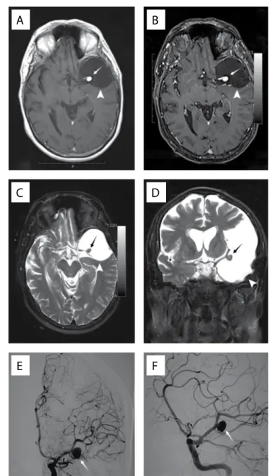
Neurological investigation was performed through brain magnetic resonance imaging (MRI),
which revealed a let temporal arachnoid cyst (Figure 1). here were no signs of intracranial
bleeding. In addition, localized vascular dilatation at the let middle cerebral artery bifurcation (inside the cyst) was noticed, which was suggestive of saccular aneurysm. Because of this, cere-bral angiography was performed, which conirmed the presence of a let middle cerecere-bral artery
aneurysm with dimensions of 9 mm x 6 mm and a neck of 3 mm (Figure 1).
Surgical treatment was proposed, consisting of let pterional craniotomy to clip the middle cerebral artery aneurysm. his procedure was implemented without complications. Fenestration
CASE REPORT | Aguiar GB, Santos RG, Paiva ALC, Silva JMA, Silva RC, Veiga JCE
2 Sao Paulo Med J. 201X; XXX(X):xxx-xxx
of the cyst was also performed to provide communication with
the basal cisternae (Figure 2). Only a single clip was needed to
achieve occlusion of the aneurysm. he patient presented good
recovery, with complete exclusion of the aneurysm from the brain circulation and cyst volume reduction. She presented with-out neurological deicits and was discharged from hospital for ambulatory follow-up.
DISCUSSION
Arachnoid cysts are congenital lesions that may cause major
neurological symptoms,1 but which generally constitute an
incidental inding. Because MRI and computed tomography (CT) have become more available, these cysts are becoming diagnosed more frequently. Regarding etiology, there are three main theories:
Figure 1. A) Axial T1-weighted non-contrasted magnetic resonance
imaging (MRI) showing hypointense lesion at the middle fossa and nodule formation inside the cyst; B) Axial T1-weighted MRI showing hypointense lesion at the middle fossa with saccular dilation inside; C) Axial T2 MRI showing the cyst and the middle cerebral artery bifurcation with saccular dilation at this location; D) Coronal T2 MRI showing the temporal cyst and middle cerebral artery bifurcation with dilation; E) Cerebral arteriography showing aneurysm at the left middle cerebral artery bifurcation; F) Oblique-incidence cerebral angiography showing the aneurysm.
A
C
E
B
D
F
Figure 2. Microsurgical views: A) Before cyst wall opening;
B) After cyst wall opening showing the aneurysm at the left middle cerebral artery bifurcation inside it.
A
B
Aguiar GB, Santos RG, Paiva ALC, Silva JMA, Silva RC, Veiga JCE
Intracranial aneurysm and arachnoid cyst: just a coincidence? A case report | CASE REPORT
Sao Paulo Med J. 201X; XXX(X):xxx-xxx 3
1. Embryonic dysgenesis during arachnoid cyst formation due to a primary defect of the mesenchyme adjacent to the neural tube;
2. Localized brain agenesis, atrophy or hypoplasia causing sec-ondary expansion of the space for cerebrospinal luid (CSF); 3. Localized disorder secondary to an inlammatory, infectious,
traumatic or hemorrhagic lesion.1
Most authors have maintained that arachnoid cysts are congeni-tal, and this theory has been accepted because of their association with other malformations such as corpus callosum agenesis, Marfan
syndrome, type 1 neuroibromatosis and polycystic kidney disease.1
Similarly, the etiology of saccular brain aneurysms is not well
established.4 here is an association between collagen diseases
and other forms of brain dysplasia, such as in Elhers-Danlos and
Marfan syndromes and in polycystic kidney disease.1
he current prevalence of brain aneurysms in patients with polycystic kidney disease ranges from 4 to 12%, which is higher
than in the general population (1-4%).3 In these cases, the risk of
rupture risk is higher: about ive times greater than in patients
without this disease.3
Romão et al.3 evaluated 92 patients with polycystic disease and
found that six of them had some form of intracranial lesion: three with aneurysms and three with arachnoid cysts. However, none
of them had both lesions.3 It is possible that arachnoid cysts and
brain aneurysms are distinct disorders relating to a single
dyse-mbryogenesis.2 hus, it can be seen that an association between
brain aneurysm and an arachnoid cyst, as in the case reported here (Figures 1 and 2), is very rare.
Arachnoid cysts of the middle fossa only rarely induce
neurological symptoms.5 These symptoms occur when there is
increased pressure on the neighboring structures.5 Neurological
signs and symptoms may originate from bleeding inside the cyst. Intracystic hemorrhages are generally due to traumatic brain
injury (TBI).1 Even mild TBI can cause subdural hematomas
or intracystic hemorrhage.1,5
Intracystic hemorrhage due to ruptured brain aneurysm is an
extremely rare condition,1 with few cases reported in the literature.
In most cases, the aneurysm is attached to the cyst wall and its rup-ture gives rise to arachnoid membrane permeation, thus causing
intracystic bleeding.1 Intracystic hemorrhage is usually caused by
rupture of aneurysms of posterior communicating arteries, inter-nal carotid or middle cerebral bifurcations and anterior
commu-nicating arteries. hese aneurysms may be adjacent to the cyst1
and may evolve with intracystic or subarachnoid hemorrhage and
subdural hematomas.2
An association between brain aneurysm and an arachnoid cyst
is a very rare condition (Table 1). A review of the literature was
performed through PubMed, searching for the terms “arachnoid cyst” and “intracranial aneurysm” and the few cases reported in the literature were found to describe patients with brain hem-orrhage. herefore, simultaneous arachnoid cyst and
non-rup-tured brain aneurysm is an even rarer situation.2 In the present
report, this diagnosis was an incidental inding. de Oliveira et
al.5 found associations between aneurysms and arachnoid cysts
through a review of the literature in which only 10 cases were reported. In most of these cases, intracranial hemorrhage was
the irst manifestation.5
There has also been one report of multiple aneurysms
asso-ciated with arachnoid cysts,2 which evolved with rupture of
the intracystic aneurysm, without typical subarachnoid
hem-orrhage.1 All of these possibilities should be borne in mind
during neuroimaging evaluations on patients with arach-noid cysts without symptoms and on those who present with intracranial hemorrhage.
CONCLUSION
here is no strong evidence in literature to correlate arachnoid cysts and brain aneurysms. However, for all patients with diag-noses of arachnoid cyst and brain hemorrhage, intracranial vas-cular investigation should be performed, because these condi-tions may be associated due to their common pathogenesis. he present unique case of non-ruptured brain aneurysm and arachnoid cyst also serves to raise awareness about the impor-tance of proper vascular investigation, even in cases without intracranial hemorrhage.
Database Search strategies Papers found Papers related (with ruptured aneurysm) Papers related (with non-ruptured aneurysm) Female Male Main neurological symptom MEDLINE (via PubMed, on March 5, 2017)
“Arachnoid cysts” [MESH] AND “Intracranial aneurysm” [MESH]
AND Case Reports[ptyp]
23 8 0 6 2 Headache
LILACS (via BVS, on June 1, 2017)
Arachnoid cysts [Palavras] and
Intracranial aneurysm [Palavras] 3 2 0 1 1 Headache
Table 1. Search of the literature in medical databases for case reports on intracranial aneurysm and arachnoid cysts
Intracranial aneurysm and arachnoid cyst: just a coincidence? A case report
CASE REPORT | Aguiar GB, Santos RG, Paiva ALC, Silva JMA, Silva RC, Veiga JCE
4 Sao Paulo Med J. 201X; XXX(X):xxx-xxx
REFERENCES
1. Kajiwara I, Tanaka T, Kan I, et al. Intracystic hematoma of middle
fossa arachnoid cyst caused by rupture of internal carotid-posterior
communicating artery aneurysm. Neurol Med Chir ( Tokyo).
2008;48(5):220-2.
2. Secer HI, Duz B, Solmaz I, Gonul E. Endoscopic clipping of a middle
cerebral artery aneurysm in a middle fossa arachnoid cyst and review
of the literature. Minim Invasive Neurosurg. 2010;53(3):132-7.
3. Romão EA, Moysés Neto M, Teixeira SR, et al. Renal and extrarenal
manifestations of autosomal dominant polycystic kidney disease. Braz
J Med Biol Res. 2006;39(4):533-8.
4. Zanini MA, Faleiros AT, Rondinelli G, Gabarra RC, Resende LA. A form
of dysplasia or a fortuitous association? A cerebral aneurysm inside
an arachnoid cyst: case report. Neurosurgery. 2007;61(3):E654-5;
discussion E655.
5. de Oliveira JG, Giudicissi-Filho M, Rassi-Neto A, et al. Intracranial
aneurysm and arachnoid cyst: a rare association between two cerebral
malformations. Br J Neurosurg. 2007;21(4):406-10.
Sources of funding: None
Conlict of interest: None
Date of irst submission: March 19, 2017
Last received: May 23, 2017
Accepted: May 29, 2017
Address for correspondence:
Aline Lariessy Campos Paiva
Faculdade de Ciências Médicas da Santa Casa de São Paulo (FMSCSP)
Rua Cesário Mota Júnior, 112
São Paulo (SP) — Brasil — CEP 01225-020
Tel. (+55 11) 2176-7000
E-mail: [email protected]
Aguiar GB, Santos RG, Paiva ALC, Silva JMA, Silva RC, Veiga JCE