MALIGNANT PARAGANGLIOMA WITH
VERTEBRAL METASTASIS
Case report
Bruno Lázaro
1, Mônica Klemz
2, Marlo Steiner Flores
3, José Alberto Landeiro
4ABSTRACT - A paraganglioma is a rare tumor, composed of chromaffin cells, groups of cells associated to the autonomous system. When the tumor occurs in the adrenal gland, it is called pheochromocitoma. The malignant paraganglioma is a very rare presentation; it is diagnosed by local recurrence after total resection of the primary mass, or findings of distant metastases. We present a case report of a 29-year-old woman with cervico-brachial pain. In 1995 she underwent a carotid body tumor resection. Magnetic resonance imaging (MRI), plain X-rays and computerized tomography scan revealed multiple lesions in C5, T5 and T12. She underwent a surgical procedure to correct the cervical lesion. The histological and immunohistochemical assays revealed a malignant paraganglioma. She received adjuvant radiotherapy, showing clinical improvement after treatment, presenting no symptoms after one year. The therapeutic approach is based on the total resection of the tumor. The treatment of distant metastases can be made with adjuvant measures such as conventional radiotherapy, I¹³¹-MIBG, or chemotherapy, especially in malignant pheochromocitomas.
KEY WORDS: paraganglioma, vertebral metastasis, spinal tumor
P PP
PParaganglioma maligno com metástase vertebral: relato de casoaraganglioma maligno com metástase vertebral: relato de casoaraganglioma maligno com metástase vertebral: relato de casoaraganglioma maligno com metástase vertebral: relato de casoaraganglioma maligno com metástase vertebral: relato de caso
RESUMO - O paraganglioma é tumor raro, composto de células cromafins, associado ao sistema nervoso autônomo. Quando localizado na glândula supra-renal, o tumor é chamado feocromocitoma. Descreve-se um caso de paciente do sexo feminino, 29 anos, que se apresentou com cervicobraquialgia e que havia sido operada em 1995 para exérese de tumor glômico da carótida cervical. RM, RX e TC revelaram múltiplas lesões acometendo o corpo vertebral de C5, T5 e T12. Foi submetida à ressecção cirúrgica radical da lesão cervical, com substituição do corpo vertebral por prótese de titânio. A histopatologia e o estudo imunohistoquímico da lesão confirmaram o diagnóstico de paraganglioma maligno. As outras lesões foram tratadas com radioterapia. Um ano após os procedimentos, a paciente apresenta-se assintomática. O tratamento destas lesões consiste na associação da ressecção cirúrgica radical do tumor e medidas complementares como radioterapia convencional, aplicação de I131-MIBG, ou quimioterapia, principalmente nos paragangliomas malignos.
PALAVRAS-CHAVE: paraganglioma maligno, metástase vertebral, tumor vertebral
Neurosurgery and Oncology Clinics of Brazilian Air Force Hospital, Rio de Janeiro RJ, Brazil: 1Student of Medicine ; 2Oncologist; 3Assistant
of Neurosurgery Clinic; 4 Head of Neurosurgery Clinic.
Received 4 October 2002, received in final form 26 December 2002. Accepted 15 January 2003. Dr. José Alberto Landeiro - Rua Monsenhor Ascaneo 591/202 - 22621-060 Rio de Janeiro RJ - Brasil
A paraganglioma is a rare tumor, composed of chromaffin cells, originated from the neural crest, a group of cells associated to the autonomous nervous system. Paragangliomas can be found in several lo-cations. When the primary location is the adrenal gland, the tumor is called pheochromocitoma; in the head and neck region, the paragangliomas can be most found in the carotid body, jugular and vagal glomus; other locations also include the retroperi-toneum, para-aortic region, bladder, filum terminale, skull, larynx, and others. The presentation of the tumor is variable: in most cases it is represented as a non-symptomatic slow-growing mass,
compres-sing the anatomic structures around it. On the other hand, the tumor can secrete catecholamines, indu-cing a typical clinical history represented by hyper-tension, paroxysms and headache. Malignant para-gangliomas are a very rare presentation, limited to less than ten cases registered between 1985-19961; the annual incidence in retrospective studies is 1/10 000 0002. It is diagnosed by local recurrence after total resection of the primary mass, or findings of distant metastases. The time intervals for recurrence may be from months to many years.
464 Arq Neuropsiquiatr 2003;61(2-B)
diagnosis and surgical removal of a carotid body tumor. She underwent a surgical procedure to correct the vertebral lesions, and adjuvant radiotherapy.
CASE
In 1995 a 29-year-old woman presented a cervical mass on her left side, which evolved to a progressive cervical pain irradiating to the left ear. The physical examination was normal, except for a decrease on the left carotid and temporal arteries pulse. All laboratorial exams were nor-mal; a MRI of the neck showed a 4.7x 3.3 x 3.1cm lobular mass with an intermediate T1-weighted sign and a high heterogeneous T2-weighted sign, enhanced with gadoli-nium, on the left carotid bifurcation, with a mass effect over the ipsilateral sub-mandibular gland, suggesting a carotid body tumor. A carotid-vertebral angiography sho-wed an encasement of the left carotid by the tumor.
A resection of the mass was performed followed by a carotid-carotid internal saphenous vein by-pass. The outcome was positive; a carotid-vertebral duplex scan showed an adequate flow, demonstrating a good functi-oning of the graft. Histopathology revealed a 4.0 x 3.0 x 2.5 cm grayish-brown mass, with necrotic areas. The mi-croscopy showed a pattern of cords and nest cells, typical of a carotid body tumor. The patient was discharged from the hospital without any symptom.
In October 2001, the patient was readmitted to the hospital presenting a cervico-brachial pain; the patient was well until the last six months. The neurological exam sho-wed a hyposthesia of the left territory from C4 to C5 (Fig 1). The erythrocyte sedimentation rate (ESR) was 37mm. Plain X-rays of the cervico-thoracic spine revealed a pa-thologic fracture of the vertebral body of C5, T5 and T12. A bone scintillography showed an uptake of the
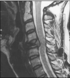
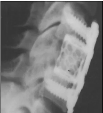
topogra-phy of C5, T5 and T12. CT scan of the cervico-thoracic spine showed a fracture of T5 and T12, with a severe lesion on vertebral body of C5 (Fig 1). Cervico-thoracic MRI examination revealed a destructive lesion of C5, with displacement of the vertebral posterior wall towards the spinal canal; bone destruction of T5 and T12, with invasion of the spinal canal on T5 level suggesting a secondary implant (Fig1 a 4). In November 2001 a surgical procedure was proposed, comprising the C5 corpectomy, identifying an hemorrhagic tumor tissue; the setting of a titanium cage filled with an osteoinductive coral graft, and the osteosynthesis of C4 to C6 using four 16-mm screws and a 16-mm titanium plate (Fig 5).
Histopathologic assessment revealed vertebral bone in-filtration with a pattern of spread cells suggestive of epithelial origin, with an early result of adenocarcinoma. A full diagnostic trial was performed, including a thoracic-abdominal-pelvic CT scan, endoscopy, colonoscopy,
mam-Fig 3. Computed tomography scan of the thoracic shows the vertebral body compromise.
Fig 1. Computed tomography scan shows the vertebral body destruction in axial cut. The coronal and sagittal 2 D reconstruction show the vertebral collapse.
mography, pelvic and intravaginal ultrasound scan, measu-rement of CEA, CA-125, CA-19.9, CA-15.3, β-HCG, α-FP, and regular laboratory trials. An endoscopy of gastrointestinal tract five revealed inactive gastric ulcers at the antrum (with normal serum gastrin, Helicobacter pylori and histopathologic analysis) and a left kidney cyst. A hypo-thesis of malignant paraganglioma was then suggested. A revised histopathologic assessment was requested; and an immunohistochemical study was performed in both primary and secondary lesions with enolase, chromo-granin, synaptofisin, S-100. Both lesions showed a brown aspect on microscopy, although the secondary lesion de-monstrated a weaker staining to enolase and S-100 pro-tein; the final revisited diagnosis was the presence of secondary implants of a malignant paraganglioma.
The patient has received adjuvant radiotherapy for cer-vico-thoracic spine 4 000cGy, with improvement in her symptomatology.
DISCUSSION
Paragangliomas can occur in several locations, usually related to mesodermal branchial archs. These tumors arise from neuro-ectodermal cells, forming neuroendocrine tumors divided in four categories: typical and atypical carcinoid, oat cell carcinoma, and giant cell neuroendocrine carcinomas3.
Neuroendocrine cells, similar to chromaffin cells, are found spread on extra-adrenal tissues, as nodules and aggregated cells, associated with the adrenal gland, composing the paraganglionic tissue. These
Fig 4. T2-weighted, unenhanced sagittal magnetic ressonance imaging reveals the vertebral body destruction with ventral compression of the dural sac.
Fig 5. Postoperative lateral cervical spine film, the patient underwent an anterior corpectomy at 5C with placement of an anterior interbody titanium cage between C4-C6 and was fused and stabilized with screws and plate (Synthes®).
extra-adrenal paraganglia are in close association with the autonomous nervous system, and can be divided in three groups, according to their anatomic location: branchiometric, intra-vagal, and aortic-sympathetic. Intra-vagal and branchiometric para-ganglia are related to the parasympathetic system; the general location is next to the great arteries and cranial nerves of the head and neck, including the carotid body. The intra-vagal paraganglia are distri-buted along the vagus nerve. The aortic-sympathetic paraganglia are related to the sympathetic ganglia, distributed along the abdominal aorta.
Some of the branchiometric paraganglia, specially the carotid body, act as chemoceptors responding to variations in oxygen tension and carbon-dioxide concentrations; therefore their other denomination: chemodectomas4.
Paragangliomas, however, are found in other lo-cations; the possible explanation for this fact is the migration of the paraganglionic tissue to unconven-tional sites such as the skull, parasellar region, pon-to-cerebellar angle and cauda equina, acting as a differential diagnosis on backache5.
466 Arq Neuropsiquiatr 2003;61(2-B)
Carotid body paraganglioma - The first description of a carotid body paraganglioma was reported in 1891. It is characterized as a mass of long duration in the lateral region of the neck, usually slow-growing and non symptomatic at the beginning. The symptoms may emerge years after the appearance of the mass7. By 1973, 500 cases had been published, 16 of which (3.2%) presented distant metastases, and other 16 cases (3.2%) presented local metastases8. But how can we define a malignant paraganglioma? It is well known that a malignant behaviour cannot be designated by histological findings such as the number of mitoses, size of the nuclei, nuclei-cytoplasm relation or others. The malignant paraganglioma is defined by findings of local recrudescence or presence of distant metas-tasis. The tumor spreads via blood stream or lym-phatics. The malignant potential of the carotid body tumor has been described in some report series, and it varies from extremely low incidence up to a 50% prevalence9. The mean ratio is about 10% and the mean age of incidence is about 44 years old1.
The most common manifestation of the malignant paraganglioma is the local recrudescence even after the “complete” resection of the tumor8. The mean time of recurrence after the initial resection is about 6 years10. The usual sites are cervical and mediastinal lymph nodes, bone, lung, liver and heart. There are no risk factors concerning gender, race or age.
Isolated vertebral metastasis is an extremely rare occurrence. Vertebral body and epidural metastases are the most common ones associated to cervical paragangliomas10. Scott et al.11 described five pati-ents with painful bony metastasis being the first in-dication of the malignant behaviour of the tumor. In three patients the earliest sign of metastatic dise-ase was a clinical and radiological finding of verte-bral collapse and spontaneous femur fracture.
Pathology - Paraganglionic tissue is composed of 2 types of cells: type 1 or chief neuroectodermal cells, bearing catecholamines granules; and type 2 or sus-tentacular cells. The histological aspect of the tu-mor on hematoxiline-eosine and reticuline stain is the classical “Zellballen” pattern, presented as nests or cords of type 1 cells surrounded by type 2, or sustentacular cell. On immunohistochemical analysis, type 1 cells contain neuron specific enolase, chro-mogranin and leu-enkephalin; type 2 cells contain S-100 protein12. In some cases, particularly those that were recurrent, locally aggressive and malignant, the organoid pattern was less apparent; central necrosis on the Zellballen was found, as nuclear pleomor-phism. But the diagnosis of malignant paraganglio-ma is only paraganglio-made by findings of local recurrence or
distant metastasis. Linnoila et al.06 described the decreased expression of neuropeptides related to malignant paraganglioma, using immunohistoche-mical assays with leu and met-enkephalin, somatos-tatin, pancreatic polipeptide and VIP. Possible expla-nations would be the decreased synthesis or elevated secretion rate or defective intracellular storage. They conclude that no matter the mechanism, the decrea-sed expression of neuropeptides is related to a worse prognosis. Kliewer et al.12 demonstrated a maximum staining intensity with enolase and chromogranin in benign paragangliomas. Malignant paraganglio-mas showed minimal staining to S-100 protein on type 2 cells.
Symptomatology, diagnosis and treatment - The clinical aspect of a paraganglioma is mainly related to the mass effect of the tumor on adjacent structures. The classical triad of headache, diaphoresis and palpitations, and other symptoms of a hyperadrenergic state, such as hypertension, paroxysms, and cardiac, gastrointestinal and metabolic manifestations are typical of a pheochromocitoma, being less apparent in a paraganglioma and rarely present in a malignant paraganglioma.
The diagnosis of a paraganglioma is based on ima-ge methods that can be associated to biochemical assays in cases that the functioning tumors are secre-ting catecholamines. The “gold-standard” method is the MRI. It shows great accuracy demonstrating the size and density of the tumor and its relation to the adjacent structures. Plain X-rays and CT are valuable methods in showing bone and vertebral metastasis. Ultrasound scan is a limited but important method in diagnosing retroperitoneal and pelvic paragangliomas, showing less sensibility in tumors located in other sites. Angiography is an important tool for the diagnosis of vascular commitment caused by the tumor, as on plan-ning pre-operative embolization.
Scintillography is an essential method of deter-mining metastatic disease. The use of I¹³¹- MIBG (meta-iodobenzylguanidine), a structural analog of guanethidine, with uptake in the adrenergic granu-les has been elucidated in some studies13,14. This me-thod can show the presence of the disseminated disease even with a non- functioning tumor, being useful on the diagnosis of primitive neural crest tu-mors, such as neuroblastomas and paraganglioma. I¹³¹- MIBG has a sensitivity of 87-91% and a specificity of 90-94%14. The use of I¹³¹- MIBG for the treatment of paragangliomas is discussed below.
le-vels and its metabolites on a 24-hour urine sample. Levels of metanephrine, vanilmandelic acid and free catecholamines can be measured. The number of samples depends upon the degree of suspicion; the presence of abnormal levels in a patient with a typical clinical history is very suggestive of the diagnosis.
The success in treating benign and malignant paragangliomas is based on the early diagnosis, com-plete resection of the tumor using an adequate cate-cholamine blockage, and an excellent anesthesia11. The strategy must be carried on the adequate study of the tumor and adjacent structures and vessels; a complete resection of the primary mass is the treat-ment of choice. Usually the tumor presents as a latent slowly growing mass, the resection of isolated se-condary masses shows a better prognosis, as de-monstrated on pulmonary metastasis of tumors of the head, trunk and extremities15.The use of I¹³¹-MIBG radiotherapy (RT) has been implicated on par-tial remission of individuals with malignant pheoch-romocitomas. The intensity in the uptake of the ra-diotracer by the tumor cells is not fundamental, as even the non-secreting tumors show an adequate response to the treatment. Although good results have been described with I¹³¹- MIBG therapy in trea-ting bony metastasis, the main role of I¹³¹- MIBG is diagnostic and palliative instead of curative16. Con-ventional RT has been described for treatment of malignant paragangliomas. A series of 84 patients with chemodectomas of the head and neck demons-trated 73% local control for 25 years with radiothe-rapy solely, compared to 54% for 15 years with sur-gery solely17. The recommended doses are 4 500-5 000cGy during 4-5 weeks. The combined use of RT and chemotherapy has been proposed in treatment of malignant pheochromocitomas, using a three-drug trial composed of Cyclophosphamide, Dacar-bazine and Vincristine; these two methods would act in sequence in different sites, with the final result being better than the isolated use of one method or the other18. The previous use of chemotherapy would be implicated in increasing sensibility and uptake of I¹³¹- MIBG by the tumor16. The treatment of choice for malignant paragangliomas with vertebral metastasis is decompressive surgery. According to the National cancer database on malignant paragangliomas of the head and neck1, the 5-year relative survival rate was 59.5% (76.8% for regionally confined carcinoma and 11.8% for distant metastasis). Among the patients who were followed-up until death, those treated with ad-juvant irradiation had a longer median survival (45 months) compared with those patients who were trea-ted with surgery solely (12 months).
In conclusion, we report a rare case of a
para-ganglioma with vertebral body metastasis in 29-year-old patient six years after the complete removal of a carotid body tumor, and it,s surgical and radiotherapy aspects. A long-term follow-up is mandatory for all patients, as previously demonstrated. The time in-tervals for local recurrence and distant metastasis can vary from months to many years after the initial diagnosis. The use of scintillography with I¹³¹- MIBG is a powerful tool for the identification of the me-tastasis, and a palliative method for treating the ma-lignant paraganglioma. The primary management of a malignant paraganglioma should be directed towards the complete surgical resection of the pri-mary tumor, regional lymph nodes and distant me-tastasis. The therapeutic proposal for our patient was the resection of secondary implants in C5 given the main destructive site at this topography, associated with adjuvant RT. The patient has shown clinical im-provement, having no symptoms after 6 months of adjuvant therapy.
REFERENCES
1. Lee JH, Barich F, Karnell LH, Robinson RA, Zhen WK, Gantz BJ, Hoffman HT. National cancer data base report on malignant paragangliomas of the head and neck. Cancer 2002;94:730-737. 2. Siddiqui MZ, Von Eyben FE, Spanos G. High-voltage irradiation and
combination chemotherapy for malignant pheochromocytoma. Cancer 1988;62:686-690.
3. Flitsch J, Schroder F, Hagel C, Papavero L. Cranial neuroendocrine carcinoma primarily diagnosed as malignant paraganglioma with rapid progress to a fatal outcome. Acta Neurochir (Wien) 2001;143:523-524. 4. Lack EE, Cubilla AL, Woodruff JM. Paragangliomas of the head and
neck region. Hum Pathol 1979;10:191-218.
5. Roche PH, Figarella-Branger D, Regis J, Peragut JC. Cauda equina paraganglioma with subsequent intracranial and intraspinal metastases. Acta Neurochir (Wien) 1996;138:475-479.
6. Linnoila RI, Lack EE, Steinberg SM, Keiser HR. Decreased expression of neuropeptides in malignant paragangliomas: an immunohistoche-mical study. Hum Pathol 198819:41-59.
7. Blades DA, Hardy RW, Cohen M. Cervical paraganglioma with subsequent intracranial and intraspinal metastasis. J. Neurosurg 1991;75:320-323. 8. Say CC, Hori J, Spratt J. Chemodectoma with distant metastasis: case
report and review of the literature. Am Surg 1973;39:333-341. 9. Capt Reese HE, Capt Lucas RN, Col Bergman PA. Malignant carotid
body tumors: report of a case. Ann. Surg. 1963;157:232-243. 10. Hamilton MA. Metastatic paraganglioma causing spinal cord
compression. Br J Radiol 2000;73:901-904.
11. Scott HW Jr, Raynolds V, Green N, Page D, Oatis JA, Robertson D. Clinical experience with malignant pheocromocytomas. Surg Gynec Obst 1982;154:801-818.
12. Kliewer KE, Wen D-Ren, Cancilla PA, Cochran AJ. Paragangliomas: Assessment of prognosis by histologic, immunohistochemical, and ultrastructural techniques. Hum Pathol 1989;20:29-39.
13. Beierwaltes WH. Update on basic research and clinical experience with metaiodobenzylguanidine. Med Pediatric Oncol 1987;15:163-169. 14. Konings JE, Bruning PF, Abeling NGGM, van Gennip AH, Hoefnagel CA.
Diagnosis and treatment of malignant pheochromocytoma with ¹³¹I-Meta-iodobenzylguanidine: a case report. Radiother Oncol 1990; 17: 103-10. 15. Gustilo RB, Lober PH, Salovich EL. Chemodectoma (carotid-body
tu-mor) metastasizing to bone. J Bone Joint Surg Am 1965;47:155-160. 16. Hartley A, Spooner D, Brunt AM. Manegement of malignant
phaeochromocytoma: a retrospective review of the use of MIBG and chemotherapy in the West Midlands. Clin Oncol 2001;13:361-366. 17. Yo L, Fleckman AM, Chadha M, Sacks E, Levetan C, Vikram B.
Radiation therapy of metastatic pheochromocytoma: case report of the literature. Am J Clin Oncol 1996;19:389-393.