Corneal Confocal Microscopy Detects
Neuropathy in Patients with Type 1 Diabetes
without Retinopathy or Microalbuminuria
Ioannis N. Petropoulos1,2☯‡, Patrick Green1☯‡, Agnes W. S. Chan3, Uazman Alam1, Hassan Fadavi1, Andrew Marshall1, Omar Asghar1, Nathan Efron4, Mitra Tavakoli1, Rayaz A. Malik1,2*
1Centre for Endocrinology and Diabetes, Institute of Human Development, University of Manchester and Central Manchester National Health System Foundation Trust, Manchester Academic Health Science Centre, Manchester, United Kingdom,2Weill Cornell Medical College Qatar, Division of Research, Qatar Foundation, Education City, Doha, Qatar,3Queen Mary's University, Bart's and London National Health System Trust, London, United Kingdom,4Institute of Health and Biomedical Innovation and School of Optometry and Vision Science, Queensland University of Technology, Brisbane, Australia
☯These authors contributed equally to this work. ‡These authors are joint first authors on this work.
Abstract
Objective
Corneal innervation is increasingly used as a surrogate marker of human diabetic peripheral neuropathy (DPN) however its temporal relationship with the other microvascular complica-tions of diabetes is not fully established. In this cross-sectional, observational study we aimed to assess whether neuropathy occurred in patients with type 1 diabetes, without reti-nopathy or microalbuminuria.
Materials and Methods
All participants underwent detailed assessment of peripheral neuropathy [neuropathy dis-ability score (NDS), vibration perception threshold (VPT), peroneal motor nerve conduction velocity (PMNCV), sural sensory nerve conduction velocity (SSNCV) and in vivo corneal confocal microscopy (IVCCM)], retinopathy (digital fundus photography) and albuminuria status [albumin: creatinine ratio (ACR)].
Results
53 patients with Type 1 diabetes with (n=37) and without retinopathy (n=16) were compared to control subjects (n=27). SSNCV, corneal nerve fibre (CNFD) and branch (CNBD) density and length (CNFL) were reduced significantly (p<0.001) in diabetic patients without retinop-athy compared to control subjects. Furthermore, CNFD, CNBD and CNFL were also signifi-cantly (p<0.001) reduced in diabetic patients without microalbuminuria (n=39), compared to control subjects. Greater neuropathic severity was associated with established retinopathy and microalbuminuria.
a11111
OPEN ACCESS
Citation:Petropoulos IN, Green P, Chan AWS, Alam U, Fadavi H, Marshall A, et al. (2015) Corneal Confocal Microscopy Detects Neuropathy in Patients with Type 1 Diabetes without Retinopathy or Microalbuminuria. PLoS ONE 10(4): e0123517. doi:10.1371/journal.pone.0123517
Academic Editor:Soroku Yagihashi, Hirosaki University Graduate School of Medicine, JAPAN
Received:October 30, 2014
Accepted:February 19, 2015
Published:April 8, 2015
Copyright:© 2015 Petropoulos et al. This is an open access article distributed under the terms of the
Creative Commons Attribution License, which permits unrestricted use, distribution, and reproduction in any medium, provided the original author and source are credited.
Data Availability Statement:All data from this study are included in the paper. All study related files are available from Figshare:http://dx.doi.org/10.6084/m9. figshare.1318406
Conclusions
IVCCM detects early small fibre damage in the absence of retinopathy or microalbuminuria in patients with Type 1 diabetes.
Introduction
Diabetes and its complications represent a growing global health burden, affecting over an esti-mated 366 million people worldwide [1]. The triad of retinopathy, nephropathy and neuropa-thy are well recognised microvascular complications, and are the leading causes of premature blindness, end-stage renal failure, foot ulceration and amputation, respectively [2]. Once estab-lished, they have a major impact on the quality of life of patients with diabetes and are associat-ed with adverse healthcare outcomes [3].
Diabetic retinopathy (DR) is strongly associated with nephropathy [4], and is one of the ear-liest microvascular complications [5]. However, recent studies have shown that early neuronal abnormalities, such as altered multi-focal electroretinogram (mfERG) responses [6], retinal nerve fibre layer thinning [7] and loss of central visual field sensitivity [8] occur before the onset of overt vascular lesions in the retina, and may be of prognostic value. Krolewski et al. have previously found a strong association between cardiac autonomic neuropathy (CAN) and proliferative DR (PDR) in patients with type 1 diabetes suggesting an underlying etiologic link [9]. Indeed, the Rochester Diabetic Neuropathy Study has shown that markers of microvessel damage such as DR and proteinuria or microalbuminuria (MA) are the strongest predictors for the severity of DPN [10].
Recently, corneal nerve morphology assessed by IVCCM has been proposed as an early sur-rogate marker for small nerve fibre damage in DPN [11,12]. Furthermore, corneal nerve fibre length correlates with clinical and electrophysiological measures of diabetic peripheral neuropa-thy [13,14] and long term glycaemic control [15,16]. Recent studies have shown a stepwise de-terioration in corneal nerve morphology in healthy subjects and patients with pre-proliferative and PDR [17,18]. The exact temporal relationship between neuropathy and retinopathy or ne-phropathy however remains unclear. The purpose of this cross-sectional, observational study was to establish whether neuropathy determined using the highly sensitive techniques of IVCCM and neurophysiology was present in patients with Type 1 diabetes without retinopathy or microalbuminuria.
Materials and Methods
Study subjects
80 subjects in total (53 with type 1 diabetes and 27 controls) were assessed in this study. Partici-pants were excluded if they had a positive history of malignant, connective tissue or infectious disease, deficiency of vitamin B12or folate, chronic renal failure, liver failure, active diabetic foot ulcers, family history of peripheral neuropathy. Participants were also excluded if they had active ocular disease (except for DR), systemic disease known to affect the cornea other than di-abetes or chronic corneal pathologies. Inclusion criteria for patients with didi-abetes mellitus were a previous clinical diagnosis of type 1 diabetes, confirmed by laboratory biochemistry, and age between 18–85 years old. Controls were confirmed not to have diabetes also by laboratory test-ing and were of the same age range.
Ethics Statement and data availability
This study adhered to the tenets of the Declaration of Helsinki and was approved by the North Manchester Research Ethics Committee. Study subjects with diabetes were recruited from the Manchester Diabetes Centre and control subjects were recruited from the community or were relatives of the subjects with diabetes. Informed written consent was obtained from all subjects prior to participation after explanation of the nature and possible consequences of the study. The full, anonymised dataset can be found at https://researchdata.ands.orf.au/longitudinal-assessment-neuropathy-markers-landmark/461294.
Clinical assessment
All study participants underwent assessment of HbA1c(%), lipid fractions [total cholesterol (TC) (mmol/l), high (HDLC) and low density lipoprotein cholesterol (LDLC) (mmol/l), tri-glycerides (mmol/l)] and albumin to creatinine ratio (ACR) (mmol/l).
Peripheral Neuropathy assessment
The modified neuropathy disability score (NDS) was assessed [19]. VPT was tested on the hal-lux using a Neuroesthesiometer (Horwell, Scientific Laboratory Supplies, Wilfrod, Nottingham, UK). Electro-diagnostic studies were undertaken using a Dantec“Keypoint”system (Dantec Dynamics Ltd, Bristol, UK) and peroneal motor and sural sensory nerves [peroneal motor nerve amplitude (PMNCV), sural sensory nerve amplitude (mV) and peroneal motor nerve conduction velocity (PMNCV), sural sensory nerve conduction velocity (SSNCV) (m/s)] were assessed in the left lower limb (calf-to-ankle) by a consultant neurophysiologist.
In-Vivo Corneal Confocal Microscopy
All study subjects were scanned with a laser IVCCM [Heidelberg Retinal Tomograph III Ros-tock Cornea Module (Heidelberg Engineering GmbH, Heidelberg, Germany)]. This IVCCM uses a 670 nm wavelength helium neon diode laser, which is a class I laser and therefore does not pose any ocular safety hazard. A 63x objective lens with a numerical aperture of 0.9 and a working distance, relative to the applanating cap (TomoCap©, Heidelberg Engineering GmbH, Heidelberg, Germany) of 0.0 to 3.0 mm was used. The size of each two-dimensional image pro-duced was 384μm x 384μm which has a 15° x 15° field of view and 10μm/pixel transverse
op-tical resolution. This type of IVCCM uses an entirely digital image capture system and all images are stored in an external hard drive.
A drop of 0.4% benoxinate hydrochloride (Chauvin Pharmaceuticals, Chefaro, UK) was used to anaesthetise each eye and Viscotears (Carbomer 980, 0.2%, Novartis, UK) were used as the coupling agent between the cornea and the applanating cap. All subjects were asked to fix-ate on an outer fixation light throughout the IVCCM scan and a CCD camera was used to image the cornea and correctly position the applanating cap onto the corneal apex. The overall examination took approximately 5 minutes for both eyes of each subject and in this study two experienced optometrists performed all IVCCM scans. All images were captured using the
Image Analysis
One examiner masked from cardiometabolic, peripheral neuropathy and retinopathy status quantified subbasal nerve morphology in 480 images of all study participants, using semi-automated, purpose-written, proprietary software (CCMetrics, M. A. Dabbah, Imaging Science Biomedical Engineering, University of Manchester, Manchester, UK). The specific parameters measured per frame were those we have previously established [12]: CNFD (no./mm2), CNBD (no./mm2) and CNFL (mm/mm2). Specific details on the definition and measurement of each parameter can be found elsewhere [21].
Study definition for retinopathy and nephropathy
The grade of retinopathy for patients with diabetes was defined as the most recent result (1 or more examinations per annum) obtained from the UK NHS Diabetic Eye Screening Pro-gramme (the full scope of the proPro-gramme can be accessed here:http://diabeticeye.screening. nhs.uk/service-specification), which is responsible for providing nationwide retinal screening for patients with diabetes mellitus (the latest DR grading criteria as set by the Royal
College of Ophthalmologists can be found at:http://www.rcophth.ac.uk/news.asp?itemid= 1016&itemTitle=DIABETIC+RETINOPATHY+GUIDELINES+ADDED§ion=
24§ionTitle=News). Patients with“background retinopathy”or greater were classified as
“with retinopathy”. To rule out retinopathy in control subjects, a standard field fundus pho-tograph was captured using a Canon CR-2 Plus digital, non-mydriatic retinal camera (Canon Healthcare Technologies, Melville, New York, USA) and the image was graded by a study cer-tified optometrist using the criteria proposed by the Early Treatment of Diabetic Retinopathy Study [22]. To be eligible for the study, control subjects (determined by assessment of their cardio-metabolic status and medical history) had to have a gradable ocular fundus image in at least one eye. If signs of retinopathy were found the participant was informed and excluded from the study. Albuminuria was determined using the albumin-to-creatinine ratio (ACR) and microalbuminuria (MA) in this study was defined as an ACR>2.5 mg / mmol in males and>3.5 mg / mmol in females. Similarly, control subjects with an abnormal ACR were in-formed and excluded from the study.
Statistical analysis
Statistical analysis was performed using SPSS for Windows XP (Version 16.05.0, IBM, NY, USA) and graphs were generated with OriginPro for Windows XP (Version 8.5.0 SR1, Origi-nLab Corporation, Northampton, MA, USA). All data are expressed as mean ± SEM. Variables were tested for normality by means of univariate analysis, histograms and the Shapiro-Wilk test. One-way analysis of variance or non-parametric Kruskal Wallis was used to the test for differences between the means. Post-hoc analysis (Tukey) was performed and after correction for multiple testing (Bonferroni) a P<0.05 was considered significant.
Results
Clinical assessment
Detailed clinical and demographic results are presented inTable 1. Briefly, diabetic patients (duration 28.6 ± 2.2 years) compared to control subjects had similar age (49.7 ± 2.1 v
49.7 ± 2.3, P>0.05) and BMI (27.0 ± 0.6 v 27.9 ± 0.9), higher HbA1c (%) (8.3 ± 0.3 v 5.6 ± 0.1, P<0.001), a significantly lower TC (4.2 ± 0.2 v 5.2 ± 0.2, P<0.001), LDLC (2.1 ± 0.1 v 3.0 ± 0.1, p<0.001), serum triglycerides (1.2 ± 0.1 v 1.7 ± 0.2, P<0.005) and comparable HDLC
Neuropathy assessment
Detailed neuropathy results for patients stratified according to their retinopathy or microalbu-minuria status are presented inTable 2.
Neuropathy vs. retinopathy
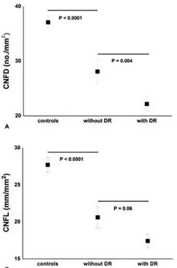
Patients without DR compared to control subjects showed a significantly lower CNFD (P = 0.0001), CNBD (P<0.0001), CNFL (P<0.0001) and SNCV (P<0.05). There was a further
Table 1. Demographic results for study participants stratified according to retinopathy and nephropathy status.
Group Age T1D Duration
(Years)
HbA1c (%) BMI (kg/ m2)
ACR (mg/ mmol) TC (mmol/ mol) HDLC (mmol/mol) LDLC (mmol/mol) Trigs (mmol/mol) Controls (n = 23)
49.7±2.1 N/A 5.6±0.1 27.9±0.9 0.8±0.1 5.2±0.2 1.5±0.1 3.0±0.1 1.7±0.2
Diabetes No DR (n = 17)
43.5±13.6 19.2±4.0 8.0±0.3† 26.5±1.3 0.9±0.2 4.3±0.3† 1.5±0.1 2.2±0.3† 1.1±0.2†
Diabetes DR (n = 36)
52.3±15.8‡ 32.7±2.4‡ 8.4±0.3† 27.1±0.7 4.0±1.4‡ † 4.1±0.1† 1.6±0.1 2.0±0.2† 1.3±0.1†
Diabetes No MA (n = 39)
46.9±2.9 25.7±2.9 8.1±0.2* 27.5±1.8 0.9±0.1 4.1±0.2* 1.6±0.1 2.0±0.8* 1.1±0.2*
DiabetesMA (n = 14)
55.9±4.8** 35.1±4.2** 8.8±0.8* **
25.3±1.4 6.1±2.0* **
4.3±0.3* 1.6±0.1 2.2±0.8 1.3±0.1*
Results are expressed as mean±SEM. For patients with diabetes stratified according to retinopathy status:
†signi
ficantly different from controls,
‡signi
ficantly different from“diabetes without DR”.
For patients with diabetes stratified according to albuminuria status:
*significantly different from controls,
**significantly different from“diabetes without MA”. AP<0.05 was considered significant.
doi:10.1371/journal.pone.0123517.t001
Table 2. Neuropathy measurements in controls and patients with Type 1 diabetes stratified according to retinopathy or microalbuminuria status.
Group NDS VPT (V) SSNA
(μV)
SSNCV (m/s)
PMNA (μV)
PMNCV (m/s)
CNFD (no./mm2)
CNBD (no./mm2)
CNFL (mm/mm2)
Controls(n = 23) 0.4±0.2 6.8±1.1 16.4±1.7 50.2±0.8 5.4±0.4 48.1±0.6 37.1±1.3 101.7±7.4 27.7±1.1
Neuropathy in patients with and without diabetic retinopathy
Diabetes No DR (n = 17)
1.8±0.7 8.7±2.1 11.8±1.5 45.9±1.6† 7.8±3.1 44.8±0.8 28.1±2.3† 56.1±6.9† 20.6±1.5†
Diabetes DR (n = 36)
3.5±0.5† 17.7±2.2† 7.3±1.0† 42.3±0.9‡ 3.3±0.7
†
39.0±1.2† 22.1±1.2‡ 49.9±4.8 17.4±0.9
Neuropathy in patients with and without microalbuminuria
Diabetes No MA (n = 39)
2.1±0.5* 10.9±1.6 9.9±1.2 44.5±1.0 5.7±1.7 42.7±1.0 26.3±1.5* 56.7±5.5* 19.9±1.7*
Diabetes MA (n = 14)
5.2±1.0
**
25.0±4.7
**
5.5±1.6* 40.5±1.7* 1.8±0.5 35.5±2.4* 17.3±2.0** 37.9±7.0** 14.3±1.7**
Results are expressed as mean±SEM. For patients with diabetes stratified according to retinopathy status:
†
significantly different from controls,
‡
significantly different from“diabetes no DR”.
For patients with diabetes stratified according to albuminuria status:
*significantly different from controls,
**significantly different from“diabetes no MA”. AP<0.05 was considered significant.
significant reduction in CNFD between patients with and without DR (P = 0.004) (Figs1and2 andTable 2). There was an inverse correlation between the retinopathy grade and CNFD (r = -0.67, P<0.001), CNBD (r = -0.58, P<0.001) and CNFL (r = -0.66, P<0.001).
Neuropathy vs. microalbuminuria
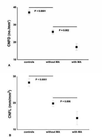
Diabetic patients without MA compared to control subjects had a significant reduction in CNFD (P<0.0001), CNBD (P = 0.001) and CNFL (P<0.0001). There was a further significant reduction for CNFD (P = 0.002), CNBD (P = 0.02), CNFL (P = 0.006) and PMNA (P<0.001) in patients with MA (Fig 3andTable 2).
Discussion
Diabetic microvascular complications result in considerable morbidity and both microalbu-minuria and the severity of DR relate to the severity of DPN [10]. However, the temporal
Fig 1. Significant and progressive loss of CNFD (A) and CNFL (B) between controls, diabetic patients
‘without DR’(n = 17) and‘with DR’(n = 36).Results are expressed as mean±SEM.
relationship for the development of the microvascular complications has been systematically assessed in very few studies. The United Kingdom Prospective Diabetes Study estimated the prevalence of DPN, defined as loss of vibration perception, at ~7% [23], DR at 35–39% [24] and MA at 7% at diagnosis of Type 2 diabetes [25]. In another population based study of
Fig 2. Fundus photograph of the central 30° with corresponding IVCCM image of the central subbasal nerves (yellow arrows) for control (non-mydriatic) (CTR) (A) and patients with diabetes and varying stages of DR (B, C and D).From left to right: (A) IVCCM image shows abundant corneal nerve axons for a control without retinopathy, B) significant decrease of subbasal nerves in a patient with diabetes‘without DR’ (No DR), C) slight progressive loss of subbasal nerves in a patient with diabetes and background DR (BDR) and D) severe axonal loss on IVCCM in a patient with diabetes and pre-proliferative DR (PDR).
doi:10.1371/journal.pone.0123517.g002
Fig 3. Significant and progressive loss of CNFD (A) and CNFL (B) between controls, diabetic patients
‘without MA’(n = 39) and‘with MA’(n = 14).Results are expressed as mean±SEM.
newly diagnosed patients with Type 2 diabetes, DPN was present in 19%, retinopathy in 5% and nephropathy in 37%-48% [26]. A major limitation of these studies is the use of variable definitions for neuropathy, which are subjective and lack sensitivity, and the use of measures which detect neuropathy at an advanced stage compared to more standardized measures of retinal and renal disease, which detect these complications at an earlier stage of disease.
Over the past decade IVCCM has emerged as a novel non-invasive technique to identify early neuropathy [11–14,27]. The loss of corneal sub-basal innervation detected using IVCCM correlates with peripheral nerve dysfunction, intra-epidermal nerve fiber [13] and retinal nerve fiber layer [7] loss. Furthermore, neuroretinal abnormalities, such as altered mfERG response waveforms, occur in the absence of retinopathy [6] and recently loss of central visual field sen-sitivity has been related to the severity of DPN [8]. Importantly, prospective evaluation of mfERG responses has shown that signal alterations can predict by precise location impending retinal vasculopathy and/or edema. Previously, the Retinopathy in the Chronic Renal Insuffi-ciency Cohort Study found a strong relationship between DR and urine protein concentration but did not assess the relationship with DPN [4].
The present study shows that IVCCM detects a significant reduction in CNFD, CNBD and CNFL in diabetic patients without DR. This is an important observation as it suggests that neu-ropathy may precede detectable retinopathy, consistent with recent findings from other centres [17,18], but also that worsening of retinopathy from no DR to PDR is paralleled by further cor-neal nerve loss and significant peripheral nerve dysfunction. Previously, a strong link has been found between CAN and PDR [9]. Of pathophysiological relevance, retinal vessels are devoid of sympathetic innervation and depend entirely on blood flow autoregulation, which has been found defective even in diabetic subjects without retinopathy [28] and is further impaired with increasing blood flow [29].
The present study also demonstrates a significant reduction in CNFD, CNFL and CNBD and significant electrophysiological evidence of neuropathy in Type 1 diabetic patients‘without MA’, but the observed associations were less strong than with retinopathy, especially in sub-jects‘with MA’. Only 14 subjects were classified as having‘MA’compared to 36‘with DR’. Moreover, these subjects with MA showed more marked alterations in electrophysiology, clini-cal testing and IVCCM than those with DR. This suggests that incipient nephropathy may rep-resent a relatively late microvascular complication, which occurs after significant neuropathy and retinopathy have developed [30]. Furthermore, differences may be attributed to the use of urine protein concentration, a measure of glomerular function as opposed to fundus photogra-phy and IVCCM both measures of structural damage.
Acknowledgments
This work was presented as an abstract at the 22ndmeeting of Neuropathy in Diabetes (NEU-RODIAB) Study Group of the European Association for the Study of Diabetes. This research was facilitated by the Manchester Biomedical Research Centre and the Greater Manchester Comprehensive Local Research Network.
Author Contributions
Conceived and designed the experiments: RAM. Performed the experiments: PG INP AWSC UA HF AM OA MT. Analyzed the data: PG INP. Contributed reagents/materials/analysis tools: AWSC UA HF OA. Wrote the paper: PG INP NE MT RAM.
References
1. Whiting DR, Guariguata L, Weil C, Shaw J. IDF diabetes atlas: global estimates of the prevalence of di-abetes for 2011 and 2030. Didi-abetes Research and Clinical Practice. 2011; 94(3):311–21. doi:10.1016/ j.diabres.2011.10.029PMID:22079683
2. Fowler MJ. Microvascular and Macrovascular Complications of Diabetes. Clinical Diabetes. 2008; 26-(2):77–82. doi:10.2337/diaclin.26.2.77
3. Candrilli SD, Davis KL, Kan HJ, Lucero MA, Rousculp MD. Prevalence and the associated burden of ill-ness of symptoms of diabetic peripheral neuropathy and diabetic retinopathy. Journal of Diabetes and its Complications. 2007; 21(5):306–14. PMID:17825755
4. Grunwald J, Alexander J, Ying G, Maguire M, Daniel E, Whittock-Martin R, et al. Retinopathy and Chronic Kidney Disease in the Chronic Renal Insufficiency Cohort (CRIC) Study. Arch Ophthalmol. 2012; 130(9):1136–44. PMID:22965589
5. Lövestam-Adrian M, Agardh E, Agardh C. The temporal development of retinopathy and nephropathy in type 1 diabetes mellitus during 15 years diabetes duration. Diabetes Research and Clinical Practice. 1999; 45(1):15–23. PMID:10499881
6. Adams AJ, Bearse MA Jr. Retinal neuropathy precedes vasculopathy in diabetes: a function-based op-portunity for early treatment intervention? Clinical and Experimental Optometry. 2012; 95(3):256–65. doi:10.1111/j.1444-0938.2012.00733.xPMID:22497728
7. Shahidi AM, Sampson GP, Pritchard N, Edwards K, Vagenas D, Russell AW, et al. Retinal nerve fibre layer thinning associated with diabetic peripheral neuropathy. Diabetic Medicine. 2012; 29(7):e106–e11. doi:10.1111/j.1464-5491.2012.03588.xPMID:22269030
8. Sampson G, Shahidi A, Vagenas D, Pritchard N, Edwards K, Russell A, et al. Visual sensitivity loss in the central 30° of visual field is associated with diabetic peripheral neuropathy. Diabetologia. 2012:1–7. doi:10.1007/s00125-012-2694-yPMID:22945305
9. Krolewski AS, Barzilay J, Warram JH, Martin BC, Pfeifer M, Rand LI. Risk of early-onset proliferative retinopathy in IDDM is closely related to cardiovascular autonomic neuropathy. Diabetes. 1992; 41-(4):430–7. PMID:1607070
10. Dyck PJ, Davies J, Wilson D, Melton L, O'Brien P. Risk factors for severity of diabetic polyneuropathy: intensive longitudinal assessment of the Rochester Diabetic Neuropathy Study cohort. Diabetes Care. 1999; 22(9):1479–86. PMID:10480512
11. Tavakoli M, Quattrini C, Abbott C, Kallinikos P, Marshall A, Finnigan J, et al. Corneal Confocal Micros-copy A novel noninvasive test to diagnose and stratify the severity of human diabetic neuropathy. Dia-betes Care. 2010; 33(8):1792–7. doi:10.2337/dc10-0253PMID:20435796
12. Malik RA, Kallinikos P, Abbott C, van Schie CHM, Morgan P, Efron N, et al. Corneal confocal microsco-py: a non-invasive surrogate of nerve fibre damage and repair in diabetic patients. Diabetologia. 2003; 46(5):683–8. PMID:12739016
13. Quattrini C, Tavakoli M, Jeziorska M, Kallinikos P, Tesfaye S, Finnigan J, et al. Surrogate markers of small fiber damage in human diabetic neuropathy. Diabetes. 2007; 56(8):2148–54. PMID:17513704
14. Ahmed A, Bril V, Orszag A, Paulson J, Yeung E, Ngo M, et al. Detection of Diabetic Sensorimotor Poly-neuropathy by Corneal Confocal Microscopy in Type 1 Diabetes A concurrent validity study. Diabetes Care. 2012; 35(4):821–8. doi:10.2337/dc11-1396PMID:22323412
16. Wu T, Ahmed A, Bril V, Orszag A, Ng E, Nwe P, et al. Variables associated with corneal confocal mi-croscopy parameters in healthy volunteers: implications for diabetic neuropathy screening. Diabetic Medicine. 2012; 29(9):e297–e303. doi:10.1111/j.1464-5491.2012.03678.xPMID:22519850
17. Nitoda E, Kallinikos P, Pallikaris A, Moschandrea J, Amoiridis G, Ganotakis E, et al. Correlation of Dia-betic Retinopathy and Corneal Neuropathy Using Confocal Microscopy. Current Eye Research. 2012; 37(10):898–906. doi:10.3109/02713683.2012.683507PMID:22632054
18. Zhivov A, Winter K, Hovakimyan M, Peschel S, Harder V, Schober HC, et al. Imaging and Quantifica-tion of Subbasal Nerve Plexus in Healthy Volunteers and Diabetic Patients with or without Retinopathy. PloS one. 2013; 8(1):e52157. doi:10.1371/journal.pone.0052157PMID:23341892
19. Young M, Boulton AJM, MacLeod A, Williams DRR, Sonksen P. A multicentre study of the prevalence of diabetic peripheral neuropathy in the United Kingdom hospital clinic population. Diabetologia. 1993; 36(2):150–4. PMID:8458529
20. Vagenas D, Pritchard N, Edwards K, Shahidi AM, Sampson GP, Russell AW, et al. Optimal image sam-ple size for corneal nerve morphometry. Optometry & Vision Science. 2012; 89(5):812.
21. Petropoulos IN, Manzoor T, Morgan P, Fadavi H, Asghar O, Alam U, et al. Repeatability of In Vivo Cor-neal Confocal Microscopy to Quantify CorCor-neal Nerve Morphology. Cornea. 2013; 32(5):e83–e9. doi:
10.1097/ICO.0b013e3182749419PMID:23172119
22. Group DRSR. A modification of the Airlie House classification of diabetic retinopathy. Report 7. Investi-gative Ophthalmology & Visual Science. 1981; 21:210–26.
23. Stratton IM, Adler AI, Neil HAW, Matthews DR, Manley SE, Cull CA, et al. Association of glycaemia with macrovascular and microvascular complications of type 2 diabetes (UKPDS 35): prospective observa-tional study. Bmj. 2000; 321(7258):405–12. PMID:10938048
24. Kohner EM, Aldington SJ, Stratton IM, Manley SE, Holman RR, Matthews DR, et al. United Kingdom Prospective Diabetes Study, 30: diabetic retinopathy at diagnosis of non-insulin-dependent diabetes mellitus and associated risk factors. Archives of Ophthalmology. 1998; 116(3):297. PMID:9514482
25. Adler AI, Stevens RJ, Manley SE, Bilous RW, Cull CA, Holman RR. Development and progression of nephropathy in type 2 diabetes: the United Kingdom Prospective Diabetes Study (UKPDS 64). Kidney international. 2003; 63(1):225–32. PMID:12472787
26. Drivsholm T, de Fine Olivarius N, Nielsen ABS, Siersma V. Symptoms, signs and complications in newly diagnosed type 2 diabetic patients, and their relationship to glycaemia, blood pressure and weight. Diabetologia. 2005; 48(2):210–4. PMID:15650820
27. Edwards K, Pritchard N, Vagenas D, Russell A, Malik RA, Efron N. Utility of corneal confocal microsco-py for assessing mild diabetic neuropathy: baseline findings of the LANDMark study. Clinical and Ex-perimental Optometry. 2012; 95(3):348–54. doi:10.1111/j.1444-0938.2012.00740.xPMID:22540156
28. Lorenzi M, Feke GT, Pitler L, Berisha F, Kolodjaschna J, McMeel JW. Defective myogenic response to posture change in retinal vessels of well-controlled type 1 diabetic patients with no retinopathy. Investi-gative Ophthalmology & Visual Science. 2010; 51(12):6770–5.
29. Rassam S, Patel V, Kohner E. The effect of experimental hypertension on retinal vascular autoregula-tion in humans: a mechanism for the progression of diabetic retinopathy. Experimental Physiology. 1995; 80(1):53–68. PMID:7734138
30. Ra H, Yoo JH, Ban WH, Song HC, Lee SS, Kim SR, et al. Predictors for diabetic retinopathy in normoal-buminuric people with type 2 diabetes mellitus. Diabetology & Metabolic Syndrome. 2012; 4(1):29. 31. Bitirgen G, Ozkagnici A, Malik R, Kerimoglu H. Corneal nerve fibre damage precedes diabetic