Diarrhoea
Sonja Bialowas1☯, Marie Hagbom1☯, Johan Nordgren1, Thommie Karlsson2,
Sumit Sharma1, Karl-Eric Magnusson2, Lennart Svensson1*
1Department of Clinical and Experimental Medicine, Division of Molecular Virology, Linköping University, Linköping, Sweden,2Department of Clinical and Experimental Medicine, Division of Medical Microbiology, Linköping University, Linköping, Sweden
☯These authors contributed equally to this work. *[email protected]
Abstract
Rotavirus (RV) has been shown to infect and stimulate secretion of serotonin from human enterochromaffin (EC) cells and to infect EC cells in the small intestine of mice. It remains to identify which intracellularly expressed viral protein(s) is responsible for this novel prop-erty and to further establish the clinical role of serotonin in RV infection. First, we found that siRNA specifically silencing NSP4 (siRNANSP4) significantly attenuated secretion of serotonin from Rhesus rotavirus (RRV) infected EC tumor cells compared to siRNAVP4, siRNAVP6and siRNAVP7. Second, intracellular calcium mobilization and diarrhoeal capac-ity from virulent and avirulent porcine viruses correlated with the capaccapac-ity to release sero-tonin from EC tumor cells. Third, following administration of serosero-tonin, all (10/10) infants, but no (0/8) adult mice, responded with diarrhoea. Finally, blocking of serotonin receptors using Ondansetron significantly attenuated murine RV (strain EDIM) diarrhoea in infant mice (2.9vs4.5 days). Ondansetron-treated mice (n = 11) had significantly (p<0.05) less diarrhoea, lower diarrhoea severity score and lower total diarrhoea output as compared to mock-treated mice (n = 9). Similarly, Ondansetron-treated mice had better weight gain than mock-treated animals (p<0.05). A most surprising finding was that the serotonin receptor antagonist significantly (p<0.05) also attenuated total viral shedding. In sum-mary, we show that intracellularly expressed NSP4 stimulates release of serotonin from human EC tumor cells and that serotonin participates in RV diarrhoea, which can be atten-uated by Ondansetron.
Introduction
Rotavirus (RV) is the leading cause of acute gastroenteritis in infants and young children worldwide and associated with significant mortality [1]. Most deaths result from an excessive loss of fluids and electrolytes through vomiting and diarrhoea. Despite its large clinical impor-tance and years of research, the knowledge on the pathophysiological mechanisms that under-pin this life-threatening disease remains limited. Several mechanisms have been proposed to account for the watery diarrhoea associated with RV infection. These include imbalance in a11111
OPEN ACCESS
Citation:Bialowas S, Hagbom M, Nordgren J, Karlsson T, Sharma S, Magnusson K-E, et al. (2016) Rotavirus and Serotonin Cross-Talk in Diarrhoea. PLoS ONE 11(7): e0159660. doi:10.1371/journal. pone.0159660
Editor:Martin E Rottenberg, Karolinska Institutet, SWEDEN
Received:February 5, 2016
Accepted:July 5, 2016
Published:July 26, 2016
Copyright:© 2016 Bialowas et al. This is an open access article distributed under the terms of the
Creative Commons Attribution License, which permits unrestricted use, distribution, and reproduction in any medium, provided the original author and source are credited.
Data Availability Statement:All relevant data are within the paper and its Supporting Information files.
Funding:This work was supported by the Swedish Research Council (LS) 320301 (http://www.vr.se/ inenglish.4.12fff4451215cbd83e4800015152.html) and the Diarrhoeal Disease Center, Linköping University (LS; K-EM). The funders had no role in study design, data collection and analysis, decision to publish, or preparation of the manuscript.
osmosis following induced loss of epithelial absorptive functions, effects of the virus-encoded enterotoxin NSP4 and/or an active role of the enteric nervous system (ENS) and neu-rotransmitters [2–7]. Moreover, a RV-infection has been shown to stimulate vagal afferent nerves to thenucleus tractus solitarii(NTS) in the brain stem, a structure in the vomiting center [3].
RV has been shown to infect mature enterocytes in the tip of the villi of the small intestine [6,8]. Recently it has also been shown that RV can infect enterochromaffin (EC) cells [3]. The EC cells are the largest enteroendocrine cell population in the small intestine. They are charac-terized by their synthesis and release of the 5-hydroxytryptamine (5-HT, serotonin)[9,10]. EC cells can“taste”and“sense”the luminal contents and release mediators such as serotonin to activate ENS, as well as extrinsic vagal afferents to the brain. They are the only neuroendocrine cells in the human body that actively synthesize serotonin in the digestive tract and small intes-tine while other cell types like epithelial cells in the lining of the intesintes-tines only store to degrade serotonin produced by EC cells [11,12]. EC cells are strategically positioned in the intestinal mucosa to release mediators of endocrine signalling from the basolateral surface activating afferent neuron endings within thelamina propria[13,14]. Upon stimulation by several fac-tors, e.g. hyperosmolarity, carbohydrates, mechanical distortion of the mucosa, cytostatic drugs and toxins like cholera toxin [14,15], EC cells mobilize intracellular Ca2+followed by release of serotonin [10]. Serotonin is involved in the regulation of gut motility, intestinal secre-tion, blood flow, several gastrointestinal (GI) disorders [16–20], illness and acute gastroenteri-tis [21,22] andStaphylococcalenterotoxin-induced vomiting [23]. Serotonin has also been shown to affect immune and inflammatory response by regulating cytokine levels [24,25]. Synthesis of serotonin in EC cells is regulated by the rate-limiting enzyme tryptophan hydroxylase (TPH) localized in the pineal gland and gut intestinal EC cells [26–28]. To prevent receptor desensitization by excess of serotonin, cells utilize the serotonin reuptake transporter (SERT) to transport serotonin across the cell membrane for internal storage and terminating serotonergic signalling [29–31]. This sodium-and chloride-coupled transporter is localized on both sides of the cell membrane [32] along the human intestine with the highest expression in the ileum [31] thereby it can play a key role in the regulation of serotonin content and availabil-ity of serotonin along the GI tract [33]. Moreover, all epithelial cells in the intestine that express SERT can take up extracellular serotonin to control extracellular serotonin levels to avoid desensitization of the 5-HT receptors [16,29,34,35]. Two clinical studies have indeed reported a reduction in serotonin reuptake due to reduced levels of SERT in patients with IBS and ulcer-ative colitis [16,36]. Furthermore, decreased SERT expression was observed in the small intes-tine of mice infected with enteropathogenicE.coli(EPEC) [37].
We have previously shown that extracellularly added NSP4 can stimulate secretion of sero-tonin from human EC tumor cells [2,3]. While RV can infect murine EC tumor cellsin vivo
and human EC tumor cellsex vivoand stimulate secretion of serotonin in a dose-and time-dependent manner [3], it remains to be determined if an intracellularly expressed viral protein is responsible for this unique neurotransmitter-stimulation property.
The serotonin receptor antagonist Ondansetron is used to attenuate illness in children with acute gastroenteritis [21,22] and diarrhoea in patients with IBS [38,39]. Furthermore, it has documented obstipation effects [40,41]. While administration of commercially available Ondansetron such as Zofran©has documented beneficial effects on various gastrointestinal ill-nesses there is no information on whether RV-induced diarrhoea can be attenuated by Ondan-setron. This question was also addressed in this study.
mobilization and diarrhoea capacity from virulent and avirulent porcine (strain OSU) viruses [42] correlated with release of serotonin from EC tumor cells. Importantly, blocking of seroto-nin receptors by Ondansetron attenuated murine rotavirus (strain EDIM) diarrhoea in infant mice and significantly (p<0.05) reduced the total viral shedding in adult mice. The objectives
with this study were to: (i) identify the intracellularly expressed viral protein with serotonin-stimulating-capacity and to (ii) further investigate the cross-talk between RV and serotonin in diarrhoea. We found that NSP4 carries serotonin-stimulating properties and that Ondansetron reduced RV-induced diarrhoea. The importance of these observations in the context of disease mechanisms is discussed.
Material and Methods
Cells, viruses and antibodies
The rhesus monkey kidney epithelial cell line MA104 (ATCC CRL 23781) was cultured in Eagle’s minimal essential medium (Eagles MEM) (D6046, Sigma-Aldrich, USA) supplemented with 10% fetal bovine serum (FBS), 2 mM L-glutamine (M11-004, GE Healthcare, Austria), 0.02 mg/mL gentamycin (P11-004, GE Healthcare), 1X MEM non-essential amino acids (M11-003, A&E Sci-entific (PAA), Belgium) and 1 mM sodium pyruvate (SH30239.01, Thermo SciSci-entific, USA) at 37°C in 5% CO2. The human EC cell line (GOT1), a tumour derived enterochromaffin cell line arising from a midgut carcinoid tumour [43] was cultivated in RPMI 1640 medium (R0883, Sigma-Aldrich) supplemented with 10% FBS, 1X MEM non-essential amino acids, 0.02 mg/mL gentamycin and 5 mM L-glutamine. Cells were tested to be free from mycoplasma using MycoA-lertTMmycoplasma detection kit. Rhesus rotavirus (RRV) and wild-type murine RV (EDIM strain) were used in most experiments. The OSU virulent (OSU-v) and attenuated (OSU-a) RV were kindly provided by Dr. Linda Saif (Ohio State University, Ohio, USA).
Monoclonal antibodies against VP7 (M60) and VP4 (HS1) were kindly provided by Dr H. B. Greenberg, Stanford University, USA. The monoclonal antibody against serotonin was pur-chased from Dako (M758, Dako Cytomation, Denmark). Rabbit polyclonal antibodies included anti-NSP4 [44] and anti-VP6 (K224). Secondary antibodies for immunofluorescence microscopy were rhodamine-labelled goat anti-rabbit IgG (111-025-045, Jackson ImmunoRe-search, USA) and fluorescein (FITC)-labelled goat anti-mouse IgG (115-095-003, Jackson ImmunoResearch). Horseradish peroxidase (HRP)-conjugated goat anti-rabbit (170–6515, Bio-Rad, USA) and goat anti-mouse (170–1011, Bio-Rad) were used as secondary antibodies while performing western blot analysis and immunoperoxidase staining.
Animals
BALB/c mice (B&K Laboratories, Sollentuna, Stockholm, Sweden) were used and housed in standard cages with free access to food and water. Pregnant females were transferred to indi-vidual cages 1 week before the expected day of birth, and offspring remained with their mother during the experimental period. Animal experiments were approved by Stockholm Norra Djurförsöksetiska nämnd (the local Ethical Committee), Stockholm, Sweden; Approval No: N289/09, N291/010 and N256/14.
Definition of diarrhoea
and weight of the mice were noted. The daily percentage of mice with diarrhoea was calculated by dividing the number of mice with diarrhoea by the number of mice in the group.
Serotonin administration and diarrhoea
EC tumor cells (1 x10⁶cells/well) cultured in a 6-well plate were infected with RRV at a MOI of 1. At 1 hours post infection (h p.i) cells were washed twice and incubated with serum-free Eagles-MEM. At 10 h p.i cell media was collected, centrifuged at 580 x g to remove cell debris and serotonin concentration determined in the supernatant. Eight 5–7 days old BALB/c mice were given intra-peritoneally 50μL cell medium containing 0.04μg (7.4μg/kg) of serotonin
and 8 mice were given 2 x 0.04μg. As control, 7 mice were administered supernatant from
infected MA104 cells. Infant mice (n = 10) also received approximately 700-fold higher dose (5 mg/kg) of serotonin (H9523, Sigma-Aldrich). Adult BALB/c mice (n = 8) were intra-peritone-ally administered 5 mg/kg of serotonin and observed for diarrhoea every 30 min for 5 h in total.
Rotavirus infection and treatment studies
Five-to seven days old and adult BALB/c mice were orally infected with 100DD50(diarrhoeal doses) of murine rotavirus (strain EDIM) 10μL/animal as previously described [3]. Mice were
sacrificed 24 and 48 h p.i and duodenum, jejunum and ileum were collected and stored at -20°C until further analyse of TPH1 and SERT mRNA levels. In another set of experiments EDIM infected infant mice where treated twice a day (10μL orally) with Ondansetron (n = 11)
(O3639, Sigma-Aldrich) (5 mg/kg) or 0.9% saline (n = 9) (mock), until 144 h p.i. As control, Ondansetron was in parallel given to uninfected infants (n = 4), in equal concentration and time points. To investigate the effect on viral shedding, independently of diarrhoea, infected adult mice were treated orally with Ondansetron (5 mg/kg) (n = 10) or mock-treated (n = 10) twice a day, starting 4 h p.i until 144 h p.i. For analysis of viral load, fecal pellets were collected at the same time point once a day from each individual animal. The fecal pellet was stored at -20°C until analysis.
siRNA transfection
siRNA sequences targeting the viral genes encoding NSP4, VP6, VP7 and VP4, as previously reported [46–49], and siGENOME Non-Targeting siRNA (Nt-siRNA) were synthesized by Thermo Scientific. MA104 and EC tumor cells were transfected with siRNA as previously described by Cuadras et al. [49]. Incubation time-points and siRNA concentrations differed, depending on each siRNA and cell line (Table 1). Briefly, 1 x 10⁶EC tumor cells/ well and 2.5 x 10⁵MA104 cells were seeded in 6-well respectively 48-well plates (Nunclon Delta Si, Den-mark). Transfection with a mixture of siRNA, Lipofectamine 2000 (Invitrogen, USA) and Opti-MEM (Invitrogen) was done at 80% confluence of the cells. After 4 h or 7 h, the transfec-tion mixture was removed and replaced with MEM containing 10% FBS. Both cell types were then incubated for another 20–24 h.
Rotavirus infection
RRV was cultivated and its titer determined as previously described [45]. Prior to the infection, RRV was activated with 10μg/mL trypsin, for 60 min at 37°C (T8353, bovine pancreas, type
gentamycin). Cells were washed, fresh media added and after 1 h supernatant was collected for serotonin estimation.
Immunofluorescence
MA104 and EC tumor cells were transfected with respective siRNA and then infected with RRV as described above. At 7 h p.i cells were trypsinized and fixed on microscope slides with ice-cold aceton (A/0520/PB17, Fisher Chemical, USA). They were further prepared for immu-nohistochemistry. Briefly, specimens were blocked with 1% bovine serum albumin (BSA) in PBS for 30 minutes at room temperature (R.T). Respective primary antibody was added to the cells and incubated in a humid chamber for 1 h at R.T. Following 3x washing with PBS, a sec-ondary rhodamine goat anti-rabbit or fluorescein (FITC) goat anti-mouse IgG was added and incubated for 1 h in a humid chamber at R.T. Following 3x washes of specimens, nuclear coun-terstaining was performed with 300 nM DAPI (D1306, Invitrogen) diluted in PBS for 10 min. After 3 more washes with PBS specimens were mounted with fluorescence mounting media (S3023, Dako Cytomation). Fluorescence was examined by fluorescence microscopy (Nikon Eclipse E600) and images were captured with a digital camera (Nikon DXM 1200F, Japan).
Double staining of serotonin and VP6 was performed on both RRV-infected and uninfected EC tumor cells, as previously described [3]. Briefly, infected cells were trypsinized 18 h p.i, dropped on microscope slides and fixed with 4% paraformaldehyde (PFA) (02176, Histolab, Sweden) in PBS on microscope slides overnight at 4°C. Cells were washed with PBS and then treated with 0.1% Triton X-100 in PBS for 15 min at R.T. Cells were washed with PBS and incu-bated for 1h with mouse anti-serotonin (M0758, Dako, Denmark) (diluted 1/50 in PBS) in a humidified chamber at R.T, washed 3x with PBS and incubated for another 1h with rabbit RV antiserum (K224) diluted 1/500. After 3 washes, a mix of rhodamine goat anti-rabbit IgG diluted 1/400 and fluorescein (FITC) goat anti-mouse IgG diluted in 1/200 was added to the cells and incubated for 1 h at R.T in a humidified chamber. Following 3 washes, 300 nM DAPI in PBS was added to the cells and incubated for another 10 min. After 3 washes, specimens were mounted with fluorescence mounting media (S3023, Dako Cytomation) and examined by confocal microscopy (Axiovert 200 M, Zeiss, Jena, Germany)
Confocal microscopy
Images were taken with Axiovert 200 M microscope stage equipped with a mercury short-arc lamp (HXP120c; Carl Zeiss), a structured illumination-aperture correlation unit (VivaTome, Zeiss) with FITC (ex; 494/20–25, em; 536/40–25), and TexasRed (ex; 575/25–25, em; 628/40–
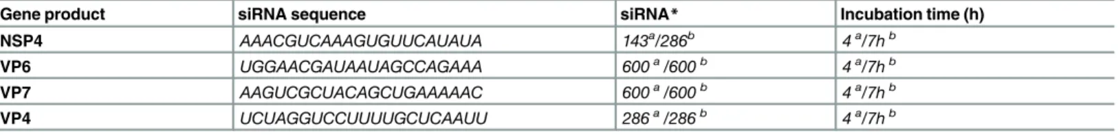
25) activations in combination with a triple band dichroic mirror (436/514/604). A 40x (NA Table 1. siRNA concentration, sequence and transfection incubation time-points in MA104 and EC tumor cells for NSP4, VP6, VP7 and VP4.
Gene product siRNA sequence siRNA* Incubation time (h)
NSP4 AAACGUCAAAGUGUUCAUAUA 143a/286b 4a/7hb
VP6 UGGAACGAUAAUAGCCAGAAA 600a/600b 4a/7hb
VP7 AAGUCGCUACAGCUGAAAAAC 600a/600b 4a/7hb
VP4 UCUAGGUCCUUUUGCUCAAUU 286a/286b 4a/7hb
All siRNA were synthesized with 3´-UU overhangs.
aMA104 cells bEC tumor cells
*(pmol/mL)
1.3; Carl Zeiss) objective was used for all images. The detector for this system was an Axiocam MRm CCD camera with a pixel size of 6.45×6.45μm.
Western blot
Semi-confluent MA104 and EC cell monolayers in 6-well plates were transfected and infected as described above. After collecting media 7 h p.i., lysis buffer containing 2% Triton X-100, 1% SDS, 0.15 M NaCl and 0.1M Tris-HCl was added to each well and freeze-thawed 3–4 times. Cell lysates were then centrifuged at 10 000 x g for 10 min and the supernatant collected and boiled for 10 min at 95°C in loading buffer (5% 2-Mercaptoethanol, 161–0710 Bio-Rad in Laemmli Sample Buffer, 161–0737 Bio-Rad) before separation with 10% polyacrylamide gel electrophoresis (PAGE). The proteins were stained with Coomassie Brilliant Blue and the relative protein concentration deter-mined. Equal relative concentration of each sample was separated on a PAGE gel and transferred (western blot assay) to a polyvinylidene difluoride (PVDF) membrane at 375 mA for 60 min. The membrane was blocked with 3% BSA in PBS-T buffer (PBS containing 0.05% Tween) for 1 h. Antibodies specific to NSP4 (dilution 1/400 in PBS-T with 1% BSA), VP4 (dilution 1/300), VP6 (dilution 1/500) and VP7 (dilution 1/30) were added and incubated for 2 h at R.T. The membranes were washed 4x with PBS-T. HRP goat anti-rabbit (170–6515, Bio-Rad) or goat anti-mouse (172–
1011, Bio-Rad) was used as secondary antibody at dilutions of 1/10000 and the membrane was incubated for 90 minutes. After washing, the reaction was developed with Bio-Rad Immun-Star HRP substrate (170–5041, Bio-Rad) and the bands were visualised with Molecular Imager1 Che-miDoc™XRS (Bio-Rad) together with Quantity One11-D analysis software (Bio-Rad).
Intracellular calcium clamping
To analyse the possible connection between intracellular calcium and release of serotonin from EC tumor cells, BAPTA/AM (B-1205, Molecular Probes Thermo Fisher, USA) (C34H40N2O18) a calcium-specific chelator was used to block intracellular calcium transients in EC tumor cells. EC tumor cells were infected with RRV for 1 h at an MOI of 0.5. Pre-toxicity testing of BABTA/AM revealed that 25μM was the most appropriate concentration for the EC tumor cells. BAPTA/AM
was added to RRV-infected cells in fresh media at 3 h p.i. The cell supernatants were collected 7 h p.i and stored at -80°C until analysis of serotonin by ELISA. Serotonin release from RRV-infected cells, with and without BAPTA/AM was compared. Uninfected cells with or without BAPTA/AM and infected cells without BAPTA/AM were used as controls.
Serotonin ELISA
A commercial serotonin ELISA kit was used (RE59121, IBL International, Germany) to deter-mine serotonin concentration according to the manufacturers instructions.
RNA extraction and reverse transcription from the small intestine
RNA in amounts of 1–0.5μg was used for reverse transcription of jejunum, ileum and the
duo-denum samples. RNA was stored at -80°C until analysis by quantitative real-time PCR.
Real-time quantitative PCR for SERT, TPH1 and GAPDH
SERT,TPH1andGAPDHprimer sequences were obtained from previous studies [50,51]. cDNA were quantified by SYBR Green based real-time PCR, withGAPDHas a reference gene. The real-time PCR reaction was performed with an ABI 7500 (Applied Biosystems, USA) with the following cycling conditions. First denaturation was done for 10 min at 95°C followed by 40 cycles of 15 s at 95°C, 1 min at 60°C, followed by melting curve analysis. Negative controls for RNA extraction, cDNA synthesis and non-template control (NTC) were included in each run. Results were analysed using theΔΔCt method and presented as normalized data.
Extraction of viral RNA in faeces
Fecal pellets were weighed and dissolved in 400μL of PBS, stored over night at 4°C and
subse-quently vortexed. Samples were centrifuged at 13 000 x g for 10 min and the supernatant trans-ferred to a new tube for extraction of viral RNA, using Magattract Viral RNA M48 kit (955235, Qiagen), according to the manufacturer’s instructions. RNA samples were stored at -80°C until reverse transcription and analysis by quantitative real-time PCR.
Quantitative PCR for detection of Rhesus rotavirus
MA104 cells grown in 24-well plates were infected with trypsin-activated Rhesus rotavirus (RRV) at a multiplicity of infection of 0.1 [52]. After 1h of infection, serum-free Eagles MEM containing 10μM Ondansetron was added. For controls, only media was used. At 48 h p.i
RNA was extracted from cell supernatant and cell-lysates and analysed with a one-step quanti-tative RV PCR [53] with external plasmid standard.
Detection of RRV-infected cells
The number of infected cells in Ondansetron-treated (10μM) and non-treated cells at 16 h p.i
was determined by immunoperoxidase staining using an anti-NSP4 polyclonal antibody (1:400). Briefly, trypsin-activated RRV (MOI 0.1) was added to confluent MA104 cells in 96-well plate. After 1 h serum-free Eagles MEM containing 10μM Ondansetron was added, while for controls
only media was used and the plate was incubated at 37°C with 5% CO2. After 16 hours of infec-tion, cells were fixed with 4% paraformaldehyde in PBS at R.T for 2 hours. After being washed with PBS, cells were permeabilized by incubation with 1% Triton X-100 in PBS for 10 min at R.T. Rotavirus-infected cells were identified by immunoperoxidase staining essentially as described [45] using a rabbit anti NSP4 antibody [44] and peroxidase-labelled goat anti-rabbit (Bio-Rad) diluted 1:1000. The reaction was developed with aminoethylcarbazole (1 mg/ml) in 0.1 M acetate buffer (pH 5.2) and stained cells (3 independent wells) were counted under a light microscope.
Determination of RRV infectious titre
Reverse transcription of fecal RNA
RNA (28μL) was mixed with 2.5μg of random hexadeoxynucleotides (pd[N[12]6) primer (27-7858-03, GE Healthcare), denatured at 97°C for 5 min and chilled on ice for 2 min. The suspen-sion was then added to 1 illustraTMReady-To-GoTMRT-PCR Bead (27-9259-01, GE Health-care) with RNase-free water to a final volume of 50μL. The RT reaction was performed for 40
min at 42°C for cDNA synthesis.
Quantitative real-time PCR for determination of viral load in faeces
Real-time PCR was performed in duplicate. TwentyμL mixtures containing 10μL of Power
SYBR Green PCR Master mix (Applied Biosystems) and 50nM of forward and reverse primers were analysed on a 7500 fast real-time PCR system (Applied Biosystems) with following cycling conditions: 95°C for 10 min followed by 40 cycles of 95°C for 15s and 60°C for 1 min. Melting curve analysis was performed after each run for specificity analysis. An EDIM positive stool sample in a 1:10 dilution series was used as standard curve, with the lowest detected dilu-tion arbitrarily set at 30 genome copies. If RV was undetected a value of 15 genes per PCR reac-tion was set. Primers used were taken from a previous study [54].
OSU-v and OSU-a infection
The virulent (OSU-v) and attenuated (OSU-a) porcine viruses have been previously character-ized [42]. The OSU-v strain, being a fecal sample, was passaged 7x (P7) in MA104 cells as toxic effect of faeces was observed on the EC tumor cells. Viral titration was performed on EC tumor cells (S1 Fig). For serotonin stimulation experiments, both viruses were activated with trypsin (10μg/mL) for 1 h at 37°C and diluted to a MOI of 1 and added to the EC tumor cells. After 1
h and 7 h p.i supernatant were collected and analysed for serotonin concentration.
To exclude the possibility that theNSP4gene of OSU-v strain had mutated during the 7 pas-sages in MA104 cells, sequencing of the NSP4 gene was performed. Briefly, RNA extraction was done from 140μL of the OSU-v P7 strain using QIAamp Viral RNA Mini Kit (Qiagen,
Germany) according the manufacturer’s instructions. Twenty-eightμL of RNA was
subse-quently mixed with 50 pmol of random hexadeoxynucleotides [pd(N)6], denatured at 97°C for 5 min, and chilled on ice for 2 min, followed by addition of one illustraTMReady-To-GoTM RT-PCR Beads (GE Healthcare) and RNase-free water to a final volume of 50μL. The RT
reac-tion was carried out for 42 min at 42°C to produce the cDNA. The NSP4 gene was amplified using GEN_NSP4F 5’- GGC TTT TAA AAG TTC TGT TCC -3’and GEN_NSP4R 5’- GGW YAC RYT AAG ACC RTT CC -3’as previously described [55] and the (750 bp) PCR product was sequenced.
Statistical analyses
The Mann-Whitney U-test was used to analyse differences in the quantification of serotonin levels and the mean fluorescent intensity from the serotonin expression studies. Differences in gene expression between uninfected and RV-infected mice were analysed with Kruskal-Wallis and Bonferroni’s multiple comparison test. Two-tailed tests were used and p<0.05 was
Results
Calcium mobilization and diarrhoea properties of virulent and attenuated
OSU virus strains correlate with the release of serotonin from human EC
tumor cells
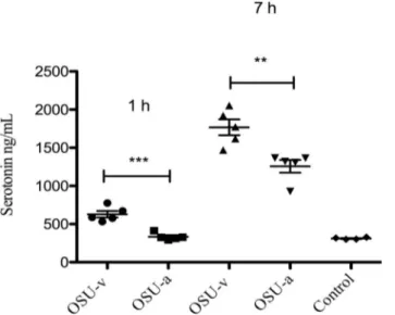
We have previously shown that RV can infect EC tumor cellsin vivoandin vitroand stimulate release of serotonin [3]. As serotonin is a known secretogue [14,56] we thus investigated if RV-stimulated release of serotonin is a contributing virulence factor. To address this question we investigated two porcine RV strains (OSU-v and OSU-a) having distinct differences in capacity to mobilize intracellular calcium and to induce diarrhoea in infant mice [42]. As shown inFig 1, both viruses stimulated release of serotonin, but the virulent OSU-v stimulated significantly more than the tissue culture-adapted OSU-a, both after 1 h p.i (p<0.001) and 7 h
p.i (p<0.01) infection. Furthermore it should be noted that attenuated OSU-a (1 h) and
con-trol (MA104 cells) had similar low effect (Fig 1). To ascertain that identical MOI was used for both viruses, careful viral titration was done on EC tumor cells (S1 Fig).S1 Figalso concludes that both viruses could replicate in EC tumor cells. To confirm that 7 passages of OSU-v in MA104 cells had not resulted in mutations during cultivation, the NSP4 gene of OSU-v was sequenced and compared to the original OSU-v strain [42], no changes in the amino acid sequence was found (S2 Fig).
Serotonin secretion from human EC tumor cells is attenuated upon
stimulation by supernatant from NSP4 silenced RV-infected MA104 cells
We have previously shown that RV infection results in secretion of serotonin from human EC tumor cellsin vitroandex vivo[3]. To identify which viral protein(s) is (are) responsible for this unique property, VP4, VP6, VP7 and NSP4 gene expression were silenced. These are pro-teins with known biological functions and which previously have been successfully silenced
Fig 1. Virulent OSU virus stimulates more release of serotonin from EC tumor cells than attenuated OSU virus.Following infection (MOI = 1) of EC tumor cells with attenuated OSU-a and virulent OSU-v virus for 1 and 7 h p.i, media was collected and the serotonin release determined with ELISA. Control: cell media from MA104 cells. Data is presented as mean±SEM.***= p˂0.001 and**= p˂0.01 with Student’s t-test;
n = 5.
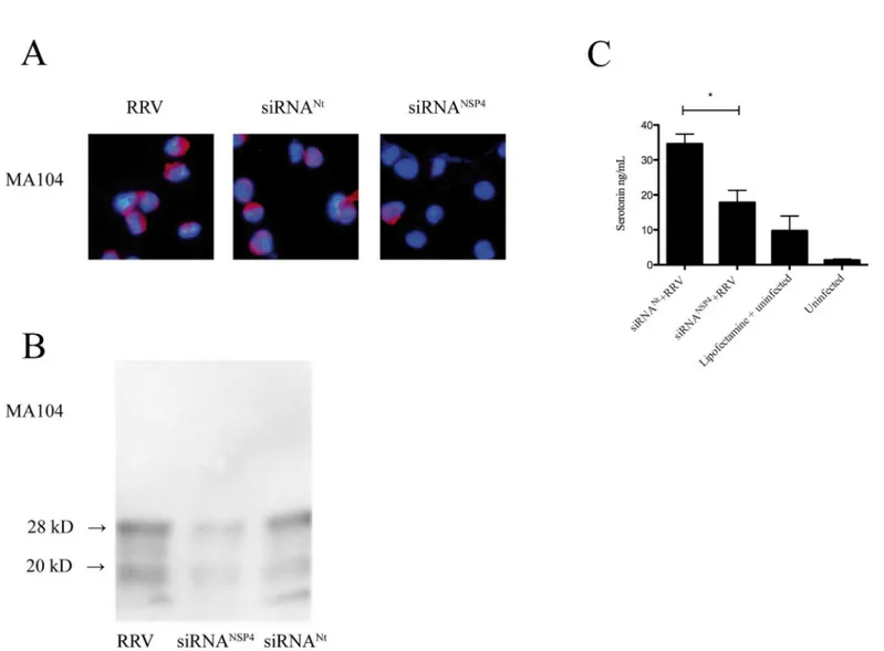
[47–49]. In a first set of experiments we investigated whether these viral proteins had an extra-cellular effect on serotonin release from EC tumor cells. To test this, we first silenced the expression of NSP4, VP4, VP6 and VP7 in RRV-infected MA104 cells. After optimization, the efficiency of siRNANSP4silencing was approximately 66% in MA104 cells (Fig 2A and 2B) and 33% for siRNAVP4, 47% for siRNAVP6and 35% for siRNAVP7. Supernatant was collected 7 h p. i and used to stimulate EC tumor cells. After 1 h, cell media was collected and serotonin con-tent determined. As illustrated inFig 2C, supernatant from MA104 cells silenced for NSP4 reduced secretion of serotonin from EC tumor cells with 48% as compared to siRNANt (p<0.05). In contrast no significant difference in serotonin secretion was observed from EC
tumor cells stimulated for 1h with supernatant from VP4, VP6 and VP7 silenced and infected MA104 cells (S3 Fig).
Fig 2. Extracellular stimulation of silenced NSP4 from MA104 cells attenuates secretion of serotonin from EC tumor cells.(A) MA104 cells were transfected with siRNANSP4. At 24 h p.i cells were infected with RRV at a MOI of 0.5 and after 7 h p.i cells were harvested and stained for NSP4 by
a rabbit anti- NSP4 and a rhodamine-conjugated goat anti-rabbit (red) conjugate. Nuclear staining was performed with DAPI (blue). (B) NSP4 western blotting of transfected and infected MA104 cells. Protein quantification performed and the relative protein concentration was determined. 28
kD = glycosylated form of NSP4, 20 kD = non-glycosylated form of NSP4. (C) EC tumor cells stimulated for 1 h with cell supernatants from MA104 infected for 7 h. Serotonin secretion was analysed by ELISA. Data is presented as means + SEM.*= p˂0.05 with Mann-Whitney U test; n = 4. siRNANt
denotes non-targeting sequence.
Intracellularly expressed NSP4 stimulates serotonin release from human
EC tumor cells
Next we investigated which of the intracellularly expressed viral proteins had effect on seroto-nin release from EC tumor cells. EC tumor cells were transfected with siRNANtor siRNANSP4/ siRNAVP4/ siRNAVP6/ siRNAVP7. After optimization, the efficiency of siRNANSP4silencing was approximately 63%, 52% for siRNAVP6, 81% for siRNAVP4and 47% for siRNAVP7(Fig 3A and 3B) in EC tumor cells. At 7 h p.i cell medium was replaced with new media and 60 min later
Fig 3. Intracellular expressed NSP4 stimulates serotonin release from human EC tumor cells.(A) EC tumor cells were transfected with siRNANSP4. At 24 h p.i cells were infected with RRV at a MOI of 0.5 and after 7 h p.i cells were harvested and
stained for NSP4 by a rabbit anti- NSP4 and a rhodamine-conjugated goat anti-rabbit (red) conjugate. (B) Silencing effect on NSP4, VP4, VP6 and VP7 expression in EC tumor cells. Following transfection and infection, cells were lysed and viral protein expression analysed by western blotting. With every western blot analysis, the amount of loaded protein was adjusted by comparing protein content in Comassie Blue-stained gels. 28 kD = glycosylated form of NSP4, 20 kD = non-glycosylated form of NSP4. (C) EC tumor cells transfected with siRNANSP4and infected. At 7 h p.i medium was changed and after 1 h supernatants
were collected and analysed for serotonin.*= p˂0.05 with Mann-Whitney U test; n = 4.
media collected and analysed for serotonin content. As shown inFig 3C, silencing of NSP4 attenuated secretion of serotonin from RRV-infected EC tumor cells by 68% compared to siR-NANt(p<0.05). No statistic difference in serotonin secretion was observed from
RRV-infected EC tumor cells silenced for VP4, VP6 and VP7 expression in comparison to RRV-infected cells transfected with siRNANt(S4 Fig).
Basal secretion of serotonin was unaffected in uninfected EC tumor cells transfected with siRNANSP4compared to uninfected, non-transfected EC tumor cells, showing that siRNA itself did not affect serotonin secretion (Fig 3C). Furthermore, no effect of Lipofectamineper sewas observed (Fig 3C). These results show that intracellularly expressed NSP4 has serotonin-stimu-lating properties.
Rotavirus re-organizes serotonin appearance in human EC tumor cells
It is known that EC cells produce, store and release serotonin [10,14] and that electron-dense serotonin granules upon specific stimulation are translocated to the cell membrane [57]. The fact that RV infection of EC tumor cells resulted in secretion of serotonin raised the question if infectionper sewould be associated with changes in appearance of serotonin-containing secre-tory granules. To address this question, the appearance and distribution of serotonin in RRV-infectedvsuninfected EC tumor cells were investigated by confocal microscopy. At 18 h p.i RRV-infected EC tumor cells showed an intense granular membrane-associated appearance of serotonin compared to a rather diffuse pattern in uninfected EC tumor cells (Fig 4A and 4B). This particular appearance is illustrated inFig 4B, with one infected cell in close vicinity to an uninfected cell. In addition, we observed a large number of granules of intense and thicker structures in the cytoplasm of RRV-infected EC tumor cells, as compared to uninfected cells, where such granules were almost absent. The appearance (intense large granularvsdiffuse pat-tern) of serotonin was determined for each infected (n = 22) and uninfected (n = 13) cell and intense granular appearance of serotonin was significantly associated (p<0.001) with infected
EC tumor cells (Fig 4C).
Rotavirus stimulation of serotonin from human EC tumor cells is
calcium-dependent
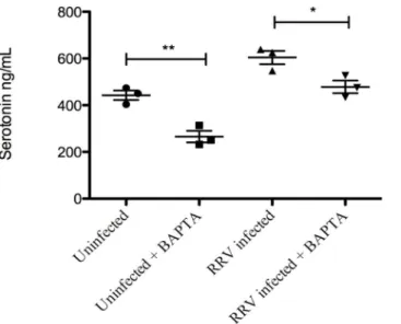
Calcium is essential for proper RV maturation in the ER [58] and NSP4 has previously been shown to affect intracellular Ca2+levels [59]. BAPTA/AM, a commonly used Ca2+chelator [60,61] was used to determine whether the release of serotonin from EC tumor cells was cal-cium-dependent. BAPTA/AM (25 uM) was added to RV-infected (MOI 0.5) EC tumor cells 3 h p.i. At 7 h p.i the supernatant was collected and serotonin content determined. As shown in
Fig 5, secretion of serotonin from infected as well as from uninfected EC tumor cells were
sig-nificantly attenuated (p˂0.05 and p˂0.01 respectively) by BABTA/AM.
SERT mRNA expression is down-regulated in ileum of rotavirus-infected
infant mice
The serotonin transporter protein SERT plays an important role in terminating and regulating the action of serotonin [30,31]. It is thus possible that the magnitude of the nerve-stimulating signal by serotonin, released by EC cells, is serotonin-concentration dependent. Hence, we investigated the expression ofSERTmRNA in RV-infected and compared to uninfected ani-mals. In general, only low amounts ofSERTmRNA were observed in duodenum and jejunum (real-time PCR Ct value>35); thus, the differences were not possible to quantify, although the
expression levels were higher and quantifiable and we observed a significant down-regulation of SERT expression in RV-infected mice (48 h p.i) compared to uninfected animals (average 3.7 fold, p<0.05). A similar down-regulation ofSERTmRNA was observed at 24 h p.i,
although not statistically significant (Fig 6).
TPH1 mRNA expression is unaffected in the small intestine of
rotavirus-infected mice
Serotonin is synthesized from L-tryptophan, and tryptophan hydroxylase (TPH1) catalyses the first step making it the rate-limiting enzyme in the biosynthesis of serotonin. TPH1 is known to be present in different cells including intestinal EC cells [28]. We expected to observe an up-regulation inTPH1mRNA levels in thein vivomodel, as serotonin release is increased during RV infectionin vitro[3]. However, there was a high variability in the mRNA levels among the animals and no significant differences inTPH1mRNA in duodenum, jejunum or ileum was observed in infant mice infected with EDIM (100DD50) compared to uninfected infant mice, neither at 24 or 48 h p.i (Fig 7A–7C).
Fig 4. Rotavirus re-organizes serotonin appearance in EC tumor cells.(A) Infected and uninfected cells were co-stained for serotonin (green) and VP6 (red) expression. Infected cells displayed re-organization of serotonin from diffuse cytoplasmic and membrane-associated appearance in uninfected cells to an intense granular membrane-associated appearance in infected cells. (B) Illustration of an uninfected EC cell in close vicinity of an infected cell (red). Note the pronounced granular appearance of serotonin near the plasma membrane of the infected cell. (C) Fluorescent intensity (arbitar units, AU) (VP6) was calculated for each infected (n = 22) and uninfected (n = 13) cell and corresponding appearance (granular vs non-granular) taken into account. Data is presented as means±SEM.***= p<0.001 with Mann-Whitney U test.
Serotonin induces rapid diarrhoea in infant but not in adult mice
Next we investigated if serotonin released from RV–infected EC tumor cells could stimulate diarrhoea in infant mice. Eight infant BALB/c mice were intra-peritoneally administered 50μL
cell supernatant from EC tumor cells containing 0.04μg (7.4μg/kg) of serotonin and 8 mice
were given 2 x 0.04μg. As control, 7 mice were administered supernatant from RRV-infected
MA104 cells. Infant mice (n = 10) were also administered a higher dose (5 mg/kg) of serotonin. Fig 5. Release of serotonin from rotavirus-infected EC tumor cells is a calcium-dependent process.
EC tumor cells were first infected with RRV (MOI = 0.5) and then at 3 h p.i BAPTA/AM was added to the cells to quench Ca2+ signals and 7 h p.i cell medium was collected for further serotonin ELISA assessment. Data is presented as means±SEM.*= p˂0.05 and**= p˂0.01 with Student’s t-test; n = 3.
doi:10.1371/journal.pone.0159660.g005
Fig 6. Rotavirus down-regulates SERT mRNA in ileum of infected mice.The small intestines of infected and uninfected mice were collected 24 and 48 h p.i and SERT and GAPDH mRNA levels quantified at each time-point by SYBR Green based real-time PCR. A significant down-regulation of SERT mRNA was found at 48 h p.i in ileum of infected compared to uninfected mice. Relative fold- change was set in relation to uninfected pups. Statistical analyses were done using Kruskal-Wallis and Bonferroni’s multiple comparison test. Data is presented as means + SEM.*= p<0.05; n = 6.
While none of the mice given supernatant from infected MA104 cells or EC tumor cells responded with diarrhoea, all mice (10/10) given the higher dose (5 mg/kg) of serotonin responded with diarrhoea within 30 min. It is well established that adult mice can be infected with RV, but do not respond with diarrhoea [62], presumably due to a block in water secretion from the small intestine [54]. To strengthen our proposed association between serotonin and RV-induced diarrhoea, adult BALB/c mice were intra-peritoneally administered 5 mg/kg of serotonin. None of the 8 adult mice responded with diarrhoea within 5 h. Thus similar as for RV these results demonstrate an age-dependent susceptibility of serotonin induced-diarrhoea in mice.
Blocking of serotonin receptors attenuates rotavirus diarrhoea in infant
mice
To address our hypothesis that serotonin is associated with RV-induced diarrhoea, infant BALB/c mice were infected with wild type EDIM (100DD50) and orally treated with Ondanse-tron twice a day with 5 mg/kg. We used the serotonin-3 receptor antagonist OndanseOndanse-tron, as this drug is commonly used to attenuate vomiting in children with acute gastroenteritis [21,
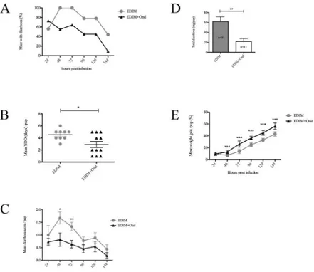
22] and have anti-diarrhoeal and obstipation properties [38–41].Fig 8Ashows that Ondanse-tron-treatment twice daily reduced the number of mice with diarrhoea, starting at 48 h p.i until 144 h p.i when the experiment was terminated. Previous studies [63] have shown that infant mice infected with EDIM have diarrhoea peak at 48 h p.i a time point when Ondansetron-treatment began to show effect. All infected mice in the Ondansetron-treated and mock-treated group had diarrhoea at any given time point.Fig 8Bshows that the mean number of diarrhoea days per pup was significantly (p<0.05) lower in the intervened group compared to
mock-treated, reducing the days from a mean of 4.5 to 2.9 days in the intervened group. To obtain more detailed information about the anti-diarrhoeal effect of Ondansetron, the severity of diar-rhoea was scored for each mouse and time-point.Fig 8Cshows that there was a significant dif-ference in the diarrhoea severity (score) between intervened (n = 11) and mock-treated (n = 9) mice at 48 (p<0.05), and 72 h p.i (p<0.01).Fig 8Dshows that mock-treated mice had
signifi-cantly (p<0.01) more diarrhoea per mouse than Ondansetron-treated mice and moreover,
mock-treated mice had significantly more total diarrhoea (mean 558.1 mg) output than inter-vened mice (mean 240.17 mg) (p<0.01). Considering the positive effect of Ondansetron on
duration and severity of diarrhoea, we also investigated whether Ondansetron-treatment could effect weight development during the course of infection. As shown inFig 8E, Ondansetron-Fig 7. TPH1 mRNA levels are unaffected in the small intestine of rotavirus-infected pups.Infant mice were infected with EDIM (100DD50) and the small intestine collected 24 h and 48 h p.i followed by real-time PCR for TPH1 and GAPDH mRNA. No statistical difference in TPH1 mRNA levels compared to GAPDH mRNA was found in duodenum (A), jejenum (B) or ileum (C) of infected compared to uninfected pups. Relative fold-change is set in relation to uninfected pups. Statistical analyses were made using Kruskal-Wallis and Bonferroni’s multiple comparison test. Data is presented as mean + SEM; n = 6.
treated mice had significantly (p<0.001) better weight gain during the course of infection
compared to mock-treated mice. Ondansetron itself did not induce diarrhoea in uninfected mice (n = 4) thus concluding that the drug itself had no effect on described factors inFig 8. Altogether, this suggests that RV infection of infant mice triggers serotonin signalling that affects duration, severity of diarrhoea and consequently weight gain and that duration and severity can be attenuated by the serotonin-3 receptor antagonist Ondansetron.
Blocking of serotonin receptors attenuates murine rotavirus shedding in
adult mice
Next we investigated if interference in serotonin signalling by a serotonin receptor antagonist could effect viral shedding independently of diarrhoea. Adult RV BALB/c mice were first infected with EDIM (100DD50) and treated orally with Ondansetron (n = 10) (5 mg/kg) twice a day or mock-treated (n = 10) and followed for up to 144 h p.i. As shown inFig 9, intervention by the serotonin receptor antagonist significantly attenuated viral shedding at any given time point, most pronounced at 72 h p.i (p<0.05). Moreover, the total amount of viral shedding
during the course of experiment was significantly lower (12365 vs 30676 RV genome copies) in Ondansetron-treated mice.
Fig 8. Serotonin antagonist Ondansetron attenuates diarrhoea in rotavirus-infected mice.(A) Prevalence of diarrhoea over time in rotavirus (EDIM) infected mice. Mice infected with EDIM were compared with mice infected with EDIM and treated with Ondansetron (5 mg/kg). Mean values are presented. (B) Number of days with diarrhoea (NDD) in mice infected with EDIM or EDIM plus Ondansetron (5 mg/kg). Data is presented as mean±SEM.*= p<0.05 with Student’s t-test. (C) Daily diarrhoea score for each day between the groups of mock-treated infected mice and infected mice treated with Ondansetron. Mock-treated infected mice received significantly more severe diarrhoea after 48 and 72 h p.i. Data is presented as mean±SEM.*= p<0.05 and**= p<0.01 with Student’s t-test. (D) Total amount (mg) of diarrhoea per
mouse up to 144 h p.i. Data is presented as mean +SEM.**= p<0.01 with Student’s t-test. (E) Comparison
of weight gain between treated and mock-treated mice over time. Treated infected mice had gained significantly more weight from 48 h p.i. in comparison to only those without treatment. Data is presented as mean±SEM.***= p<0.001 with Student’s t-test. EDIM (n = 9), EDIM + Ondansetron (n = 11).
The fact that RV RNA shedding was significantly reduced in Ondansetron-treated and infected mice led us next to investigate if the reduced shedding was due to an antiviral effect. To address this question we investigated if Ondansetron had anyin vitroeffect on (i) number of infected cells, (ii) viral RNA synthesis or (iii) production of infectious virus. Briefly, MA104 cells were infected with Rhesus rotavirus (strain RRV) and at 1 h p.i media was replaced with serum-free media containing 10μM Ondansetron (highest non-toxic concentration
deter-mined by trypan blue staining). To determine if Ondansetron affected the number of infected cells, cells were fixed at 16 h p.i and the number of infected cells determined by staining for the intracellular expressed non-structural NSP4 protein, by a rabbit anti NSP4 antibody [44]. The experiment revealed no difference in number of infected cells between Ondansetron-treated (10μM) and mock-treated MA104 cells (148 infected cells per 1.04 cm2in infected and treated
cells vs 171 infected and mock-treated cells) Furthermore, no inhibitory effect of Ondansetron was observed on viral RNA synthesis by quantitative qPCR at 48 h p.i (S5 Fig). Next, we inves-tigated if Ondansetron had any effect on progeny virus. Ondansetron-treated (10μM) and
mock-treated RRV infected (MOI 1) MA104 cells (n = 3) were freeze-thawed at 16 h p.i. and virus titres were determined. We found similar infectious titre among Ondansetron-treated (mean titre 4.1x10⁵pfu/ml) and mock-treated (mean titre 4.8x10⁵pfu/ml) cells, suggesting that Ondansetron did not have any detectable antiviral effect on rotavirus.
Discussion
In this work it is shown that intracellularly expressed NSP4 is associated with the release of the neurotransmitter serotonin from human EC tumor cells. By employing virulent and attenuated porcine RV it is evident that virulence factors such as the capacities to induce diarrhoea in infant mice and to mobilize intracellular calcium [42] correlate with the ability to stimulate release of serotonin from human neuroendocrine cells. Ondansetron significantly attenuated murine RV diarrhoea in infant mice and that the treated mice gained weight better than mock-treated animals. A surprising finding is also that the serotonin receptor antagonist attenuated viral shedding. The later observation may support a novel therapeutic strategy for intervention of RV illness.
Fig 9. Ondansetron treatment reduces virus shedding.RV shedding as determined by real-time PCR in faeces of mice mock-treated or treated with Ondansetron. Mice were treated twice-a-day with Ondansetron (5 mg/kg). Total amount of RV shedding was significantly reduced in Ondansetron-treated mice with 30676 vs 12365 genome copies for mock-treated and Ondansetron-treated mice respectively. Data is presented as mean±SEM.*= p<0.05 with Mann-Witney U test; n = 10 in each group.
By inhibiting the expression of RV capsid proteins VP4, VP6, VP7 and the non-structural protein NSP4 by siRNA silencing, it was shown that intracellularly expressed NSP4 did stimu-late release of serotonin, which is to the best of our knowledge the first intracellularly expressed viral protein with a neurotransmitter-stimulating property. Since the experiments did not include all RV proteins it cannot be excluded that other non-investigated proteins have similar properties. By employing previously described siRNA sequences and methodologies; NSP4 expression was silenced by 63% in EC tumor cells. This is similar to those of previous studies using other cell lines [48,49,64]. Expression of VP6 was reduced by approximately 52%, VP4 by 81% and VP7 47% in EC tumor cells. The silencing efficiency depend on a wide range of fac-tors including cell lines and type of cells, siRNA concentration, incubation times and transfec-tion agents being used.
Ca2+signalling is known to play a key role in diverse cell functions and pathological mecha-nisms including RV infections and replication [60,64–66]. There are also studies reporting changes in Ca2+homeostasis coupled to the NSP4 enterotoxin [59,61,67,68]. Using the cell-permeable Ca2+chelator BAPTA/AM, we found that secretion of serotonin from EC tumor cells was Ca2+-dependent (Fig 5), an observation in line with the proposed model for seroto-nin-induced secretion from EC cells [57,69].
It is well-known that EC cells synthesize serotonin through hydroxylation and decarboxyl-ation of tryptophan in the cytoplasm of the EC cell [14]. The reason for studying EC cells in this work is that they form the largest enteroendocrine cell population in the small intestine and the only enteroendocrine cell that can synthesise serotonin. Upon stimulation, these cells are believed to account for the larger part of extracellularly released serotonin [11,12]. Newly synthesized serotonin is subsequently transported into and stored in secretory granules by a vesicular monoamine transporter (VMAT) [70,71] or SERT [16,29,34,35] and/or degraded to maintain a normal extracellular serotonin level. Upon specific stimulation, these secretory granules are transported to the cell membrane and their content, including serotonin is released from the granules after exocytosis. In this way, extracellular serotonin may possibly reach lamina propria, where it can stimulate nerve terminals. The importance of serotonin in the GI lumen [72] is well documented and together with earlier findings on RV and serotonin [3] we hypothesized EC cells and serotonin to be the sensors during RV infection. First, we investigated, the distribution of serotonin following a RV-infection and compared it with unin-fected cells. We found distinct differences with intense granular appearance in the periphery of the infected cells in contrast to more diffuse serotonin localization in uninfected cells (Fig 4A
and 4B). Moreover the infected cells had intense, thick structures in the cytoplasm. Putatively,
the infection of EC tumor cells did promote translocation of serotonin from the cytoplasm to secretory granules and further extracellular release via exocytosis [14],in vivopresumably to
lamina propriaand nerve endings of the ENS.
It has previously been shown that RV infection induces changes in cytoskeleton and cell morphology. Cytoskeleton modifications, are sensitive to changes in calcium concentration, which have been associated with the action of NSP4 [61], suggesting, but do not proving, that NSP4-induced changes in Ca2+homeostasis may result in actin filament re-arrangement and thus altered appearance of the serotonin-containing secretory granules.
study showed decreased mRNA levels of SERT [77], while in others there was an increase or no difference [73,76]. Camilleri and co-workers [73] collected RNA from rectal and sigmoid colon mucosal biopsies in IBS patients and analysedSERTmRNA expression and found the levels to be normal. Hence, there are contradictory results on SERT expression in GI inflamma-tory diseases. Here we observed a significant down-regulation ofSERTmRNA in the ileum of infected mice at 48 h p.i compared to uninfected mice (Fig 6). The modest down-regulation at 24 h p.i might reflect that the most pronounced diarrhoea and enteric lesions with the murine RV strain EDIM occurs at 48 h p.i [63]. The levels of SERT mRNA in duodenum and jejunum were generally down-regulated but too low to allow for any firm conclusions. The finding that the highestSERTmRNA expression in uninfected animals was found in ileum corroborates with a previous human study [31]. Furthermore, the down-regulation of SERT mRNA in ileum of infected animals might be due to a more preferential localization to this intestinal segment and as a consequence more extensive effects on host protein synthesis. A previous study showed indeed that during an infection the mouse ileum contained 100-fold more RV RNA than jejunum or duodenum [54].
A question is whether the increased serotonin secretion upon the RV infection of EC tumor cells was a result of an accumulation of pre-made serotonin in secretory granules, or of increased TPH1 transcription and translation. Serotonin is synthesized from L-tryptophan by tryptophan hydroxylase (TPH1) catalysis. TPH1 is known to be present in different cells, including intestinal EC cells [28] and one might assume that TPH1 expression would correlate with SERT expression. This is however not always the case, in one study there was no differ-ence in theTPH1mRNA expression in duodenum of IBS patients [76], whereas the same type of patients demonstrated reduced expression ofTPH1mRNA in the large intestine [16,76]. Hence, contradictory results have been reported not only for SERT but also TPH1 expression in IBS and UC patients. Some of the discrepancies could however be related to the intestinal segment being investigated. Similar to Kerckhoffs and co-workers who observed no alternation in theTPH1mRNA expression in duodenum of IBS patients [76], we did not find any statisti-cal difference for it in the small intestine of RV-infected mice (Fig 7A–7C). This can suggest that in either case the release of serotonin primarily occured from pre-made rather than from newly synthesised serotonin. This could also explain the rapid release of serotonin following a RV challenge.
Since serotonin was capable to induce rapid (<30 min) diarrhoea in infant mice (10/10),
we aimed to determine whether differences in viral virulence could be associated with capacity to stimulate release of serotonin from EC tumor cells. To address this question we compared the effects of virulent and avirulent porcine OSU viruses with known virulence differences in calcium mobilization and diarrhoea [42]. Not only can OSU-v virus mobilize more intracellu-lar calcium and cause more diarrhoea in infant mice than OSU-a [42], but it also elicited stron-ger serotonin release from EC tumor cells than attenuated OSU-a virus (Fig 1). It is likely that Ca2+mobilization is a key regulator of the secretion of serotonin, since clamping with BAPTA/ AM, reduced both the basic and RV-induced release of serotonin (Fig 5), which corroborates previous findings [3].
Infant mice were treated with the serotonin receptor antagonist Ondansetron with the objective to further test our hypothesis that serotonin is participating is RV-induced diarrhoea. This receptor antagonist is rather frequently used to attenuate illness in children with acute gastroenteritis [21,22], to attenuate diarrhoea in patients with inflammatory bowel disease [38,
significantly more total diarrhoea output than intervened mice and (iii) Ondansetron-treat-ment resulted in better weight gain (Fig 8A–8E). The latter finding should be put in the light of observations that serotonin help control food intake [78,79] and that feeding behaviour in fast-ing mice may be related to C-fos activity in the hypothalamus and brainstem [80]. It should also be noted that RV infection activates C-fos in CNS [3], which suggests a common signalling pathway that may result in less appetite during RV illness.
To assess whether Ondansetron could affect viral shedding independent of diarrhoea, adult mice were infected and non-or Ondansetron-treated and the viral load determined by real-time PCR. A most surprising effect was that the drug significantly attenuated total viral shed-ding (Fig 9). To better understand the unexpected effect of Ondansetron on viral shedding in mice,in vitrostudies were performed. These studies revealed that Ondansetron neither had any effect on the number of infected cells, nor did it impair RV RNA synthesis at 48 h p.i up to concentrations that are toxic to cells (S5 Fig). Furthermore, no effect of Ondansetron was observed on viral progenies with mean (n = 3) titre of 4.1x10⁵pfu/ml after Ondansetron treat-ment and 4.8x10⁵pfu/ml after mock treatment (n = 3), thus it remains unresolved how Ondan-setron attenuates RV shedding in mice.
It is interesting to mention that serotonin participate in immune activation and inflamma-tion. It has been proposed that serotonin released from EC cells in response to stimuli, such as toxins and microbes, can act on innate immune cells such as macrophages and dendritic cells, to activate a proinflammatory cytokine response and thereby influence interaction between innate immune and adaptive immune cells [81,82]. The presence of EC cells in contact or very close proximity to CD3+and CD20+lymphocytes [24] suggests existence of such an interaction between EC cells and immune cells.
Serotonin is a potent secretagogue of electrolytes and fluids in the intestines of all species studied, including humans [14,56]. The serotonin-induced secretion mechanism is consistent with Cl-/HCO3-secretion, which presumably occurs from crypt cells [83]. Pathways by which serotonin evokes intestinal secretion are likely both neuronal and non-neuronal and include at least activation of the serotonin-3 receptor and a neural reflex, which is tetrodotoxin sensitive [14]. Tetrodotoxin-sensitivity is most interesting, as it has been previously shown that RV-induced electrolyte and water secretion can be attenuated by tetrodotoxin [4].
In conclusion, we show that intracellularly expressed NSP4 carry serotonin neurotransmit-ter-stimulating properties and that serotonin participates in RV–induced diarrhoea, which can be attenuated by the commonly used serotonin-3 receptor antagonist Ondansetron. This new information can be added to the proposed gut-nerve-brain cross-talk axis in RV illness.
Supporting Information
S1 Fig. Viral titration of OSU-a and the virulent OSU-v P7 virus on EC tumor cells.EC tumor cells infected with attenuated OSU-a virus and the virulent OSU-v P7 virus and stained for VP6 expression to evaluate rates of infection and equal amount of cells infected. EC tumor cells were infected with MOI = 1 with respective viruses and 18 hours post infection fixed and stained with specific antibodies against VP6 (red expression), as described in the Material and Methods. Virus titration was previously performed on MA104 cells and the calculations were applied on EC tumor cells.
(PDF)
Material and Methods. (PDF)
S3 Fig. No difference in serotonin secretion was observed from EC tumor cells stimulated for 1h with supernatant from VP4, VP6 and VP7 silenced and infected MA104 cells.EC tumor cells stimulated for 1 h with cell supernatants from silenced and infected MA104 cells. Serotonin secretion was analysed by ELISA. Data is presented as means + SEM with Mann-Whitney U test; n = 4. siRNANtdenotes non-targeting sequence and ns denotes not significant. (PDF)
S4 Fig. No difference in serotonin secretion was observed from RRV-infected EC tumor cells silenced for VP4, VP6 and VP7 expression in comparison to infected cells transfected with siRNANt.EC tumor cells transfected with siRNAVP4, siRNAVP6, siRNAVP7and siRNANt and infected with RRV. At 7 h p.i medium was changed and after 1 h serotonin secretion was analysed. Data is presented as means + SEM. Statistics were made using Mann-Whitney U test; n = 4. siRNANtdenotes non-targeting sequence and ns denotes not significant.
(PDF)
S5 Fig. No difference in RV gene copy number was observed between RRV-infected Ondan-setron-treated and mock-treated MA104 cells.Quantitfication of RV genes 48 h p.i as deter-mined by real-time PCR in supernatants and cell lysates of RRV-infected Ondansetron-treated (10μM) and mock-treated MA104 cells. Data is presented in a log2 scale with geometric mean
values and 95% confidence interval. No significant differences between the groups; n = 3. (PDF)
Acknowledgments
This work was supported by Swedish Research Council (LS) 320301 and the Diarrhoeal Disease Center, Linköping University (L.S; K-E M).
Author Contributions
Conceived and designed the experiments: LS K-EM SB MH. Performed the experiments: SB MH JN TK SS. Analyzed the data: SB MH JN TK SS K-EM LS. Contributed reagents/materials/ analysis tools: SB MH JN TK SS K-EM LS. Wrote the paper: SB MH JN SS K-EM LS.
References
1. Parashar UD, Gibson CJ, Bresee JS, Glass RI. Rotavirus and severe childhood diarrhea. Emerg Infect Dis. 2006; 12(2):304–6. Epub 2006/02/24. doi:10.3201/eid1202.050006PMID:16494759; PubMed Central PMCID: PMC3373114.
2. Hagbom M, Sharma S, Lundgren O, Svensson L. Towards a human rotavirus disease model. Curr Opin Virol. 2012; 2(4):408–18. Epub 2012/06/23. doi:10.1016/j.coviro.2012.05.006PMID:22722079.
3. Hagbom M, Istrate C, Engblom D, Karlsson T, Rodriguez-Diaz J, Buesa J, et al. Rotavirus stimulates release of serotonin (5-HT) from human enterochromaffin cells and activates brain structures involved in nausea and vomiting. PLoS Pathog. 2011; 7(7):e1002115. Epub 2011/07/23. doi:10.1371/journal. ppat.1002115PPATHOGENS-D-11-00407 [pii]. PMID:21779163; PubMed Central PMCID: PMC3136449.
4. Lundgren O, Peregrin AT, Persson K, Kordasti S, Uhnoo I, Svensson L. Role of the enteric nervous sys-tem in the fluid and electrolyte secretion of rotavirus diarrhea. Science. 2000; 287(5452):491–5. Epub 2000/01/22. PMID:10642552.
5. Estes MK, editor. The rotavirus NSP4 enterotoxin: Current status and challenges. Amsterdam: Else-vier; 2003.
7. Greenberg HB, Estes MK. Rotaviruses: from pathogenesis to vaccination. Gastroenterology. 2009; 136 (6):1939–51. Epub 2009/05/22. doi:10.1053/j.gastro.2009.02.076PMID:19457420; PubMed Central PMCID: PMC3690811.
8. Ramig RF. Pathogenesis of intestinal and systemic rotavirus infection. Journal of virology. 2004; 78 (19):10213–20. doi:10.1128/JVI.78.19.10213–10220.2004PMID:15367586; PubMed Central PMCID: PMC516399.
9. Erspamer V, Asero B. Identification of enteramine, the specific hormone of the enterochromaffin cell system, as 5-hydroxytryptamine. Nature. 1952; 169(4306):800–1. PMID:14941051.
10. Cetin Y, Kuhn M, Kulaksiz H, Adermann K, Bargsten G, Grube D, et al. Enterochromaffin cells of the digestive system: cellular source of guanylin, a guanylate cyclase-activating peptide. Proc Natl Acad Sci U S A. 1994; 91(8):2935–9. PMID:8159683; PubMed Central PMCID: PMC43489.
11. Gershon MD, Tack J. The serotonin signaling system: from basic understanding to drug development for functional GI disorders. Gastroenterology. 2007; 132(1):397–414. doi:10.1053/j.gastro.2006.11. 002PMID:17241888.
12. Mawe GM, Coates MD, Moses PL. Review article: intestinal serotonin signalling in irritable bowel syn-drome. Alimentary pharmacology & therapeutics. 2006; 23(8):1067–76. doi:10.1111/j.1365-2036. 2006.02858.xPMID:16611266.
13. Bertrand PP, Kunze WA, Furness JB, Bornstein JC. The terminals of myenteric intrinsic primary affer-ent neurons of the guinea-pig ileum are excited by 5-hydroxytryptamine acting at 5-hydroxytryptamine-3 receptors. Neuroscience. 2000; 101(2):459–69. PMID:11074168.
14. Hansen MB, Witte AB. The role of serotonin in intestinal luminal sensing and secretion. Acta Physiol (Oxf). 2008; 193(4):311–23. doi:10.1111/j.1748-1716.2008.01870.xPMID:18462271.
15. Spiller R. Serotonin and GI clinical disorders. Neuropharmacology. 2008; 55(6):1072–80. doi:10.1016/ j.neuropharm.2008.07.016PMID:18687345.
16. Coates MD, Mahoney CR, Linden DR, Sampson JE, Chen J, Blaszyk H, et al. Molecular defects in mucosal serotonin content and decreased serotonin reuptake transporter in ulcerative colitis and irrita-ble bowel syndrome. Gastroenterology. 2004; 126(7):1657–64. PMID:15188158.
17. Belai A, Boulos PB, Robson T, Burnstock G. Neurochemical coding in the small intestine of patients with Crohn's disease. Gut. 1997; 40(6):767–74. PMID:9245931.
18. Bearcroft CP, Perrett D, Farthing MJ. Postprandial plasma 5-hydroxytryptamine in diarrhoea predomi-nant irritable bowel syndrome: a pilot study. Gut. 1998; 42(1):42–6. PMID:9505884.
19. Dunlop SP, Coleman NS, Blackshaw E, Perkins AC, Singh G, Marsden CA, et al. Abnormalities of 5-hydroxytryptamine metabolism in irritable bowel syndrome. Clin Gastroenterol Hepatol. 2005; 3 (4):349–57. Epub 2005/04/12. PMID:15822040.
20. Gershon MD. Review article: roles played by 5-hydroxytryptamine in the physiology of the bowel. Ali-ment Pharmacol Ther. 1999; 13 Suppl 2:15–30. Epub 1999/08/03. PMID:10429737.
21. Freedman SB, Steiner MJ, Chan KJ. Oral ondansetron administration in emergency departments to children with gastroenteritis: an economic analysis. PLoS Med. 2010; 7(10). Epub 2010/10/23. doi:10. 1371/journal.pmed.1000350PMID:20967234; PubMed Central PMCID: PMCPMC2953527.
22. Levine DA. Oral ondansetron decreases vomiting, as well as the need for intravenous fluids and hospi-tal admission, in children with acute gastroenteritis. Evid Based Med. 2012; 17(4):112–3. Epub 2011/ 12/24. doi:10.1136/ebmed.2011.100355PMID:22193568.
23. Hu DL, Zhu G, Mori F, Omoe K, Okada M, Wakabayashi K, et al. Staphylococcal enterotoxin induces emesis through increasing serotonin release in intestine and it is downregulated by cannabinoid recep-tor 1. Cell Microbiol. 2007; 9(9):2267–77. Epub 2007/05/23. CMI957 [pii] doi:10.1111/j.1462-5822. 2007.00957.xPMID:17517065.
24. Cloez-Tayarani I, Changeux JP. Nicotine and serotonin in immune regulation and inflammatory pro-cesses: a perspective. J Leukoc Biol. 2007; 81(3):599–606. doi:10.1189/jlb.0906544PMID: 17108054.
25. Arreola R, Becerril-Villanueva E, Cruz-Fuentes C, Velasco-Velazquez MA, Garces-Alvarez ME, Hur-tado-Alvarado G, et al. Immunomodulatory effects mediated by serotonin. Journal of immunology research. 2015; 2015:354957. doi:10.1155/2015/354957PMID:25961058; PubMed Central PMCID: PMC4417587.
26. Patel PD, Pontrello C, Burke S. Robust and tissue-specific expression of TPH2 versus TPH1 in rat raphe and pineal gland. Biol Psychiatry. 2004; 55(4):428–33. Epub 2004/02/13. doi:10.1016/j. biopsych.2003.09.002PMID:14960297.
28. Legay C, Faudon M, Hery F, Ternaux JP. 5-HT metabolism in the intestinal wall of the rat-I. The mucosa. Neurochem Int. 1983; 5(6):721–7. Epub 1983/01/01. PMID:20488002.
29. Wade PR, Chen J, Jaffe B, Kassem IS, Blakely RD, Gershon MD. Localization and function of a 5-HT transporter in crypt epithelia of the gastrointestinal tract. J Neurosci. 1996; 16(7):2352–64. Epub 1996/ 04/01. PMID:8601815; PubMed Central PMCID: PMC3327288.
30. Takayanagi S, Hanai H, Kumagai J, Kaneko E. Serotonin uptake and its modulation in rat jejunal enter-ocyte preparation. J Pharmacol Exp Ther. 1995; 272(3):1151–9. Epub 1995/03/01. PMID:7891327.
31. Gill RK, Pant N, Saksena S, Singla A, Nazir TM, Vohwinkel L, et al. Function, expression, and charac-terization of the serotonin transporter in the native human intestine. Am J Physiol Gastrointest Liver Physiol. 2008; 294(1):G254–62. Epub 2007/11/10. doi:10.1152/ajpgi.00354.2007PMID:17991706.
32. Martel F, Monteiro R, Lemos C. Uptake of serotonin at the apical and basolateral membranes of human intestinal epithelial (Caco-2) cells occurs through the neuronal serotonin transporter (SERT). J Pharma-col Exp Ther. 2003; 306(1):355–62. doi:10.1124/jpet.103.049668PMID:12682218.
33. Miwa J, Echizen H, Matsueda K, Umeda N. Patients with constipation-predominant irritable bowel syn-drome (IBS) may have elevated serotonin concentrations in colonic mucosa as compared with diar-rhea-predominant patients and subjects with normal bowel habits. Digestion. 2001; 63(3):188–94. Epub 2001/05/15. 51888. PMID:11351146.
34. Chen JJ, Li Z, Pan H, Murphy DL, Tamir H, Koepsell H, et al. Maintenance of serotonin in the intestinal mucosa and ganglia of mice that lack the high-affinity serotonin transporter: Abnormal intestinal motility and the expression of cation transporters. The Journal of neuroscience: the official journal of the Soci-ety for Neuroscience. 2001; 21(16):6348–61. PMID:11487658.
35. Chen JX, Pan H, Rothman TP, Wade PR, Gershon MD. Guinea pig 5-HT transporter: cloning, expres-sion, distribution, and function in intestinal sensory reception. The American journal of physiology. 1998; 275(3 Pt 1):G433–48. PMID:9724254.
36. Bellini M, Rappelli L, Blandizzi C, Costa F, Stasi C, Colucci R, et al. Platelet serotonin transporter in patients with diarrhea-predominant irritable bowel syndrome both before and after treatment with alose-tron. Am J Gastroenterol. 2003; 98(12):2705–11. Epub 2003/12/23. doi:10.1111/j.1572-0241.2003. 08669.xPMID:14687821.
37. Esmaili A, Nazir SF, Borthakur A, Yu D, Turner JR, Saksena S, et al. Enteropathogenic Escherichia coli infection inhibits intestinal serotonin transporter function and expression. Gastroenterology. 2009; 137 (6):2074–83. Epub 2009/09/15. doi:10.1053/j.gastro.2009.09.002PMID:19747920; PubMed Central PMCID: PMC3727418.
38. Garsed K, Chernova J, Hastings M, Lam C, Marciani L, Singh G, et al. A randomised trial of ondanse-tron for the treatment of irritable bowel syndrome with diarrhoea. Gut. 2014; 63(10):1617–25. doi:10. 1136/gutjnl-2013-305989PMID:24334242; PubMed Central PMCID: PMCPMC4173656.
39. Talley NJ, Phillips SF, Haddad A, Miller LJ, Twomey C, Zinsmeister AR, et al. GR 38032F (ondanse-tron), a selective 5HT3 receptor antagonist, slows colonic transit in healthy man. Dig Dis Sci. 1990; 35 (4):477–80. PMID:2138532.
40. Bryson JC. Clinical safety of ondansetron. Semin Oncol. 1992; 19(6 Suppl 15):26–32. PMID:1485179.
41. Smith RN. Safety of ondansetron. Eur J Cancer Clin Oncol. 1989; 25 Suppl 1:S47–50; discussion S1-4. PMID:2533899.
42. Zhang M, Zeng CQ, Dong Y, Ball JM, Saif LJ, Morris AP, et al. Mutations in rotavirus nonstructural gly-coprotein NSP4 are associated with altered virus virulence. J Virol. 1998; 72(5):3666–72. Epub 1998/ 04/29. PMID:9557647; PubMed Central PMCID: PMC109587.
43. Kolby L, Bernhardt P, Ahlman H, Wangberg B, Johanson V, Wigander A, et al. A transplantable human carcinoid as model for somatostatin receptor-mediated and amine transporter-mediated radionuclide uptake. Am J Pathol. 2001; 158(2):745–55. doi:10.1016/S0002-9440(10)64017-5PMID:11159212; PubMed Central PMCID: PMCPMC1850312.
44. Mirazimi A, Nilsson M, Svensson L. The molecular chaperone calnexin interacts with the NSP4 entero-toxin of rotavirus in vivo and in vitro. J Virol. 1998; 72(11):8705–9. Epub 1998/10/10. PMID:9765412; PubMed Central PMCID: PMC110284.
45. Ruggeri FM, Johansen K, Basile G, Kraehenbuhl JP, Svensson L. Antirotavirus immunoglobulin A neu-tralizes virus in vitro after transcytosis through epithelial cells and protects infant mice from diarrhea. J Virol. 1998; 72(4):2708–14. Epub 1998/04/03. PMID:9525588; PubMed Central PMCID: PMC109713.