ABSTRACT
preosteoblast behavior within interconnected
porous hydroxyapatite ceramics -
in vit ro
analysis
Mohammad Zeshaan RAHMAN, Hideo SHIGEISHI, Kazuki SASAKI, Akira OTA, Kouji OHTA, Masaaki TAKECHI
Hiroshima University, Institute of Biomedical and Health Sciences, Department of Oral and Maxillofacial Surgery, Hiroshima, Japan.
Corresponding address: Hideo Shigeishi - Department of Oral and Maxillofacial Surgery, Institute of Biomedical and Health Sciences, Hiroshima University - 1-2-3 Kasumi - Minami-ku - Hiroshima - Japan - 734-8553 - Phone: +81-82-257-5673, FAX: +81-82-257-5671 - e-mail: [email protected]
6XEPLWWHG-DQXDU\0RGL¿FDWLRQ)HEUXDU\$FFHSWHG0DUFKst, 2016
O
bjective: Biocompatible materials such as interconnected porous hydroxyapatiteceramics (IP-CHA) loaded with osteogenic cells and bioactive agents are part of
in vit ro study was undertaken to examine the relationship between these two bioactive
agents and their combinatory effects on osteoblastic activity and mineralization in vit ro.
Material and Methods: Mouse preosteoblast cells (MC3T3-E1) were seeded and cultured within cylindrical type of CHA block (ø 4x7 mm) by vacuum-assisted method. The
IP-alkaline phosphatase enzyme activity (ALP), mRNA expressions of late bone markers, namely Osteocalcin (OCN) and Osteopontin (OPN), and Alizarin Red staining were examined
cells within the IP-CHA constructs. MEL mainly induced the mRNA expression of late bone markers (OCN and OPN) and showed increased ALP activity of MC3T3 cells cultured
osteogenic activity within the IP-CHA construct in terms of cell proliferation, upregulated expressions of OCN and OPN, increased ALP activity and mineralization with Alizarin Red.
showed increased osteogenic activity in MC3T3-E1 cells cultured within IP-CHA constructs.
with biocompatible materials to attain augmented osteogenic activity and mineralization.
Ke yw or ds: Hydroxyapatite. Fibroblast growth factor-2. Melatonin.
I N TROD UCTI ON
Bone grafting plays an essential role in craniofacial surgery performed for both reconstructive and aesthetic purposes, which has led to discovery of different biomaterials, including hydroxyapatite (HAp), a member of the non-resorbable calcium phosphate group of biomaterials23. HAp has been formed into a variety of shapes and dimensions, and shown both biocompatibility and osteoconductivity since the discovery of its similarities with natural bone23. Porous type HAp ceramics are expected to facilitate bone formation and become integrated with
while its porous architecture also provides optimum compressive strength of up to 12 MPa, similar to cancellous bone21. It has been suggested that IP-CHA may have an additional osteoinductive advantage if the porous architecture could be utilized to transplant osteoinductive agents or osteogenic cells or both.
member of the 23-polypeptide growth factor family13. It has been found to participate in a variety of biological processes, such as angiogenesis, hematopoiesis, cell growth and bone development7,13,16
been reported to stimulate osteoblast proliferation rather than differentiation in immature cells19. In
proliferation and growth of osteoblastic MC3T3-E1 cells5, suggesting that
in cell growth of osteoblasts.
Melatonin (MEL) is a pineal hormone that is also synthesized from other human cells and organs, such as the retina, bone marrow, and gastrointestinal tract2,10,18. Roth, et al.17 (1999) demonstrated the direct effects of MEL on differentiation of rat preosteoblast cells, while it has also been reported to inhibit RANKL-induced bone resorption and thereby promote bone formation14. These observations imply
proliferation of osteoblasts and MEL is thought to promote bone formation. The combination
increased activity of osteoblast. We have previously
in osseointegration around titanium implants in animal models20. However, their combined effects on osteoblast cell growth and bone formation have not been fully elucidated in vit ro. Also, their combination within useful biomaterials, such as HAp, remains undocumented. Hence, we investigated the
preosteoblast cells when cultured within an IP-CHA construct.
M ATERI AL AN D M ETH OD S
Ce ll cu lt u r e & I P- CH A
MC3T3-E1 mouse preosteoblast cells were
(Sigma-Aldrich, St. Louis, MO, USA) supplemented with 10% fetal bovine serum (FBS) (Biowest, Miami, FL, USA), 1% penicillin-streptomycin, and
2 at 37°C. We used a cylindrical type of porous IP-CHA block (NEOBONE®, MMT, Osaka, Japan) that was 4 mm in diameter, 7 mm in height, with 75% porosity. The mean pore diameter was 150 μm and the pores interconnections were 40 μm. Prior to cell seeding, IP-CHA blocks were pre-coated with cell-free
medium to enhance cell adhesion in the interior of the scaffold. Medium was trickled onto the block and then it was subjected to vacuum, which moved the air out of the porous IP-CHA and drew medium in.
determined by trypan blue staining. Next, 1x10⁵ viable cells were resuspended in 130 μl of expansion medium and concentrated cell suspensions were pipetted onto the IP-CHA in a 24-well plate. To ensure cell penetration within the IP-CHA construct, each one was subjected to vacuum of 100 mmHg for 100 milliseconds4. The samples were then placed in an incubator for 1 hour to allow the cells to adhere to the interior of the construct. An additional 1.5 ml of expansion medium was added later to the IP-CHA/MC3T3 composite to aid proliferation within
and/or MEL.
Sca n n in g e le ct r on m icr oscope
glutaraldehyde in PBS for 30 minutes. After washing the scaffolds with PBS and distilled water, these were subjected to dehydration with ethanol series (50%, 60%, 70%, 85%, 90%, 95% and 100%). The presence of MC3T3-E1 cells 3.5 mm deep by horizontal section inside IP-CHA was examined by scanning electron microscope (SEM) (VE-8800, Keyence, Osaka, Japan) at 15 kV of accelerating voltage after a gold coating.
FGF- 2 a n d M EL t r e a t m e n t
We determined the optimum concentration
previous reports15,24
(Sigma-Aldrich) was used to evaluate the optimum
therefore the samples were subjected to different μ 24. MEL (Sigma-Aldrich) was used to evaluate the optimum concentration of MEL needed for cell differentiation, therefore the samples were subjected to different concentrations of MEL (50, 200, 1000 nM)15.
Pr olife r a t ion a ssa y
treatment was evaluated using an MTS Assay (Aqueous One Cell Proliferation Assay, Promega, Madison, WI, USA) after 1, 3, and 5 days. The principle behind the MTS assay is the formation of formazan crystals by dehydrogenase enzyme in functionally active cell mitochondria. The amount of purple formazan formed is directly proportional to the number of viable cells. The method was performed according to the manufacturer’s protocol.
plate. MTS solution (100 μl per 1 ml of expansion medium) was added to the composite and subjected to vacuum at 100 mmHg for 100 milliseconds to ensure that the MTS solution entered the core. Next, the composite was allowed to incubate for 2 hours, after which medium in the wells was gently aspirated and discarded. Finally, 750 μl of dimethyl sulfoxide was added for dissolving the formazan crystals formed by the cells in the composite and 250 ml of this solution was transferred to a 96-well plate, and absorbance at 490 nm was measured using a microplate reader (Bio-Rad, Hercules, CA, USA). Results are expressed as the mean±SD of 3 independent experiments.
Qu a n t it a t ive RT- PCR a n a lysis
RNA was extracted using an RNAeasy micro kit
into cDNA was performed using SuperScript III First Strand Synthesis Supermix (Invitrogen,
was carried out using Eppendorf Master Cycler and
The reaction mixture consisted of 1.0 μg of cDNA,
μ μ
the reference mRNA control. The PCR protocol was as follows: initial melting at 95°C for 10 minutes, followed by 40 cycles at 95°C for 15 seconds, 60°C for 30 seconds, and 72°C for 40 seconds. Reverse transcribed Human Total Reference RNA (Stratagene, Cheshire, UK) was used to plot a standard curve. Results are expressed as the mean±SD of 3 independent experiments.
ALP e n z ym e a ct ivit y
ALP enzyme activity was determined using an ALP assay measurement kit (TRACP & ALP Assay
MC3T3 composites after 3, 5, and 7 days of culture
PBS, then homogenized in the provided extraction solution and sonicated for 3 minutes. Cell lysates were then collected by centrifugation at 16,000 g for
phosphate) provided in the assay kit. The solution was then incubated at 37°C for 1 hour before measuring absorbance at 405 nm (Bio-Rad). Next, we calculated the ratio of absorbance of each sample in relation to the control sample at day 3. Results are expressed as the mean±SD of 3 independent experiments.
$OL]DULQ5HGVWDLQLQJDQGTXDQWL¿FDWLRQ Extracellular calcium deposits were examined by
was freshly prepared by dissolving 2 g of Alizarin
Red (Sigma-Aldrich) in 100 ml of deionized distilled water, then pH was incrementally adjusted to 4.1-4.3 using 0.1% NH4OH solution. Both monolayer cultures and treated IP-CHA/MC3T3 composites
in enough 10% neutral buffered formalin (Sigma-Aldrich) to submerge the cells or composite. After 30 minutes, formalin was gently aspirated and the cells were washed with deionized distilled water. Finally, prepared Alizarin Red solution was added to cover the cells and incubated at room temperature in the dark for 45 minutes, after which the monolayer cells were examined under a microscope. Later, both the monolayers and treated IP-CHA/MC3T3 composites
IP-CHA/MC3T3 composites were submerged in 20% methanol and 10% acetic acid solution in water. After substantial vortexing, readings were obtained using a spectrophotometer at 450 nm of absorbance. Results are expressed as the mean±SD of 3 independent experiments.
St a t ist ica l m e t h ods
Data obtained were analyzed using one-way analysis of variance (ANOVA) and the results are presented as the mean±standard deviation. At least 3 independent IP-CHA blocks were used for each experiment in statistical analysis. Results showing
Figure 1- Proliferation assay of MC3T3-E1 cells at different concentrations of FGF-2. Cell growth of MC3T3-E1 cells was examined on monolayer culture using MTT assay at
different concentrations of FGF-2 (0, 2, 20,100 μg/ml).
The high proliferative potential of MC3T3-E1 cells was
RESULTS
Opt im u m con ce n t r a t ion of FGF- 2 a n d M EL To determine the optimum concentration of
were cultured and treated accordingly. Firstly, we
for the growth of MC3T3-E1 cells by MTT assay. The high proliferative potential of MC3T3-E1 cells on monolayer culture was observed in the presence
μ μ
study (Figure 1).
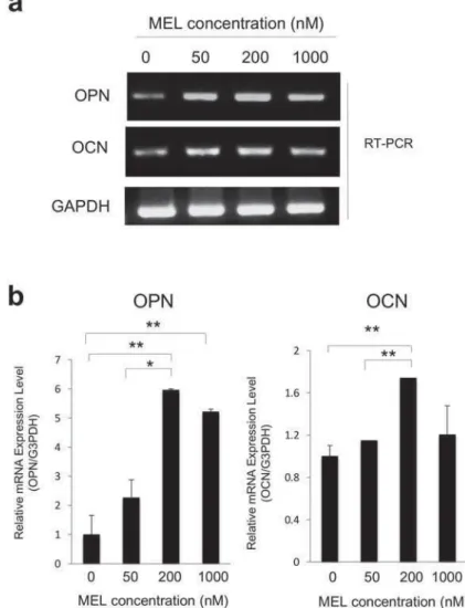
S e c o n d l y, w e d e t e r m i n e d t h e o p t i m u m concentration of MEL on monolayer cultures. Osteopontin (OPN) and osteocalcin (OCN) are considered to be late osteogenic markers, and have roles in the onset of the mineralization phase of osteoblast lineage3. Therefore, we examined OPN and OCN mRNA expression to determine the
induced OPN (Figure 2a) and OCN mRNA (Figure 2b) at 200 nM compared to the controls and 50 nM treatments. Therefore, 200 nM could be considered the lowest concentration of MEL that can elicit an osteoblastic response for our study.
SEM a n a ly sis o f I P- CH A/ M C3 T 3 - E1 ce lls com posit e s
CHA, IP-CHA/MC3T3-E1 composites were examined by SEM after 3 days of culture. We found the seeded MC3T3-E1 cells in the interior walls of porous IP-CHA, 3.5 mm deep from the surface (Figures 3a and 3b).
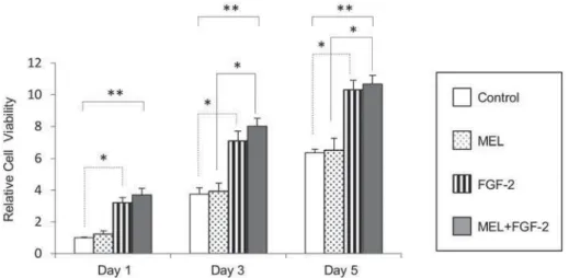
FGF- 2 in du ce d pr olife r a t ion of M C3 T3 ce lls w it h in I P- CH A con st r u ct
MEL on the proliferative potential of MC3T3-E1 cells within an IP-CHA construct, IP-CHA/MC3T3-E1 composites were examined in the presence of 20 μg/
induced growth of MC3T3-E1 cells compared to the
control on day 5 (Figure 4), whereas MEL alone
M EL m a i n co n t r i b u t o r t o i n d u ce m R N A e x pr e ssion of la t e bon e m a r k e r s w it h in I P-CH A con st r u ct
Figure 5 shows OPN and OCN expressions.
expression of these markers from day 5, though
5). On the other hand, combined treatment had an even greater effect on increased OCN and OPN expression (Figure 5).
M EL m a i n co n t r i b u t o r t o e n h a n ce A LP a ct ivit y w it h in I P- CH A con st r u ct
Figure 6 shows the relative ALP enzyme activities of MC3T3-E1 cells cultured within IP-CHA constructs
enzyme activity is known to be closely associated
independently induced ALP activity from day 3, though induction by the latter was more prominent
greater ALP enzyme activity compared to the individual treatments (Figure 6).
Com bin a t ion of FGF- 2 a n d M EL e n h a n ce d m in e r a liz a t ion
Figure 7a shows Alizarin Red Staining after MC3T3-E1 cells were treated for 2 weeks with MEL
calcium deposition in both monolayer cultures and treated IP-CHA/MC3T3 composites (Figures 7a and 7b). Those in combination resulted in more intense
(Figures 7a and 7b). These results indicate that )LJXUH Scanning electron microscopy analysis of
IP-CHA/MC3T3-E1 cells composites. a- The seeded MC3T3-E1 cells were observed in the interior walls of porous IP-CHA, 3.5 mm deep from the surface; b- Higher
the porous walls
4-augmentation of mineralization.
D I SCUSSI ON
HAp is a biocompatible material and possesses the advantage of protein adhesion whereby it can facilitate osteoblastic cell binding, proliferation, and differentiation, leading to matrix organization12. Fully interconnected porous HAp is thought to be a suitable candidate for transplantation of both osteoinductive agents and osteoblastic cells. In this study, we found that mouse preosteoblastic cells can penetrate and grow inside the construct. In addition, attachment of MC3T3-E1 cells to porous hydroxyapatite was well documented in a study
presented by Smith, et al.19 (2006). These results suggest that IP-CHA blocks are suitable scaffold for osteoblastic cells.
Bone formation is a cascade of events that occur in the initial proliferation phase, followed by the mineralization phase marked by OPN and OCN expressions14
eminent growth factor which is more favorable to cell proliferation than differentiation11, which was also shown in our study using IP-CHA constructs.
OCN highlight its role in the proliferative phase of osteoblast activity in contrast to differentiation11. This phenomenon is of particular interest, because Figure
population within the IP-CHA constructs in our study, ensuring that more cells were available for entering the maturation phase of bone formation.
MEL has been reported to positively stimulate bone formation by suppressing RANKL-mediated osteoclast formation and resultant bone resorption in the bone remodeling cycle of MC3T3-E1 cells6. In addition, Roth, et al.17 (1999) have reported that MEL can induce differentiation of MC3T3-E1 cells and mineralization of matrix in culture. These
role in bone formation. Although MEL had no
study, as anticipated, its differentiation potential was largely highlighted by the mRNA expressions of the late osteogenic markers OPN and OCN. Furthermore, we found that MEL induced mineralization, as shown
differentiation and mineralization.
MEL may induce increased cellular activity and differentiation because of its activity as an inherent free radical scavenger22. MC3T3 cells expel various free radicals during proliferation and growth8, while buildup of a large amount of free radicals hampers the natural activity of MC3T3 cells, leading to inhibition of mineralization9. Therefore, we speculate that MEL assists MC3T3 cellular activity and mineralization by neutralizing free radicals. In the present study, the
OCN, and also increased mineralization compared to
treatment with each one alone. We concluded that
number of cells for MEL to induce to differentiation into mature osteoblasts and therefore positively regulate mineralization.
other growth factors, such as bone morphogenic proteins (BMPs), and shown to significantly stimulate cell proliferation, while BMPs alone significantly stimulated differentiation and in 3.
study may operate in a similar manner, in which
help to consider delayed administration of MEL in a future in vivo model. We found that MEL induced differentiation of preosteoblasts into mature bone
20 (2008),
in which superior osseointegration was achieved by use of titanium screws in rat tibias after systemic
CON CLUSI ON S
of MC3T3-E1 cells within IP-CHA constructs by targeting different phases of the osteoblast lineage. Figure 6-
2. The combination of MEL and FGF-2 markedly increased that activity. The ratio of absorbance of each sample in relation
be a reasonable adjunct to biomaterials for use in eminent craniofacial surgery.
ACKN OW LED GM EN TS
Education, Culture, Sports, Science and Technology, Japan (No. 23592993).
REFEREN CES
1- Ayers RA, Simske SJ, Nunes CR, Wolford LM. Long-term bone ingrowth and residual micro hardness of porous block hydroxyapatite implants in humans. J Oral Maxillofac Surg. 1998;56(11):1297-301
2- Conti A, Conconi S, Hertens E, Skwarlo-Sonta K, Markowska M, Maestroni JM. Evidence for melatonin synthesis in mouse and human bone marrow cells. J Pineal Res. 2000;28(4):193-202.
3- Crockett JC, Rogers MJ, Coxon FP, Hocking LJ, Helfrich MH. Bone remodelling at a glance. J Cell Sci. 2011;124(Pt 7):991-8. 4- Dong J, Uemura T, Kojima H, Kikuchi M, Tanaka J, Tateishi T. Application of low-pressure system to sustain in v iv o bone formation in osteoblast/porous hydroxyapatite composite. Mater Sci Eng C. 2001;17(1-2)37-43.
induces downregulation of TAZ protein in osteoblastic MC3T3-E1 cells. Biochem Biophys Res Commun. 2008;366(2):471-5. 6- Koyama H, Nakade O, Takada Y, Kaku T, Lau KH. Melatonin at pharmacologic doses increases bone mass by suppressing resorption through down-regulation of the RANKL-mediated osteoclast formation and activation. Bone Miner Res. 2002;17(7):1219-29.
expression and its action in human leukemia and lymphoma cell lines. Leukemia. 2003;17(4):818-20.
8- Kwak EJ, Lee Y, Choi EM. Effect of magnolol on the
2012;2012:829650.
9- Lee YS, Chen X, Anderson JJ. Physiological concentrations of genistein stimulate the proliferation and protect against free radical-induced oxidative damage of MC3T3-E1 osteoblast-like cells. Nutrition Res. 2001;21(9):1287-98.
Figure
10- Liu C, Fukuhara C, Wessel JH 3rd
Localization of Aa-nat mRNA in the rat retina: melatonin
in sit u hybridization and laser capture microdissection. Cell Tissue Res. 2004;315(2):197-201. 11- Mansukhani A, Bellosta P, Sahni M, Basilico C. Signaling
induces apoptosis in osteoblasts. J Cell Biol. 2000;149(6):1297-308.
E, Ransjö M. I n vit ro study of the biological interface of Bio-Oss: implications of the experimental setup. Clin Oral Implants Res. 2013;24(3):329-35.
13- Ornitz DM, Marie PJ. Fibroblast growth factor signaling in
86.
Endokrynol Pol. 2010;61(1):117-23.
15- Park KH, Kang JW, Lee EM, Kim JS, Rhee YH, Kim M, et al. Melatonin promotes osteoblastic differentiation through the BMP/ ERK/Wnt signaling pathways. J Pineal Res. 2011;51(2):187-94.
growth factor-2 selectively stimulates angiogenesis of small vessels in arterial tree. Arterioscler Thromb Vasc Biol. 2000;20(5):1250-6
osteoblast differentiation and bone formation. J Biol Chem. 1999;274(31):22041-7.
18- Singh M, Jadhav HR. Melatonin: functions and ligands. Drug Discov Today. 2014;19(9):1410-8.
19- Smith IO, McCabe LR, Baumann MJ. MC3T3-E1 osteoblast attachment and proliferation on porous hydroxyapatite scaffolds fabricated with nanophase powder. Int J Nanomedicine. 2006;1(2):189-94.
20- Takechi M, Tatehara S, Satomura, K, Fujisawa K, Nagayama
histomorphometric study. J Mater Sci Mater Med. 2008;19(8):2949– 52.
21- Tamai N, Myoui A, Tomita T, Nakase T, Tanaka J, Ochi T, et al. Novel hydroxyapatite ceramics with an interconnective porous structure exhibit superior osteoconduction in vivo. J Biomed Mater Res. 2002;59(1):110-7
22- Tan DX, Reiter RJ, Manchester LC, Yan M, El-Sawi M, Sainz RM, et al. Chemical and physical properties and potential mechanisms: melatonin as a broad spectrum antioxidant and free radical scavenger. Curr Top Med Chem. 2002;2(2):181-97.
23- Woodard JR, Hilldore AJ, Lan SK, Park CJ, Morgan AW, Eurell JA, et al. The mechanical properties and osteoconductivity of hydroxyapatite bone scaffold with multi-scale porosity. Biomaterials. 2007;28(1):45-54.
24- Yuan Q, Kubo T, Doi K, Morita K, Takeshita R, Katoh S,