ABSTRACT
KWWSG[GRLRUJ
I n vivo
m odel for m icrobial invasion of t oot h root
dent inal t ubules
Jane L. BRITTAN1, Susan V. SPRAGUE1, Emma L. MACDONALD1, Robert M. LOVE2, Howard F. JENKINSON1, Nicola X. WEST1
1- University of Bristol, Department of Oral and Dental Sciences, Bristol, United Kingdom.
2- University of Otago, Department of Oral Diagnostic and Surgical Sciences, Dunedin, New Zealand.
Corresponding address: Howard F. Jenkinson - School of Oral and Dental Sciences - University of Bristol - Lower Maudlin - Street - Bristol BS1 2LY - United Kingdom - Phone: +44-117-342-4424 - e-mail: [email protected]
6XEPLWWHG2FWREHU0RGL¿FDWLRQ-DQXDU\$FFHSWHG-DQXDU\
O
bj ect ive: Bact erial penet rat ion of dent inal t ubules via exposed dent ine can lead t oroot caries and prom ot e infect ions of t he pulp and root canal syst em . The aim of t his work was t o develop a new experim ent al m odel for st udying bact erial invasion of dent inal t ubules wit hin t he hum an oral cavit y. Mat erial and Met hods: Sect ions of hum an root dent ine were m ount ed int o lower oral appliances t hat were worn by four hum an subj ect s for 15
G5RRWVZHUHWKHQ¿[HGVHFWLRQHGVWDLQHGDQGH[DPLQHGPLFURVFRSLFDOO\IRUHYLGHQFHRI
bact erial invasion. Levels of invasion were expressed as Tubule I nvasion Fact or ( TI F) . DNA
ZDVH[WUDFWHGIURPURRWVDPSOHVVXEMHFWHGWRSRO\PHUDVHFKDLQUHDFWLRQDPSOL¿FDWLRQRI 6U51$JHQHVDQGLQYDGLQJEDFWHULDZHUHLGHQWL¿HGE\FRPSDULVRQRIVHTXHQFHVZLWK *HQ%DQNGDWDEDVH5HVXOWV$OOURRWGHQWLQHVDPSOHVZLWKSDWHQWWXEXOHVVKRZHGHYLGHQFH
of bact erial cell invasion ( TI F value range from 5.7 t o 9.0) t o dept hs of 200 Pm or m ore.
$VSHFWUXPRI*UDPSRVLWLYHDQG*UDPQHJDWLYHFHOOPRUSKRW\SHVZHUHYLVXDOL]HGDQG PROHFXODUW\SLQJLGHQWL¿HGVSHFLHVRIGranulicat ella, St rept ococcus, Klebsiella, Ent erobact er,
Acinet obact er, and Pseudom onas as dent inal t ubule resident s. Conclusion: A novel in vivo PRGHOLVGHVFULEHGZKLFKSURYLGHVIRUKXPDQURRWGHQWLQHWREHHI¿FLHQWO\LQIHFWHGE\RUDO
m icroorganism s. A range of bact eria were able t o init ially invade dent inal t ubules wit hin exposed dent ine. The m odel will be useful for t est ing t he effect iveness of ant isept ics, irrigant s, and pot ent ial t ubule occluding agent s in prevent ing bact erial invasion of dent ine.
Ke yw or ds: 'HQWLQH5RRWFDULHV0LFURELRORJ\%LR¿OPV
I N TROD UCTI ON
Root caries, pulpit is, and dent ine hypersensit ivit y ar e becom ing incr easingly m or e pr oblem at ic as t h e den t at e h u m an popu lat ion ages1 7 *LQJLYDO recession can lead t o exposure of dent ine and t oot h wear can result in opening of dent inal t ubules on t he exposed surface. When dent ine becom es exposed as a result of gingival recession, or t hrough dent al caries, cracks, or m icroleakage around rest orat ions, m icr oor ganism s ar e able t o gain access t o t he t ubules15. More m icroorganism s are found in t he dent ine adj acent t o per iodont al pocket s t han in healt hy radicular dent ine, and m ore bact eria are IRXQG LQ VXSHU¿FLDO URRW GHQWLQH WKDQ LQ PLGGOH dent ine1. Bact eria can also lat erally invade t he root surface along t he increm ent al lines of cem ent um
an d t h en in f ilt r at e t h e d en t in e2 1. Bact er ia can penet rat e t hrough hypom ineralized enam el int o t he dent ineand cont ribut e t o pulpal pain sym pt om s of t eet h wit h m olar incisor hypom ineralizat ion10.
Bact erial persist ence wit hin t he dent inal t ubules m ay exacerbat e developm ent of root caries, and t he form at ion of com plex m icrobial com m unit ies wit hin deeper dent ine or root canal space26 m ay SOD\ D VLJQL¿FDQW SDUW LQ HQGRGRQWLF WUHDWPHQW failure18. Evidence suggest s t hat t he bact eria t hat init ially invade dent inal t ubules ar e oft en fr om t h e g en er a En t er ococcu s an d St r ep t ococcu s1 5.
and irrigant s12. The m odels have provided valuable inform at ion on t he m echanism s involved in growt h and penet rat ion of dent ine5, and t he pot ent ial for various agent s, such as phot odynam ic t herapy29 t o help wit h cont rolling infect ion. However, t he various lab or at or y m od els u su ally in cor p or at e d en t in e VDPSOHVWKDWDUHH[SRVHGWRDQDUWL¿FLDOQXWULHQW env ir onm ent in or der t o achiev e infect ion w it h relevant m icroorganism s. Under nat ural condit ions, dent ine would be exposed t o salivary com ponent s, gin giv al f lu id, im m u n e sy st em m olecu les, an d pot ent ially hundreds of different m icroorganism s19. Most in v it r o den t in e in f ect ion m odels em ploy condit ions t hat are quit e different from t he nat ural
in vivo infect ion environm ent .
Dent ine st udies in vit ro have also included t est ing various com pounds for abilit y t o occlude t ubules as d esen sit izin g ag en t s2. Valu ab le in f or m at ion h as b een o b t ai n ed ab o u t t h e p r o p er t i es an d effect iveness of such agent s27, but t hese are only j ust beginning t o be t est ed under suit able in vivo
con dit ion s. For ex am ple, West , et al.2 8 ( 2 0 1 1 ) det erm ined t he abilit ies of desensit izing t oot hpast e t echnologies t o occlude pat ent dent inal t ubules in a clinical environm ent . Healt hy subj ect s wore lower in t raoral applian ces r et ain in g den t in e sam ples, and t hese were analyzed aft er 4 d of t reat m ent for degree of occlusion28. We have ut ilized t he basis of t hat st udy t o develop a m odel for m icroorganism invasion of dent inal t ubules in vivo. This will provide a suit able plat form by which t o invest igat e bact erial invasion of dent ine wit hin a clinical environm ent , and t o t est for effect iveness of t ubule- occluding or ant im icrobial agent s t o prevent bact erial invasion of dent ine.
M ATERI AL AN D M ETH OD S
Root de n t in e
Non- carious, unrest ored hum an canine or pre-m olar t eet h wit h single root canals were obt ained from ort hodont ic ext ract ions. Teet h were obt ained wit h inform ed consent and t he st udy was approved by Cent ral and Sout h Br ist ol Et hics Com m it t ee ( REC r ef. 0 4 / Q2 0 0 6 / 5 0 ) . Follow in g ex t r act ion , t eet h w er e soak ed in 2 % sodiu m hy poch lor it e ( NaOCl) for 48 h and any soft t issue rem aining was rem oved. Prior t o sect ioning, root s were washed in copious am ount s of wat er, and rinsing was repeat ed following sect ioning t o ensure no t races of NaOCl rem ained. Teet h were st ored in st erile dist illed H2O at 4° C unt il required. Root s were sect ioned using a wat er cooled st eel bladed cut t ing m achine ( I som et Saw, Buhler Lt d., Evanst on, I L, USA) . I n brief, t he crown and root t ip were rem oved, t he rem aining root was cut int o 0.5 cm lengt hs, and t he cervical segm ent s w er e longit udinally sect ioned in such a way t hat t he root canal was exposed. The root
sect ions were t hen aut oclaved ( 121° C, 20 m in) in dist illed H2O, which did not visually affect t ubule st ruct ures5, and st ored at 4° C.
Pr e pa r a t ion of in t r a - or a l a pplia n ce s
For each subj ect , a lower alginat e im pression was r ecor ded in a per forat ed st ock t ray. Wit hin PLQ WKH LPSUHVVLRQV ZHUH SRXUHG LQ .DI¿U ' d en t al st on e an d su b seq u en t ly t w o low er - or al appliances were const ruct ed from Forest acryl® self curing acrylic ( Pearson Dent al Supply Co., Sylm ar, &$86$$GDPVFULEVZHUHFRQVWUXFWHGWR¿WWKH PDQGLEXODU¿UVWPRODUVWRDLGUHWHQWLRQDQGZLUH loops were const ruct ed in an ant erior and post erior t rench region t o hold t he dent ine sam ples in place ( Figure 1) . The cervical region root sect ions were m ount ed int o t he appliances in such a way t hat WKH EXFFDO IDFLQJ VXUIDFH ZDV ÀXVK ZLWK RU MXVW below, t he level of t he surrounding acrylic surface. Before placem ent in t he appliance, t he root sect ions were dipped in st erile dist illed wat er, t he face t o be in cont act wit h t he appliance was dried and t he sam ple m ount ed ont o a sm all drop of m olt en st icky wax wit hin t he t rench of t he appliance. Once all four sect ions were in place t hey were t hen furt her secured in posit ion wit h t he wire loop t hat was built int o t he appliance ( Figure 1) . The appliances were st ored overnight at 4° C in a st erile airt ight cont ainer cont aining dam p t issue t o prevent t hem drying out .
Ex pe r im e n t a l de sign
M icr oscopic a n a lysis of ba ct e r ia l in va sion
Six pieces of dent ine from t he appliances were ¿[HGLQQHXWUDOEXIIHUHGIRUPDOLQIRUGEHIRUH being dem ineralized in 10% form ic acid cont aining 2% form alin for 7 d. Sam ples were t hen dehydrat ed ( 70% I MS- denat ur ed alcohol x 2, 90% I MS x 2, ,06[[\OHQH[SDUDI¿QZD[[EHIRUH being blocked in wax. Fift een t ransverse sect ions, 6 m m t hick and 60 m m apart from t he next sect ion, were cut from each dent ine sam ple, m ount ed on SRO\O\VLQHVOLGHVDQGKHDW¿[HGSULRUWRVWDLQLQJ using t he Brown and Brenn m et hod6.
Pe n e t r a t i o n o f b a ct e r i a i n t o d e n t i n e w a s YLVXDOL]HGE\OLJKWPLFURVFRS\DW[PDJQL¿FDWLRQ 7KHFHQWUDOSRLQWRIWKHURRWVHFWLRQZDVLGHQWL¿HG DQG¿YH¿HOGVRIYLHZUDGLDWLQJRXWIURPWKLVSRLQW were exam ined for each of t he 15 root sect ions. The ext ent of invasion was init ially expressed as t he t ubule invasion index ( TI )16, w her e 1 t o 20 t ubules (per¿HOGLQYDGHGVFRUHGWXEXOHV
invaded scored 2; and > 50 t ubules invaded scored 3. These scor es w er e t hen conver t ed t o Tubule
I nvasion Fact or ( TI F) t hat t ook dept h of invasion of t ubules int o account5. The TI F was obt ained by m ult iplying t he TI by t he invasion dept h score: x1, ZKHUHLQYDVLRQGHSWKZDVPP[ZKHUH t ubules per¿HOGVKRZHGLQYDVLRQGHSWK!PP
DQG[ZKHUHWXEXOHVSHU¿HOGZHUHLQYDGHG
WRGHSWKRIPPDVSUHYLRXVO\GHVFULEHG 5.
,GHQWL¿FDWLRQRIEDFWHULD
Two root pieces from each appliance were rinsed in st erile dist illed wat er and st ored at - 80° C. One of t he specim ens in each case was used t o opt im ize WKHPHWKRGVIRU'1$H[WUDFWLRQDQGDPSOL¿FDWLRQ Once t his had been achieved, t he second sam ple from each subj ect was t ransferred int o a m icrofuge t u be con t ain in g 0 . 1 m L st er ile 1 0 % EDTA ( pH 6.5) , vigor ously vor t ex- m ixed, and incubat ed at room t em perat ure for 30 m in t o part ially decalcify. Sam ples were t hen t ransferred int o 0.1 m L 2 M cit r ic acid ( pH 1 . 6 ) an d in cu bat ed for 3 0 m in . Sam ples were ext ensively rinsed in st erile dist illed H22 DQG WUDQVIHUUHG WR WXEHV FRQWDLQLQJ *HQH
Releaser ( Cam bio, Cam br idge, Cam bs, UK) for DNA ex t ract ion accor ding t o t he m anufact ur er ’s inst ruct ions.
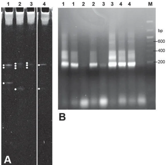
The DNA ext ract s were used as t em plat es in 3RO\PHUDVH &KDLQ 5HDFWLRQ 3&5 DPSOL¿FDWLRQ ZLWKXQLYHUVDO6U51$JHQHSULPHUV'**() ¶&*&&&*&&*&*&*&**&***&****&*****& $&******&&7$&***$**&$*&$*ZLWKWKH*& FODPSIRU'HQDWXULQJ*UDGLHQW*HO(OHFWURSKRUHVLV '**(2 5 DQG 5 ¶$77$&&*&**&7*&7** t o am plify a product of 160 bp. The presence of FRUUHFWVL]HGIUDJPHQWVZDVFRQ¿UPHGE\DJDURVH gel elect rophoresis. Subsequent ly, aliquot s ( 6 m L) RI3&5SURGXFWVZHUHVXEMHFWHGWR'**( denat urant gradient ) and t he separat ed bands were et hidium brom ide- st ained and visualized under UV light ( 344 nm ) ( Figure 2A) . Bands were excised from t he gel lanes, t ransferred t o t ubes cont aining 0.3 m L TE buffer ( 5 m M Tris- HCl, 10 m M EDTA, pH 7.5) and t he DNA was allowed t o elut e from t he gel fragm ent s for 16 h at 4° C.
Ea c h o f t h e e l u t e d g e l b a n d s w a s t h e n su b j ect ed t o f u r t h er PCR am p l i f i cat i o n u si n g
SULPHUV)¶&&7$&***$**&$*&$*DVDERYH PLQXV *& FODPS DQG 5 DERYH 3UHVHQFH RI DPSOL¿HGIUDJPHQWVZDVFRQ¿UPHGE\DJDURVHJHO elect rophoresis ( Figure 2B) . The fragm ent s were JHOSXUL¿HG4,$TXLFN3&53XUL¿FDWLRQ.LW4LDJHQ Man ch est er, Lan cs, UK) , lig at ed in t o p lasm id p CR2 . 1 ( I n v it r og en , Th er m o Fisch er Scien t if ic I nc., Walt ham , MA, USA) and t ransfor m ed int o
Escherichia coli XL1- Blue by st andard procedures. Plasm ids were ext ract ed from t ransform ant colonies using QI Aprep Spin MiniPrep Kit ( Qiagen) , checked by agar ose gel elect r ophor esis, and t he 160- bp LQVHUWVZHUHGLGHR[\VHTXHQFHG*HQHVHUYLFH/WG Cam bridge, Cam bs, UK) . The part ial 16S rRNA gene sequences w er e t hen com par ed w it h 16S r RNA JHQH VHTXHQFHV LQ *HQ%DQN XVLQJ WKH VWDQGDUG nucleot ide NCBI / BLAST program .
RESULTS
M icr oscopic a n a lysis of ba ct e r ia l in va sion
The t oot h r oot dent ine pieces w er e m ount ed int o appliances as shown in Figure 1. The sam ples
w er e designat ed A- D ( low er r ight ) ( Figur e 1 B) and E- H ( lower left ) . Aft er 15 d in vivo, t he root pieces were rem oved and processed as described in Mat erial and Met hods for m icroscopic analyses ( A-F) . All sam ples cont aining pat ent dent inal t ubules showed high levels of bact erial invasion, wit h TI F values in t he range of 5 t o 9 ( Figur e 3) . Thr ee dent ine sam ples ( Figure 3) could not be assessed for invasion because of disint egrat ion of int ernal dent ine st ruct ure.
7KHKLVWRFKHPLFDOVWDLQLQJSUR¿OHVZLWKUHVSHFW t o t ype of organism s present wit hin t he dent inal t ubules, pat t er n of t ubule penet rat ion, dept h of LQYDVLRQ DQG VXUIDFH DGKHVLRQ ELR¿OP YDULHG en o r m o u sl y a cr o ss t h e d en t i n e sa m p l es. We H[DPLQHGDOOGHQWLQHVDPSOHVWRFRQ¿UPEDFWHULDO invasion, but in t he following descript ions we have included only represent at ive m icrographs exhibit ing dist inct feat ures of t he invasion processes.
For subj ect 1, root sam ple D, t here was invasion RISXUSOHVWDLQHG*UDPSRVLWLYHFRFFL)LJXUH$ DUURZHGDDQGSLQNVWDLQHG*UDPQHJDWLYHURGV ( Figure 4A, arrowed b) t o dept hs of > 100 Pm . Root sam ple F, on t he ot her hand, seem ed t o be ent irely perm eat ed by sm all cocci bact eria. These st ained *UDPSRVLWLYH LQ DUHDV RI GHQVHU FRORQL]DWLRQ )LJXUH%DUURZHGDRU*UDPQHJDWLYHLQUHJLRQV of deeper ( ~ 150 Pm ) invasion ( Figure 4B, arrowed E7KLV*UDPYDULDEOHVWDLQLQJZDVVHHQSUHYLRXVO\ in laborat ory st udies of dent ine invasion by pure
cult ures of st rept ococci16.
I n subj ect 2, sam ple A carried a dense invasive ELR¿OP RI *UDPSRVLWLYH VWDLQHG PDWHULDO DW WKH su r face ( Figu r e 5 A) an d t h er e w as invasion of t ubules > 150 PPE\*UDPSRVLWLYHFRFFL$VLPLODU pat t ern was seen for root sam ple D ( Figure 5D) , ZKLOHVDPSOH&IURPVXEMHFWVKRZHG*UDPSRVLWLYH EDFWHULDDWWKHVXUIDFHDQGGHHSSHQHWUDWLRQ
PP E\ VPDOO *UDPQHJDWLYH RUJDQLVPV %ORFN % from t his subj ect was one of t he sam ples t hat could not be properly analyzed, as t he int ernal dent ine st ruct ure was disint egrat ed ( Figure 5B) .
Figure 6 shows sect ions from blocks C, D, and E from subj ect 3. Sect ions t hrough C ( Figure 6A) FRQWDLQHG VPDOO *UDPQHJDWLYH URGV ZLWKLQ WKH GHQWLQDOWXEXOHV$EDQGRI*UDPQHJDWLYHURGVZDV present a sm all dist ance away from t he surface of t he dent ine sam ple, perhaps having been present on t he dent ine surface prior t o sect ioning ( Figure $6DPSOH'FRQWDLQHG*UDPSRVLWLYHDQG*UDP negat ive r ods ( ~ 5 Pm lengt h) in w ell- separat ed WXEXOHVDQGSHQHWUDWLQJPm ( Figure 6B) . I n sam ple E, individual t ubules cont ained deep lines of LQYDGLQJ*UDPSRVLWLYHDQG*UDPQHJDWLYHEDFWHULD ( Figure 6C) .
From subj ect 4, sam ple D showed invasion by *UDPSRVLWLYHFRFFLDQG*UDPQHJDWLYHURGVDQG DGHQVHELR¿OPRQWKHVXUIDFHFRPSULVHGRI*UDP posit iv e cocci and m at r ix m at er ial ( Figur e 7A) . Sam ple E showed dist inct penet rat ion of t ubules by
JURXSVRI*UDPSRVLWLYHFRFFL)LJXUH%
These r esult s dem onst rat ed t hat t he dent ine sa m p l e s m o u n t e d o n t o t h e a p p l i a n ce s w e r e all r eadily su scept ible t o in f ect ion by inv adin g m icr oor ganism s. The t echnique t her efor e was a very effect ive m ean of achieving invasion of dent inal t ubules by a variet y of different oral bact eria.
0ROHFXODULGHQWL¿FDWLRQRILQYDGLQJEDFWHULD
Wit hin one of t he dent ine sam ples select ed at UDQGRPIURPVXEMHFW'**(EDQGV7DEOH ZH LGHQWL¿HG SULQFLSDOO\ *UDPQHJDWLYH EDFWHULD in clu d in g Kleb siella p n eu m on iae, En t er ob act er
species, Ent erobact er horm aechei, and sequences sim ilar t o t hose from som e uncult ivat ed bact eria from faeces ( Table 1) . There was 100% sequence m at ch over 160 bp t o E. horm aechei. Subj ect 2 VDPSOHFRQWDLQHGRQO\*UDPQHJDWLYHKlebsiella -OLNH EDFWHULD ZKLOH VXEMHFW WKUHH '**( JHO
b an d s) p r ov id ed seq u en ces 1 0 0 % id en t ical t o
Acinet obact er and St rept ococcus dat abase ent ries, an d 9 9 % t o En t er ob act er sp p. ( Tab le 1 ) . Th e r oot sam ple fr om subj ect 4 pr ovided sequences ZLWK PDWFKHV WR *UDPSRVLWLYH EDFWHULD
Granulict aella, St rept ococcus m it is, St rept ococcus or al i s, an d S. g or d on i i, an d t o Pseu d om on as
species and uncult ivat ed organism s. Overall, t hese analyses showed a diversit y of bact erial infect ion t o DGHJUHHVLPLODUWRPRUSKRORJLFDOYDULHWLHVRI*UDP QHJDWLYHDQG*UDPSRVLWLYHRUJDQLVPVYLVXDOL]HG m icroscopically ( Figures 4- 7) .
D I SCUSSI ON
I n t his st udy we have prepared dent ine sam ples in a m an n er sim ilar t o t h at d on e f or in v it r o
invasion invest igat ions16,22 of dent ine infect ion by pure cult ures of bact eria such as E. faecalis and
St r ept ococcu s species. Th ese appr oach es h av e been undert aken t o st udy t he m echanism s involved in dent inal t ubule infect ion, and t o inv est igat e t he effect s of var ious ant isept ics, ir r igant s, and a n t i m i cr o b i a l s i n p r e v e n t i n g d e n t i n a l t u b u l e infect ion. Perhaps one lim it at ion of such in vit ro
analyses is t hat t hey have been undert aken under condit ions t hat are quit e different from t hose t hat would be encount ered in vivo. These include, for ex am ple, t he pr esence of w hole saliva, salivar y ÀRZ VKHDU DQG DEUDVLRQ DQG QXWULHQW SXOVHV Our st udies here show t hat it is possible t o readily achieve dent ine infect ion in vivo t o t he levels and ext ent t hat can be obt ained in vit ro16. This m odel t herefore would be useful for t est ing t he effect s of n ew den t in al t u bu le occlu din g com pou n ds2 7 or agent s for prevent ing root caries30 in order t o com plem ent t he in vit ro experim ent s t hat have been previously em ployed.
8QGHUODERUDWRU\FRQGLWLRQV*UDPSRVLWLYHFRFFL readily penet rat e dent inal t ubules. Hist orically, E. faecalis has been considered as a m aj or invader of dent ine13,15, but m ore recent m olecular st udies t hat do not em ploy cult ivat ion m et hods suggest t hat
E. faecalis m ay not be so prevalent as generally believed23. I nvasions of dent ine have been shown WRFRQWDLQDFRPSOH[PLFURELRWDRI*UDPSRVLWLYH DQG PDLQO\ *UDPQHJDWLYH EDFWHULD8. Penet rat ion RIGHQWLQHE\*UDPQHJDWLYHEDFWHULDin vit ro has
not been invest igat ed in such det ail. I nt erest ingly, p er iod on t al b act er ia Por p h y r om on as g in g iv alis
were found t o be unable t o invade dent ine unless co- cult ured wit h St rept ococcus16. I n t his present art icle we have dem onst rat ed m icroscopically, and by m olecular m eans, t hat dent ine in vivo can be LQYDGHG E\ *UDPQHJDWLYH EDFWHULD SULQFLSDOO\ *UDPQHJDWLYH URGV 6RPH RI WKH RUJDQLVPV LGHQWL¿HGHJEnt erobact er, Klebsiella, seem ed on ¿UVWLPSUHVVLRQWRSHUKDSVEHXQXVXDO+RZHYHU
En t er obact er an d Klebsiella species h av e been LGHQWL¿HGZLWKLQWKHVXEJLQJLYDOPLFURELRWD8. More recent ly, KlebsiellaZDVLGHQWL¿HGLQGHHSFDULRXV
lesions underneat h rest orat ions20 and E. horm aechei w a s cu l t i v a t e d f r o m h u m a n a t h e r o scl e r o t i c t issue24. Mem bers of t he Ent erobact eriaceae and Pseudom onadaceae are also found on t he hum an t ongue9. Our work t hus provides furt her evidence WKDWWKHVH*UDPQHJDWLYHRUJDQLVPVDUHIRXQGLQ t he oral cavit y and have t he abilit y t o penet rat e dent ine.
A range of bact erial species were present wit hin a sm all n u m b er of d en t in e sam p les an aly zed . Thr ee sam ples show ed disint egrat ion of t ubule st r u ct u r e, m ost lik ely ar isin g fr om t h e len gt hy SUHSDUDWLRQ SURFHVV ¿[DWLRQ GHPLQHUDOL]DWLRQ dehydrat ion, sect ioning) . Only a lim it ed num ber of specim ens were em ployed here because we were LQWHUHVWHGLQ¿UVWHVWDEOLVKLQJDPRGHOV\VWHP7KH
Figure 5- Transverse sections of human roots after 15 days incubation in situ in subject 2. Sections were prepared as described in Material and Methods, and stained by Brown & Brenn method. Panels: A, sample A, Gram-positive cocci invading to a depth of >150 Pm and showing a 10 PPGHSWKGHQVHELR¿OPDWWKHVXUIDFHDUURZHG%VDPSOH% disintegration of internal dentine structure meant that sections from this sample could not be analyzed; C, sample C,
Gram-SRVLWLYHEDFWHULDDWWKHVXUIDFHDQGGHHSSHQHWUDWLRQE\VPDOOHU*UDPQHJDWLYHEDFWHULDPm (arrowed); sample D,
*UDPSRVLWLYHDQG*UDPQHJDWLYHEDFWHULDSHQHWUDWLRQZLWKDFFXPXODWLRQRI*UDPSRVLWLYHFRFFLELR¿OPDWWKHVXUIDFHRI
result s suggest t hat t he m odel can be applied t o fut ure st udies of dent ine hypersensit ivit y agent s, GHWHUPLQLQJWKHLUFOLQLFDOHI¿FDF\DQGWKHLUDELOLW\ t o occlude t ubules and block bact erial invasion27. I t is ack now ledged t hat t he m olecular m et hods
used here do not different iat e bet ween live or dead bact eria. However, it m ight be possible t o ut ilize dent ine discs, fract ure t hem , and st ain t he int ra-t ubular bacra-t eria wira-t h LI VE/ DEAD sra-t ain. This m era-t hod has recent ly been described in st udies evaluat ing
in vit ro t he ant im icrobial effect of a com m ercial product on residual bact eria in dent inal t ubules11.
One of t he sam ples in t he st udy described here ZDVLQYDGHGE\VHYHUDOVSHFLHVRI*UDPSRVLWLYH cocci, w hich cor r oborat es t he not ion t hat t hese RUJDQLVPV DUH RIWHQ VRPH RI WKH ¿UVW WR LQYDGH dent ine15. However, E. faecalis was not found in our DQDO\VHV:HLGHQWL¿HGGranulicat ella, S. oralis, S. m it is, and S. gordonii which, wit h t he except ion of
Granulicat ella, have been previously im plicat ed in t ubule invasion15. I n addit ion, all of t hese bact eria including Granulicat ella are organism s t hat have been linked wit h infect ive endocardit is. Therefore, t here could pot ent ially be an associat ion bet ween ab ilit y t o in v ad e d en t in e an d ab ilit y t o cau se
Figure 6- Transverse sections of human roots after 15 days incubation in situ in subject 3. Sections were prepared as described in Material and Methods, and stained by Brown & Brenn method. Panels: A, sample C, invasion by negative rod-shaped bacteria, with a strip of Gram-negative rods ~30 Pm from the surface (arrowed); B, sample D, larger Gram-positive and Gram-negative rods (~5 PPOHQJWKZHOOVHSDUDWHGEXWSHQHWUDWLQJPm; C, sample E, individual tubules appear to show long lines of invading Gram-positive and Gram-negative bacteria. TIF scores for specimens are shown in Figure 3
Figure 7- Transverse sections of human roots after 15 days incubation in situ in subject 4. Sections were prepared as described in Material and Methods, and stained by Brown & Brenn method. Panels: A, sample D, shows invasion by Gram-positive cocci and Gram-negative rods, together
ZLWKDWKLFNELR¿OPRQWKHVXUIDFHDUURZHGFRPSULVHGRI
endocardial or int ravascular infect ions7.
:H KDYH WKXV LGHQWL¿HG RUJDQLVPV WKDW ZHUH pr esent w it hin dent inal t ubules t hat hav e been exposed t o m any hundr eds of differ ent bact er ia
i n v i v o1 9. I n t h i s st u d y w e o n l y u t i l i zed f o u r dent ine sam ples t o ident ify bact er ia t y pes t hat could invade t he specim ens under t he condit ion XVHG )XWXUH FOLQLFDO VWXGLHV IRU WHVWLQJ HI¿FDF\ of com pounds or pr oduct s in occluding t ubules DQGSUHYHQWLQJEDFWHULDLQYDVLRQZRXOGGH¿QLWHO\ em ploy m any m or e subj ect s t o pr ovide suit able power. However, t he m olecular st udies cannot be dir ect ly r elat ed t o t he m or phological st udies at t his st age. We have est ablished t hough t hat it is IHDVLEOHWRH[WUDFWEDFWHULDO'1$IURPGHFDOFL¿HG dent ine. Our m et hodology would t end t o ident ify t he m ost prevalent m icroorganism s t hat were present w it hin t he dent ine sam ples analyzed. We w ould like t o develop t hese st udies furt her in such a way t hat we could visualize and ident ify, by m olecular t echniques, t he bact er ia t hat have invaded t he sam e dent ine sam ple. This could be achieved by
ex t r act in g bact er ial DNA, or by det ect in g DNA XVLQJÀXRUHVFHQWLQVLWXK\EULGL]DWLRQ),6+IURP adj acent sect ions t o t hose hist ochem ically st ained.
CON CLUSI ON
I n sum m ary, t his st udy has est ablished a novel in vivo m odel for st udying t he infect ion of dent ine by oral m icroorganism s. Dent ine specim ens exposed t o t he hum an oral environm ent becom e infect ed wit h m icroorganism s t o sim ilar ext ent and dept h t o dent ine infect ed in vit ro under laborat ory condit ions. I n addit ion t o st rept ococci, bact eria from t he genera
Ent er obact er, Klebsiella and Pseudom onas w er e LGHQWL¿HGDVSULPDU\LQYDGLQJRUJDQLVPV7KLVin
vivoPRGHOVKRXOGSURYLGHWKHPHDQVWRFRQ¿UP
in v it r o ex per im en t al r esu lt s on t h e ef f ect s of ant isept ics, irrigant s, or t ubule occluding agent s on dent ine invasion by oral bact eria.
Subject DGGE band GenBank description1 No. 100% matches2,4 GenBank entry
H[DPSOH
1 U1 Klebsiella pneumoniae 0 KP297466
Enterobacter hormaechei KP027682
U2 Enterobacter spp. 499 KP091277
Enterobacter hormaechei KF516241
L2 Uncultivated from faeces >500 KF841982
2 U1 Klebsiella oxytoca 499 CP004887
3 U1 Acinetobacter ursingii 69 LC014147
Uncultured from skin KF083053
L1 Uncultured Streptococcus from skin/ nasopharynx
248 KF505347
L2 Enterobacter hormaechei 4 KF516241
4 U1 Granulicatella spp. >500 KJ575555
U2 Uncultured Pseudomonas spp. 7 AY191342
Pseudomonas putida 0 KP114213
L1 Streptococcus gordonii (from infective endocarditis)
>500 KJ170416
L2 Streptococcus mitis >500 KP233800
Streptococcus oralis LN589729
L4 Uncultured human mouth 333 JQ457994
Streptococcus sanguinis AY944229
1 Representative entries from the match listing
2 Number of BLAST sequences with 100% match (160 bp)
3 GenBank Accesion numbers
4 0 indicates 99% match (159/160)
Table 1-0LFURRUJDQLVPVLGHQWL¿HGIURPZLWKLQGHQWLQDOWXEXOHVIROORZLQJ'1$H[WUDFWLRQ3&5'**(DQG6U'1$
ACKN OW LED GM EN TS
We would like t o t hank Valeria Soro and Maria Davies for t heir excellent assist ance. We are m ost grat eful t o t he Universit y of Brist ol Dent al School Technicians for t he skilled preparat ion of clinical DSSOLDQFHV7KLVVWXG\ZDV¿QDQFHGLQSDUWE\WKH Universit y of Brist ol Ent erprise Developm ent Fund.
REFEREN CES
1- Adriaens PA, De Boever JA, Loesche WJ. Bact erial invasion in root cem ent um and radicular dent in of periodont ally diseased t eet h in hum ans. A reservoir of periodont opat hic bact eria. J Periodont ol. 1988; 59: 222- 30.
2- Arnold WH, Prange M, Naum ova EA. Effect iveness of various t oot hpast es on dent ine t ubule occlusion. J Dent . 2015; 43: 440- 9. 3- Art hur RA, Mart ins VB, Oliveira CL, Leit une VC, Collares FM, Magalh ães AC, et al. Ef f ect of ov er - t h e- cou n t er f lu or idat ed pr odu ct s r egim en s on r oot car ies in h ibit ion . Ar ch Or al Biol. 2015; 60: 1588- 94.
4 - Bor g es FM, Melo MA, Lim a JP, Zan in I C, Rod r ig u es LK. Ant im icrobial effect of chlorhexidine digluconat e in dent in: in vit ro
and in sit u st udy. J Conserv Dent . 2012; 15: 22- 6.
5- Brit t an JL, Sprague SV, Hunt ley SP, Bell CN, Jenkinson HF, Love RM. Collagen- like pept ide sequences inhibit bact erial invasion of root dent ine. I nt Endod J. 2015; doi: 10.1111/ iej .12474. Epub ahead of print .
6- Brown JH, Brenn L. A m et hod for t he different ial st aining of
*UDPSRVLWLYHDQG*UDPQHJDWLYHEDFWHULDLQWLVVXHVHFWLRQV%XOO
Johns Hopkins Hosp. 1931; 48: 69- 73.
&DUJLOO-66FRWW.6*DVFR\QH%LQ]L'6DQGRH-$Granulicat ella
i n f e ct i o n : d i a g n o si s a n d m a n a g e m e n t . J Me d Mi cr o b i o l . 2012; 61: 755- 61.
8- Chen L, Qin B, Du M, Zhong H, Xu Q, Li Y. et al. Ext ensive descript ion and com parison of hum an- supragingival m icrobiom e in root caries and healt h. PLoS One. 2015; 10: e0117064. 9- Cont i S, Sant os SS, Koga- I t o CY, Jorge AO. Ent erobact eriaceae and pseudom onadaceae on t he dorsum of t he hum an t ongue. J Appl Oral Sci. 2009; 17: 375- 80.
)DJUHOO 7* /LQJVWU|P 3 2OVVRQ 6 6WHLQLJHU ) 1RUpQ -*
Bact erial invasion of dent inal t ubules beneat h apparent ly int act but hypom ineralized enam el in m olar t eet h wit h m olar incisor hypom ineralizat ion. I nt J Paediat r Dent . 2008; 18: 333- 40. 11- Ham am a HH, Yiu CK, Burrow MF. Effect of silver diam ine
ÀXRULGH DQG SRWDVVLXP LRGLGH RQ UHVLGXDO EDFWHULD LQ GHQWLQDO
t ubules. Aus Dent J. 2015; 60: 80- 7.
12- Hancock HH 3r d, Sigur dsson A, Tr ope M, Moiseiw it sch J. Bact eria isolat ed aft er unsuccessful endodont ic t reat m ent in a Nort h Am erican populat ion. Oral Sur Oral Med Oral Pat hol Oral Radiol Endod. 2001; 91: 579- 86.
13- Love RM. Ent erococcus faecalis - m echanism for it s role in endodont ic failure. I nt Endod J. 2001; 34: 399- 405.
14- Love RM, Chandler NP, Jenkinson HF. Penet rat ion of sm eared or nonsm ear ed dent ine by St r ept ococcus gor donii. I nt Endod J.1996; 29: 2- 12.
15- Love RM, Jenkinson HF. I nvasion of dent inal t ubules by oral bact eria. Crit Rev Oral Biol Med. 2002; 13: 171- 83.
16- Love RM, McMillan MD, Park Y, Jenkinson HF. Coinvasion of dent inal t ubules by Porphyrom onas gingivalis and St rept ococcus gordoniiGHSHQGVXSRQELQGLQJVSHFL¿FLW\RIVWUHSWRFRFFDODQWLJHQ
I / I I adhesin. I nfect I m m un. 2000; 68: 1359- 65.
17- Marcenes W, Kassebaum NJ, Bernabé E, Flaxm an A, Naghavi
0/RSH]$HWDO*OREDOEXUGHQRIRUDOFRQGLWLRQVLQ
a syst em at ic analysis. J Dent Res. 2013; 92: 592- 7.
1DLU 31 6M|JUHQ 8 .UH\ * .DKQEHUJ .( 6XQGTYLVW * ,QWUDUDGLFXODU EDFWHULD DQG IXQJL LQ URRW¿OOHG DV\PSWRPDWLF
hum an t eet h wit h t herapy- resist ant periapical lesions: a long-t er m lighlong-t and eleclong-t r on m icr oscope follow- up slong-t udy. J Endod. 1990; 16: 580- 8.
1DVLG]H , /L - 4XLQTXH ' 7DQJ . 6WRQHNLQJ 0 *OREDO GLYHUVLW\ LQ WKH KXPDQ VDOLYDU\ PLFURELRPH *HQRPH 5HV
2009; 9: 636- 43.
20- Neelakant an P, Rao CV, I ndram ohan J. Bact eriology of deep carious lesions underneat h am algam rest orat ions wit h different pulp- capping m at erials – an in vivo analysis. J Appl Oral Sci. 2012; 20: 39- 45.
21- Nyvad B, Fej erskov O. An ult rast ruct ural st udy of bact erial invasion and t issue breakdown in hum an experim ent al root- surface caries. J Dent Res. 1990; 69: 1118- 25.
22- Ørst avik D, Haapasalo M. Disinfect ion by endodont ic irrigant s and dressings of experim ent ally infect ed dent inal t ubules. Endod Dent Traum at ol. 1990; 6: 142- 9.
23- Ozok AR, Persoon I F, Huse SM, Keij ser BJ, Wesselink PR, Crielaard W, et al. Ecology of t he m icrobiom e of t he infect ed root canal sy st em : a com par ison bet w een apical and cor onal r oot segm ent s. I nt Endod J. 2012; 45: 530- 41.
24- Raffert y B, Dolgilevich S, Kalachikov S, Morozova I , Ju J, Whit t ier S, et al. Cult ivat ion of Ent erobact er horm aechei from hum an at herosclerot ic t issue. J At herscler Throm b. 2011; 18: 72-81.
6KHI¿HOG9&&R['5/HUPDQ/60\HUV50$WWDFKPHQWRI DEDVHSDLU*&ULFKVHTXHQFH*&FODPSWRJHQRPLF'1$
fragm ent s by t he polym erase chain react ion result s in im proved det ect ion of single- base changes. Pr oc Nat Acad Sci U S A. 1989; 86: 232- 6.
26- Siqueira JF Jr, Rôças I N. Diversit y of endodont ic m icrobiot a revisit ed. J Dent Res. 2009; 88: 969- 81.
27- Wang R, Wang Q, Wang X, Tian L, Liu H, Zhao M, et al. Enhancem ent of nano- hydroxyapat it e bonding t o dent in t hrough
D FROODJHQFDOFLXP GXDODI¿QLWLYH SHSWLGH IRU GHQWLQDO WXEXOH
occlusion. J Biom at Appl. 2014; 29: 268- 77.
28- West NX, MacDonald EL, Jones SB, Claydon NC, Hughes N, Jeffery P. Random ized in sit u clinical st udy com paring t he abilit y of t wo new desensit izing t oot hpast es t echnologies t o occlude pat ent dent in t ubules. J Clin Dent . 2011; 22: 82- 9.
29- Xhevdet A, St ublj ar D, Kriznar I , Jukic T, Skvarc M, Veranic P, et
DO7KHGLVLQIHFWLQJHI¿FDF\RIURRWFDQDOVZLWKODVHUSKRWRG\QDPLF
t herapy. J Lasers Med Sci. 2014; 5: 19- 26.