J Bras Pneumol. 2007;33(1):113-115 113
Case Reports
Mediastinal teratoma mimicking pleural effusion on chest X rays*
Miguel Angelo Martins de Castro Junior1, Nelson Perelman Rosemberg2,
Miguel Angelo Martins de Castro3, Angela Potter de Castro4,
Cacio Wietzycoscki5, Cleiton Mespaque5
Abstract
Teratomas account for 8–13% of all mediastinal tumors. A 27-year-old patient presented with chest pain and dyspnea of prolonged evolu-tion. A chest X ray revealed near total opacification of the right hemithorax, raising the suspicion of pleural effusion. On a tomography scan of the chest, a collection of heterogeneous fluid, with regular borders and 10.1 x 11.7 cm in size, was seen in the pleura of the lower
two-thirds of the right hemithorax but was not encroaching upon any of the adjacent structures. Based on the hypothesis that these findings represented a benign cystic mediastinal teratoma, an exploratory thoracotomy was performed, during which such a teratoma was found and completely excised. The post-operative evolution was favorable. The atypical presentation and considerable growth of the tumor hindered the pre-operative diagnosis.
Keywords: Teratoma; Mediastinal neoplasms; Pleural effusion; Radiography, thoracic.
*Study carried out in the Thoracic Surgery Unit, Department of Surgery, Fundação Universidade Federal de Rio Grande (FURG, Federal University of Rio Grande Foundation) Hospital Nossa Senhora da Conceição – Rio Grande (RS), Brazil.
1. Masters in Medicine from the Universidade Federal do Rio Grande do Sul (UFRGS, Federal University of Rio Grande do Sul) – Porto Alegre (RS), Brazil. 2. Full member of the Colégio Brasileiro de Cirurgiões (CBC, Brazilian College of Surgeons) – Rio de Janeiro (RJ), Brazil.
3. PhD in Medicine from the Universidade Federal de Minas Gerais (UFMG, Federal University of Minas Gerais) – Belo Horizonte (MG), Brazil. 4. Masters from the Universidade Federal do Rio Grande do Sul (UFRGS, Federal University of Rio Grande do Sul) – Porto Alegre (RS), Brazil.
5. Medical Student at the Fundação Universidade Federal de Rio Grande (FURG, Federal University of Rio Grande Foundation) – Rio Grande (RS), Brazil. Correspondence to: Miguel Angelo Martins de Castro Junior. Rua Duque de Caxias, 585, Centro, CEP 96200-020. Rio Grande, RS, Brasil.
Phone 55 53 3231-6215. E-mail: [email protected] Submitted 14/11/05. Accepted, after review 7/4/06.
Introduction
Mediastinal teratomas are rare occurrences and account for 8-13% of all mediastinal tumors. Teratomas prima-rily occur in the anterior mediastinum and affect young adults.(1,2) Mediastinal teratomas originate from pluripotent
germ cells and comprise a wide variety of tissues originating from the three embryonic layers.(2,3) They may contain several
types of tissues, such as epithelial and muscle tissues, as well as cartilage, and even teeth. Benign teratomas usually grow slowly and are asymptomatic for long periods or present minimal symptoms, which are very often disregarded not only by the patient but also by the physician. They are frequently detected on routine chest X rays. In general, they appear as well circumscribed masses and are usually excised without much difficulty.(2-4)
Case report
A 27-year-old, previously healthy female patient, a native and resident of Taquara (located in the state of Rio Grande
114 Castro Junior MAM , Rosemberg NP, Castro MAM, Castro AP, Wietzycoscki C, Mespaque C
J Bras Pneumol. 2007;33(1):113-115
In view of these findings, an exploratory thora-cotomy was performed. An incision was made at the right lung base on the midaxillary line. The opening of the chest cavity revealed a large encapsulated lesion containing yellowish secretion. The pleural cavity was opened, and there were no signs of fluid within. Due to the characteristics of the lesion, a hypothetical diagnosis of a benign cystic mediastinal teratoma was made, and this led to the complete excision of the lesion. The origin of the cyst was the anterosuperior mediastinum, and it had grown inferolaterally to the right, compressing the lung (Figure 2). The anatomopathological examination of the sample confirmed the presence of a benign mediastinal teratoma. The post-operative evolution was favorable. The patient was asymptomatic and presented re-expansion of the right lung. Therefore, she was discharged.
Discussion
The first case of mediastinal teratoma was described in 1823, and, since then, a variety of atypical presentations of this condition have been reported.(4) These tumors are often clinically
mani-fested by their complications, the most common being the compression of the intrathoracic struc-tures due to the tumor growth.(2) Perforation of the
pleural cavity, pericardial sac, and bronchi, as well as invasion of the lung tissue and massive hemop-tysis, have also been reported, although malignancy is rare.(2,5)
We discuss this case placing emphasis on the atypical presentation of the benign cystic medias-tinal teratoma, which mimicked pleural empyema, while remaining attentive to the possibility of enhanced tumor growth, which hinders the defini-tive pre-operadefini-tive diagnosis. In reviewing the literature, we observed that most physicians make an initial diagnosis of pleural empyema when faced with a benign cystic mediastinal teratoma of considerable volume. Although such misdi-agnosis is understandable due to the similarity of the clinical and radiological characteristics, it delays the diagnosis and makes the treatment more expensive. Therefore, we realize the importance of suspecting this disease in a patient presenting a large, well-defined opacification of one hemith-orax and having no history of pulmonary infection or any other condition that might predispose to
Figure 1 - Computed tomography scan of the chest revealing a heterogeneous mass with regular borders occupying a large portion of the right hemithorax, causing pulmonary collapse (arrows) and contralateral deviation of the mediastinum.
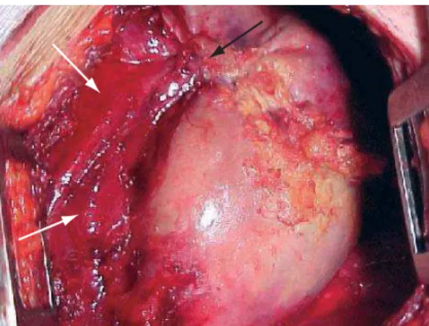
Figure 2 - Intraoperative image of the benign cystic mediastinal teratoma revealing a capsule containing a great quantity of yellowish secretion. Origin of the cyst in the anterosuperior mediastinum (black arrow) and subsequent collapse of the lung (white arrows).
pleural empyema. Surgery should be performed in order to clarify the diagnosis or whenever compli-cations such as pulmonary atelectasis, adhesion to/compression of adjacent structures, and malig-nant transformation are probable. The results after surgical resection are excellent.(1,2,6) Radiotherapy
Mediastinal teratoma mimicking pleural effusion on chest X rays
J Bras Pneumol. 2007;33(1):113-115 115
References
1. Verhaeghe W, Meysman M, Noppen M, Monsieur I, Lamote J, Op De Beeck B, et al. Benign cystic teratoma: an uncommon cause of anterior mediastinal mass. Acta Clin Belg. 1995;50(2):126-9.
2. Savas B, Kucukarslan N, Gurkok S, Özcan A, Harun H. Surgical treatment of a benign mature teratoma localised in anterosuperior mediastinum. Internet J Thorac Cardiovasc Surg [serial on the Internet]. 2005;7(1) [cited 2005 Nov
12]. Available from:. http://www.ispub.com/ostia/index. php?xmlFilePath=journals/ijtcvs/vol7n1/teratoma.xml 3. Cocks CE, Kamdar HH. Mediastinal teratoma: two case
reports. East Afr Med J. 1994;71(1):63-6.
4. Wagner RB. The history of mediastinal teratoma. Chest Surg Clin N Am. 2000;10(1):213-22, xi.
5. Sakamoto K, Kase M, Mo M, Kurata H. Mediastinal mature teratoma perforated into the pericardial sac: a case report. Kyobu Geka. 2000; 53(1):74-7. Japanese.