Published online August 21st, 2010 © http://www.ijav.org
Case Report
International Journal of Anatomical Variations (2010) 3: 141–143
Introduction
It is well known that the external iliac artery gives off two branches just proximal to the inguinal ligament; the inferior epigastric artery medially and the deep circumflex iliac artery laterally. As the external iliac artery crosses deep to the inguinal ligament, it continues in the thigh as the femoral artery which in turn gives off three branches; the superficial external pudendal, the superficial circumflex iliac and the superficial epigastric arteries. Corresponding veins of these superficial arteries join the great saphenous vein before the later pierces the cribriform fascia [1].
External pudendal arteries are usually classified as 2 but some reports mentioned that external pudendal artery was a single vessel in 55% of cases, double in 30% and with a common trunk with the superficial epigastric artery in 15% [2].
Traditional anatomy textbooks mentioned that the obturator artery is a branch of the anterior division of the internal iliac artery. It enters the obturator foramen below the obturator nerve and above the obturator vein [1]. However, it was reported that obturator artery originated from the common iliac or anterior division of internal iliac in 41.4% of cases, from inferior epigastric in 25% of cases, and in 10% from superior gluteal, in 10% from inferior gluteal/internal pudendal trunk, in 4.7% from inferior gluteal, in 3.8% from internal pudendal and in 1.1% from external iliac artery [3]. In very rare occasions,
Manal E. El-SAWAF
Department of Human Anatomy & Embryology, Faculty of medicine, Tanta University, Tanta, EGYPT.
Manal E. El-Sawaf, MD
Department of Human Anatomy & Embryology Faculty of Medicine
Tanta University Tanta, EGYPT.
+20 187319509 [email protected]
Received February 22nd, 2010; accepted August 20th, 2010
ABSTRACT
During routine dissection of a female cadaver for teaching purposes, the vessels in the ilioinguinal region in both sides showed some anatomical variations. In the right side, the external iliac artery gave off the obturator artery and a common trunk for both external pudendal artery and inferior epigastric artery. The obturator vein followed the variant obturator artery while the external pudendal vein showed a normal course. Meanwhile, the obturator artery in the left side originated from the inferior epigastric artery and the obturator vein drained into the external iliac vein. These anatomical findings may have important clinical implications. © IJAV. 2010; 3: 141–143.
Key words [external iliac vessels] [obturator vessels] [external pudendal vessels] [anatomical variations] [ilioinguinal region]
eISSN 1308-4038
Unrecorded origin of external pudendal artery with variant obturator vessels
it may come from the posterior division of the internal iliac artery [4].
Case Report
During routine dissection of bodies for teaching purposes, a female cadaver showed rare variations of the vessels in the ilioinguinal region in both sides. The external iliac artery was ligated at its proximal and distal ends, injected with red ink and its branches were traced. Corresponding veins of the variant arteries were also followed and photographs were taken and labeled.
142 El-Sawaf
canal revealed four structures passing through it. Namely; the variant obturator vein, the variant obturator artery, the obturator nerve and a normal obturator vein (Figure 3). The left side showed other type of variations. The variant obturator artery originated as a branch from the inferior epigastric artery while the variant obturator vein drained into the external iliac vein (Figure 4). The variant vessels entered the obturator canal below the obturator nerve with the artery in anterior position to the vein (Figure 5).
Discussion
Obturator artery attracted the attention of pelvic surgeons, anatomists and radiologists because of the high frequency of its variations [4]. This artery has been documented to be arising from all possible neighboring arteries, i.e. common iliac, external iliac, from any branch of internal iliac in either sex [5]. It is currently accepted that the anomalies that affect the arterial patterns of the limbs are based on an unusual selection of channels from primary capillaries. The most appropriate channel enlarges whilst others retract and disappear thereby establishing the final arterial pattern [6]. In this report, the obturator artery
Figure 1. Dissection of right ilioinguinal region. (EIA: external iliac artery; EIV: external iliac vein; VOA: variant obturator artery; VOV: variant obturator vein; IEA: inferior epigastric artery; EPA: variant external pudendal artery; DCI: deep circumflex iliac artery; ON: obturator nerve; IL: inguinal ligament)
EIV
IL
ON
DCI EPA
IEA
VOV
VOA
EIA
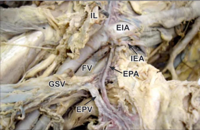
Figure 2. Dissection of right iliofemoral region. (EIA: external iliac artery; IEA: inferior epigastric artery; EPA: external pudendal artery; EPV: variant external pudendal vein; GSV: great saphenous vein; FV: femoral vein; IL: inguinal ligament)
EPA IEA
EPV
EIA IL
FV
GSV
Figure 3. Dissection of right obturator canal. (VOV: variant obturator vein; VOA: variant obturator artery; ON: obturator nerve; OV: normal obturator vein)
VO V
OV ON VOA
Figure 4. Dissection of left ilioinguinal region. (EIA: external iliac artery; EIV: external iliac vein; VOA: variant obturator artery; IEA: inferior epigastric artery; DCI: deep circumflex iliac artery; RA: rectus abdominis muscle; IL: inguinal ligament)
EIV
IL RA
DCI
IEA EIA
VOA
Figure 5. Dissection of left obturator canal. (VOA: variant obturator artery; VOV: variant obturator vein; ON: obturator nerve)
ON VOV
VOA
143 Variant origin of external pudendal artery
interventions. It may compress the external iliac vein and can result in venous stagnation in the lower limb [7]. Besides, it may be an additional source of bleeding in cases of hemorrhage secondary to pelvic fracture [8]. In this report, the superficial external pudendal artery (SEPA) in the right side originated in a common trunk with the inferior epigastric artery. To the best of my knowledge, no similar cases were recorded in the literature. Previous reports have mentioned that although an accurate knowledge of the anatomy of SEPA is of maximal importance for designing an axial pattern flap, little studies were found in the literature describing
its anatomy. They concluded that, SEPA always arises from the medial side of the femoral artery at the level of the sapheno-femoral junction [9]. Many surgeons used SEPA flap in vulvar [10] and penile reconstruction [11]. Some authors attributed the use of the SEPA flap to the diameter and length of the pedicle vessels and their constant anatomy [2]. So the variant SEPA recorded in this report must be considered.
In conclusion, these anatomical variations must be taken in consideration during surgical operations. We must throw the light on the unusual origin of SEPA recorded in this case.
References
[1] Standring S, Ellis H, Healy JC, Johnson D, Williams A, Collins P, Wigley C. Gray’s Anatomy: The anatomical basis of clinical practice. 39th Ed., London, Elsevier Churchill Livingstone. 2005; 1101, 1361.
[2] Castro M, Brenda E, Marques A, Pereira MD. Anatomic study of the external pudendal vessels to the anterior scrotal region. Eur J Plast Surg. 1998; 21: 86–90.
[3] Bergman RA, Thompson SA, Afifi AK. Compendium of human anatomic variations. Munich, Urban and Schwarzenberg. 1988; 84.
[4] Kumar D, Rath G. Anomalous origin of obturator artery from the internal iliac artery. Int J Morphol. 2007; 25: 639–41.
[5] Arey LB. The development of peripheral blood vessels. In: Orbison JL, Smith DE, eds. The peripheral Blood Vessels. Baltimore, Williams and Wilkins. 1963; 1–16.
[6] Fitzerald MJT. Human Embryology. New York, Harper International. 1978; 38–56.
[7] Nayak B, Soumya KV. Variant obturator vessels. International Journal of Anatomical Variations (IJAV). 2009; 2: 111–112.
[8] Daeubler B, Anderson SE, Leunig M, Triller J. Hemorrhage secondary to pelvic fracture: coil embolization of an aberrant obturator artery. J Endovasc Ther. 2003; 10: 676–680.
[9] La Falce OL, Ambrosio JD, Souza RR. The anatomy of the superficial external pudendal artery: a quantitative study. Clinics (Sao Paulo). 2006; 61: 441–444.
[10] Mayer AR, Rodriguez RL. Vulvar reconstruction using a pedicle flap based on the superficial external pudendal artery. Obstet Gynecol. 1991; 78: 964–968.