Isolated ruptured aneurysm of the superficial femoral artery:
case report
Aneurisma isolado de artéria femoral superficial roto contido: relato de caso
Daiane Cristina Ferreira Damasceno1
*
, Júlio Beserra Evaristo1, Geraldo Felipe Júnior1, André Luiz Guimarães Câmara1,
Cláudio Eluan Kalume1, Alcides José Araújo Ribeiro1, Leonardo Pires de Sá Nóbrega1
Abstract
Isolated true aneurysms of the supericial femoral artery (SFA) are rare events. hey mostly manifest in elderly men and are frequently seen in conjunction with other aneurysms. hey have varied etiology and are usually detected when they complicate with thrombosis or distal embolization, or, more rarely, when they rupture. he present case report describes a patient with an aneurysm of the SFA that was ruptured and contained and who had no other aneurysms. Color Doppler ultrasound of the arteries revealed the rupture and angiotomography showed a saccular aneurysm of the SFA measuring 11.4 × 8.8 cm, with a large mural thrombus. Arteriography was used to plan revascularization and showed the distal bed with outlow via the anterior tibial artery. he patient was treated with conventional elective distal femoropopliteal surgical revascularization with the ipsilateral saphenous vein inverted, which was successful. Recovery was complicated by a postoperative surgical site infection. Microbiology tests were negative and the anatomopathological study conirmed a true aneurysm of the SFA.
Keywords: supericial femoral artery; ruptured aneurysm; surgery.
Resumo
Aneurismas verdadeiros isolados da artéria femoral supericial (AFS) são eventos raros. Manifestam-se principalmente em homens idosos e frequentemente estão associados a outros aneurismas. Possuem etiologia variada e costumam ser detectados quando apresentam complicações como trombose, embolização distal ou, mais raramente, ruptura. O presente caso refere-se a um paciente cujo aneurisma de AFS se apresentou roto contido e sem associações com outros aneurismas. Foram realizados eco-Doppler colorido arterial, que diagnosticou a ruptura, e angiotomograia, que evidenciou aneurisma sacular de AFS medindo 11,4 × 8,8 cm, com grande trombo mural. Uma arteriograia foi utilizada para programação de revascularização, e detectou-se leito distal via artéria tibial anterior. O paciente foi submetido a revascularização cirúrgica convencional eletiva em artéria femoropoplítea distal com veia safena ipsilateral invertida, com sucesso. Apresentou como complicação pós-operatória infecção de sítio cirúrgico. A pesquisa microbiológica teve resultado negativo, e o estudo anatomopatológico conirmou aneurisma verdadeiro da AFS.
Palavras-chave: artéria femoral supericial; aneurisma roto; cirurgia.
1 Hospital de Base do Distrito Federal – HBDF, Unidade de Cirurgia Vascular e Angiologia – UCIVASA, Brasília, DF, Brazil.
Financial support: None.
Conlicts of interest: No conlicts of interest declared concerning the publication of this article. Submitted: August 28, 2017. Accepted: November 11, 2017.
early detection of aneurysms less likely, and the irst symptom is very often rupture.8 In contrast, common
femoral or popliteal artery aneurysms rarely rupture.5 When rupture does occur, clinical presentation may include distal ischemia, a pulsating mass in the thigh or a pulsating and painful mass.4
The major complications of SFA aneurysms are thrombosis, distal embolization or, more rarely, rupture.1,2,4,5 Resection and revascularization should be performed because of the high incidence of complications. It is also obligatory to screen for additional arterial aneurysms in other locations.4
This case report describes a ruptured SFA aneurysm in an elderly patient who was treated with conventional surgery.
unremarkable. Examination of the right lower limb found all arterial pulses were present and examination of the left lower limb revealed considerable swelling of the thigh, reaching 21 × 21 cm at its largest diameters, hardened and without thrill or pulse (Figure 1). The patient had palpable and normal femoral and popliteal pulses, but distal pulses were absent.
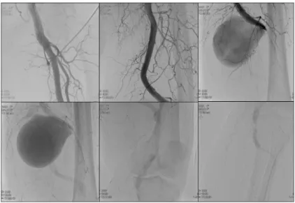
Laboratory tests were ordered, but hemogram, electrolytes, renal function, and coagulogram were all normal. Investigation was initiated with color Doppler ultrasound of the arteries, which showed a voluminous saccular aneurysm in the mid and distal thirds of the left SFA, with dissection and rupture of the walls and a lumen measuring 5.8 × 4.7 cm at its largest diameters (Figure 2). There was also
Figure 1. Preoperative: (A) anterior view, with increased volume of the left thigh; (B) medial view, with external diameters of the
bidirectional low inside the sac and through its communication with the SFA (Figure 2). Color Doppler ultrasound of the veins showed an echogenic image in the lumen of the popliteal vein, which had entirely lost its compressibility, characteristic of deep venous thrombosis (DVT).
Since the patient was hemodynamically stable and had COPD but was not in regular follow-up, and also because the distal bed had not been identiied in the workup examinations, the decision was taken to conduct a more detailed investigation of the case and plan elective surgery for a later date. Therapeutic anticoagulation was initiated in the emergency unit, because of the DVT in the left popliteal vein.
As part of the supplementary investigation, computed tomography angiography of the lower limbs was conducted (Figure 3), showing a saccular aneurysmal dilatation in the distal third of the left SFA, measuring 11.4 × 8.8 cm at its largest transverse axes, with a large mural thrombus, and the remaining lumen measuring 6.6 cm at the largest visible axis.
The patient was examined with color Doppler ultrasound to screen for other aneurysms in the aorta, iliac arteries, and arteries of the right lower limb, but no aneurysms were detected in any of these areas. Arteriography of the left lower limb was conducted to aid in planning surgery (Figure 4), showing a large aneurysm of the distal third of the left SFA, with a short neck and occlusion of the popliteal artery, which was reilled below the aneurysm by scant collateral circulation and outlowed to the anterior tibialis artery.
The patient underwent elective surgery, with an incision in the medial surface of the left thigh for proximal and distal control of the aneurysm. A voluminous encapsulated hematoma was found in the left thigh
(Figure 5), conirming the diagnosis of contained
ruptured aneurysm of the SFA. Aneurysmectomy was performed, followed by reconstruction of the vascular segment with the left great saphenous vein inverted. Fragments of dried tissues were sent for histopathology and microbiology. No microorganisms grew and the tissue biopsy result was true aneurysm of the left SFA.
The patient recovered well during the postoperative period: the left lower limb was warm, with peripheral perfusion < 3 s, femoral, popliteal, anterior tibialis and pedal pulses were all present, palpable, and normal, and there were no hematoma or signs of infection of the surgical site. The patient was discharged from hospital on the ifth day after the operation in good clinical condition, with a 6-month prescription for Rivaroxaban to treat the left popliteal vein DVT.
Fifteen days after the operation the patient returned to the emergency unit because of hyperemia and secretions from the surgical site. He was admitted to hospital for venous antibiotic treatment lasting 10 days.
At the 3-month clinical follow-up consultation
(Figure 6), the surgical site was healing well and
the edema of the lower limb was in regression. Color Doppler ultrasound of the arteries of the left lower limb showed that the graft was patent, with no signs of thrombosis or stenosis of the graft or at
the anastomoses.
Figure 2. Color Doppler ultrasound of the arteries: (A) longitudinal image of the saccular aneurysm communicating with the left
Figure 4. Arteriography showing saccular aneurysm of the distal third of the supericial femoral artery with occlusion of the popliteal artery. Scant collateral circulation. Reilling of the popliteal artery below the aneurysm and outlow via the anterior tibialis artery.
Figure 5. Intraoperative image: dissection of the aneurysm sac.
DISCUSSION
Isolated true atherosclerotic aneurysms of the SFA are rare.1 This condition occurs in the elderly
population and affects men more often than women (3:1).9 In 18% of case they are bilateral6 and they are
often seen in conjunction with aneurysms in other sites (27-69% of cases).10
Peripheral aneurysms of arteries in the limbs are generally easily palpated, but aneurysms of the SFA are unlikely to be detected early,8 because
their medial and distal portions lie below the fascia musculature and between the sartorius, adductor longus, and vastus medialis muscles. They therefore remain undiagnosed until they cause complications.7
The incidence of rates of concomitant aortoiliac aneurysms, popliteal artery aneurysms, and other peripheral aneurysms are, respectively, 40-69%, 54%, and 27%.11 Therefore, once an SFA aneurysm has
been diagnosed, investigation to screen for additional arterial aneurysms in other sites is obligatory.4
Aneurysms of peripheral arteries can be linked to a variety of etiologic factors, such as syphilis, immunological disorders (Behçet’s disease), inlammatory conditions (Wegener’s granulomatosis), connective tissue diseases (Ehlers-Danlos or Marfan Syndrome), or even to secondary factors (ibrodysplasia or malignancy).7,11-14 In the absence of clear etiologic factors, a large proportion of aneurysms are classiied
as atherosclerotic, even when there is little or no evidence of atherosclerosis in other vessels.9
According to Rigdon and Monajjem9, complications
are seen in 65% of cases of SFA aneurysm and include rupture (35%), thrombosis (18%), and distal embolic events (12%). These complications occur with less frequency in comparison with patients with popliteal artery aneurysms.3 Nevertheless, symptomatic
aneurysms, with diameters greater than 2.5 cm or more than twice the normal caliber of the artery should be repaired to prevent complications that threaten the viability of the limb.15,16
Early diagnosis of SFA aneurysms is extremely important to avoid complications affecting the limb, in addition to allow for planning of elective surgery, with lower morbidity and mortality. Color Doppler ultrasound of the arteries, angiotomography, and magnetic resonance angiography are useful for diagnosis, for assessing anatomic relationships between the aneurysm and adjacent structures, and for planning surgery. Arteriography can be reserved for assessing the distal bed for revascularization, if necessary.
Conventional surgical treatment remains the gold standard for peripheral aneurysms,16,17 with
end-to-end anastomosis, venous grafting (preferably with an autologous vein), or prosthetic grafts.3
In elective cases, patency rates with vein grafts are approximately 80% at 2 years, compared with 65% for
early or late thrombosis of the graft, stenosis of the graft, false anastomotic aneurysm and lymphedema.20 Postoperative follow-up varies depending on the methods of arterial revascularization employed. When a venous graft is constructed, color Doppler ultrasound is used to examine the arteries 30 days after the procedure; three-monthly for 1 year after the irst assessment; six-monthly for the following 2 years; and annually thereafter. If a prosthetic graft has been used, follow-up should be decided on a case-by-case basis, because there is no scientiic evidence demonstrating the cost-beneit relationship of such surveillance.20
In view of the above, it is concluded that an SFA aneurysm is a rare event and that it most often presents as a rupture. It may be seen in combination with other aneurysms and screening for aneurysms of other sites is obligatory. Conventional surgical treatment is still the best option because of the durability of grafts, but involves the risks inherent to surgery. Patients should be followed-up to monitor the graft and avoid late postoperative complications.
REFERENCES
1. Brito CJ. Aneurismas periféricos. In: Maffei FHA, editor. Doenças vasculares periféricas. Rio de Janeiro: Guanabara Koogan; 2016. 1541 p.
2. Oliveira FA, Oliveira H Fo. Aneurisma de artéria femoral superficial roto: relato de caso e revisão de literatura. J Vasc Bras. 2009;8(3):285-8. http://dx.doi.org/10.1590/S1677-54492009000300019. 3. Rutherford RB. Superficial femoral artery aneurysm. In: Rutherford
RB, editor. Vascular surgery. Philadelphia: WB Sauders; 2005. p. 1538-39.
4. Jarrett F, Makaroun MS, Rhee RY, Bertges DJ. Superficial femoral artery aneurysms: an unsusual entity? J Vasc Surg. 2002;36(3):571-4. PMid:12218983. http://dx.doi.org/10.1067/mva.2002.125841.
org/10.1016/S1078-5884(05)80178-9.
9. Rigdon EE, Monajjem N. Aneurysms of superficial femoral artery: a report of two cases and review of the literature. J Vasc Surg. 1992;16(5):790-3. PMid:1433668. http://dx.doi. org/10.1016/0741-5214(92)90235-Z.
10. Farinon AM, Rulli F, Muzi M. Ruptured aneurysm of superficial femoral artery. Panminerva Med. 1995;37(3):155-8. PMid:8869374. 11. Duhalde I, Berga C, Arrebola M, Pañella F, Rodríguez N, Admetller X. Aneurisma de arteria femoral superficial: reporte de un caso y revisión de la patología. Cuad Cir. 2004;18:48-51. http://dx.doi. org/10.4206/cuad.cir.2004.v18n1-08.
12. Carmona-Berriguete S, López-Quero D, Martín-Álvarez A, et al. Aneurisma femoral bilateral en síndrome de Behcet: a propósito de un caso. Angiol. 2008;60(2):155-9. http://dx.doi.org/10.1016/ S0003-3170(08)02012-9.
13. Luebke T, Aleksic M, Brunkwall J. Superficial femoral artery aneurysm: a rare complication of wegener granulomatosis. Vascular. 2009;17(4):213-7. PMid:19698302. http://dx.doi. org/10.2310/6670.2009.00016.
14. Giordanengo F, Beretta L, Galimberti M, Ferrero S. A rare case of aneurysm of the superficial femoral artery with dysplasic etiology. Minerva Chir. 1989;44(7):1173-7. PMid:2664565.
15. Shortell CK, DeWeese JA, Ouriel K, Green RM. Popliteal artery aneurysms: a 25-year surgical experience. J Vasc Surg. 1991;14(6):771-6. PMid:1960807. http://dx.doi.org/10.1067/mva.1991.33214. 16. Corriere MA, Guzman RJ. True and false aneurysms of the femoral
artery. Semin Vasc Surg. 2005;18(4):216-23. PMid:16360579. http:// dx.doi.org/10.1053/j.semvascsurg.2005.09.008.
17. Arendt AL, Amaral RM, Vieira MS, Ribeiro RN, Argenta R. Aneurisma verdadeiro roto de artéria femoral superficial. J Vasc Bras. 2013;12(4):315-9. http://dx.doi.org/10.1590/jvb.2013.048. 18. Dighe S, Thomas P. Ruptured superficial femoral artery aneurysm
treated by simple ligation. Singapore Med J. 2008;49(6):151-2. PMid:18581007.
19. Diethrich EB. Endoluminal grafting in the treatment of iliac and superficial femoral artery disease. Tex Heart Inst J. 1997;24(3):185-92. PMid:9339506.
*
Correspondence
Daiane Cristina Ferreira Damasceno Hospital de Base do Distrito Federal – HBDF, Unidade de Cirurgia Vascular e Angiologia – UCIVASA SGAN 91, Módulo F, Condomínio Green Park, Bloco C, apto. 122 - Asa Norte CEP 70790-110 - Brasília (DF), Brazil Tel.: +55 (61) 98273-9426 E-mail: [email protected]
Author information
DCFD and JBE - Resident physicians, Programa de Cirurgia Vascular e Angiologia, Hospital de Base do Distrito Federal (HBDF). GFJ, ALGC, CEK and AJAR - Vascular surgeons, Unidade de Cirurgia Vascular e Angiologia, Hospital de Base do Distrito Federal (HBDF). LPSN - Full member, Sociedade Brasileira de Angiologia e de Cirurgia Vascular (SBACV); Vascular surgeon and preceptor, Programa de Cirurgia Vascular e Angiologia, Hospital de Base do Distrito Federal (HBDF).
Author contributions
Conception and design: DCFD, JBE, GFJ, ALGC, CEK, AJAR, LPSN Analysis and interpretation: DCFD, AJAR, LPSN Data collection: DCFD, GFJ, ALGC, LPSN Writing the article: DCFD Critical revision of the article: DCFD, AJAR, LPSN Final approval of the article*: DCFD, JBE, GFJ, ALGC, CEK, AJAR, LPSN Statistical analysis: N/A. Overall responsibility: DCFD, LPSN