LETTER TO THE EDITOR
1. University Center of João Pessoa - Department of Biological Sciences and the Health- João Pessoa, Paraíba/ Brazil
2. Department of Orthopedics, Medical School, São Paulo University, São Paulo, SP, Brazil.
3. Medic Center Kawano- São Paulo – SP/Brazil E-mail: [email protected]
PAROSTEAL LIPOMA OF THE FEMUR WITH
HYPEROSTOSIS: CASE REPORT AND LITERATURE
REVIEW
Rosalvo Zosimo Bispo Junior1,2, Alexandre Vieira Guedes3
INTRODUCTION
Lipomas may be defined as benign lesions of mature adipose tissue without evidence of cellular atypia.1
Lipo-mas are the most common soft tissue lesions1,2 and
surpris-ingly are among the rarest bone neoplasias.1-3
Osseous lipomas have been classified according to their site of origin: either within bone (intraosseus) or on its sur-face (juxtacortical). Sursur-face osseous lipomas are subdivided into parosteal and subparosteal lipomas.4 Parosteal lipomas
often induce a periosteal reaction.1 The most frequently
af-fected sites are the diaphysis5 and metaphysealregions4 of
long bones.
The parosteal type is a rare tumor accounting for 0.3% of all lipomas6, usually asymptomatic,3 and affecting
mainly adults aged over 40.7 The most frequent complaints
are a tumoral convexity presenting as a visible or palpable mass8 or a mild-intensity pain.
A literature review in the English language MEDLINE database revealed that 12 new cases have been described since the first detailed report of a parosteal lipoma of the femur with hyperostosis by Kenin et al9 in 1959.
The present article describes a rare case of parosteal lipoma located in the femur, with extensive hyperostosis, and reviews this entity in the English language medical lit-erature.
CASE REPORT
A 55-year-old female patient noticed an increase in the volume of her left thigh, without associated pain, approxi-mately 8 years ago. She reported a related local trauma af-ter a ground-level fall at the time. In 2003 she was admit-ted to our Service, and the clinical evaluation revealed a
nonpulsatile mass in the anteromedial face of the distal two-thirds of the left thigh that had a tender consistency and regular contour adhered to the deep planes and that was painless upon diffuse palpation.
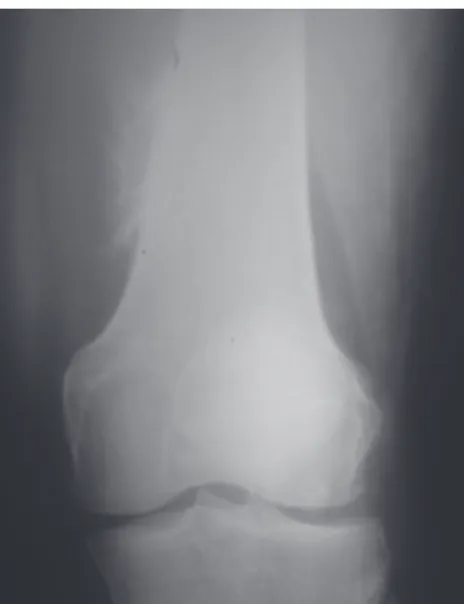
Conventional radiographs of the left thigh showed the presence of a spiculated periosteal bone formation in the anteromedial face of the distal region of the femur and an increase in the amount of soft tissue with a radiodensity characteristic of fatty tissue (radiolucent image) (Figure 1).
Computed tomography of the left thigh showed a het-erogeneous calcification image in the periosteal topogra-phy located in the medial region of the distal third of the femur in the metadiaphyseal junction, without rupture of the cortical or bone marrow invasion. In the muscular plane, the density was similar to that of subcutaneous tissue in the intermediate vastus muscle topography, suggesting fat replacement (Figure 2).
After the evaluations were completed, only in January 2005 did the patient return to the outpatient facility with a small increase in the size of the tumor.
Due to the early differential diagnosis of the liposar-coma, abdominal ultrasonography and thoracic computed tomography were requested in order to discard the possi-bility of synchronic mass of the retroperitoneum or pulmo-nary metastatic process.
In the MRI of the left thigh, we observed an expansive process measuring 19 x 8 x 9 cm with hypersignal in T1 around the distal femur under the intermediate vastus mus-cle as well as hyperostosis in the metadiaphyseal region of the femur. In the T2 weighted images, as well as in the region of the hyperostosis, the tumor mass was revealed by hyposignal (Figure 3).
We performed an incisional biopsy of the tumor in March 2005 for anatomopathological study; it was mature adipose tissue without cellular atypia.
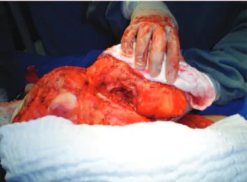
In the following month the patient underwent surgical intervention for tumor resection. During the surgery, it was confirmed that the lesion had adhered to the femur in the region of the hyperostosis (periosteal reaction) (Figure 4).
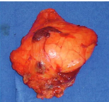
The tumor specimen weighed 437 g and had a lobu-lated appearance, with an intact, thin, and translucent pseudocapsule (Figure 5). Microscopic examination showed that the neoplasia was adhered to the strip of skeletal mus-cular tissue in its most superficial portion by fibrous tis-sue, with mature bone metaplasia in the deep portion with no evidence of malignancy (Figure 6). The results showed a lipoma with periosteal reaction.
There has been no recurrence during the postoperative period, and the (ambulatory) patient is periodically fol-lowed-up (for 24 months now) with no signs of recurrence.
DISCUSSION
The World Health Organization (WHO) currently de-fines bone lipoma as a benign neoplasia of adipose tissue that is formed inside the medullary cavity, the cortex, or the bone surface.8
The original description of this neoplasia was published in the German medical literature by Seerig* in 1836 (as reported inFleming et al6). The subject was approached in
Figure 2 - Computed tomography: window of soft parts (axial view) where an intramuscular expansive process can be seen around the femur, with the radiodensity of fat. Note the hyperostosis in the cortical medial area of the bone without medullary invasion.
Figure 3 - Magnetic resonance image: coronal T1-weighted image where a solid expansion process can be noticed with hypersignal adjacent to the femur. Note the hyperostosis in the medial cortical bone with signal intensity similar to medullary intensity.
Figure 4 - Intraoperative photograph showing resection of a tumor of large volume.
the English medical literature in 1866 by Smith.10
How-ever, the term parosteal lipoma was introduced by Power11
in 1888 and is still used.
Bone lipomas are among the rarest neoplasias of the skeleton, accounting for less than 0.1% of primary bone tumors8 and 15% of all osseous lipomas,4 often affecting
the femur.6,12 However, the real incidence seems to be much
greater than previously thought, because many lipomas re-main undiagnosed.4
In spite of the close relationship of bone lipoma with the periosteum,3,6 the bone may be normal.6 Occasionally,
these rare lesions are associated with reactive changes in the adjacent bone.3,6,13 These changes include bone
deform-ity, cortical erosion, and overproduction of the cortical bone (hyperostosis).3,6-8 More than half of parosteal lipomas
pre-senting bone reactions are associated with hyperostosis.6
Several authors consider that cortical hyperostosis can be explained by the contact of the lipoma with the bone,3,14
being presumably caused by stimulation or irritation of the periosteum.15 However, it is not clear why some lipomas
located near the periosteum cause this reaction while oth-ers do not.15
Although many cases of parosteal lipomas have been reported over the years, the data have not been appropri-ately described, either due to incomplete clinical history and/or reproduction of imaging evaluations, or lack of anatomopathological diagnosis in some cases.
Fleming et al6 reviewed the world literature between
1918 and 1962, having identified only 16 cases of parosteal lipoma with a well-defined radiolucent area in association with reactive bone changes; the radiological characteris-tics of those changes found in 11 of the 16 cases were com-patible with overproduction of cortical bone (hyperostosis). Prominent hyperostosis promotes a typical appearance that, when combined the radiolucence of the soft parts of the tumor, has been described as pathognomonic for parosteal lipoma.6,15 However, the amount of cortical hyperostosis can
vary from a small area of the thickened cortical to exuber-ant exostoses.6,13
Of the 11 cases identified in the study by Fleming et al,6 9 affected the femur, but only 2 were published in
Eng-lish (Tables 1A and B).
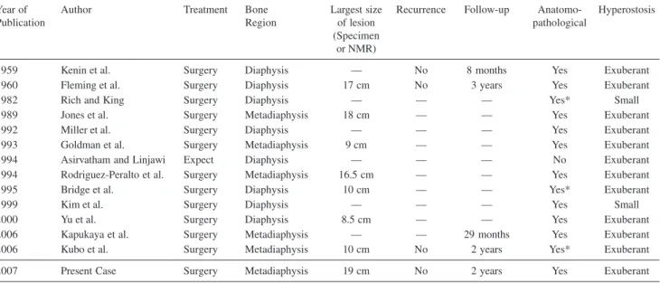
Since the review by Fleming et al6 up to now, 12 new
cases of parosteal lipoma of the femur with hyperostosis, including ours, have been published in English, totaling 14 in almost 90 years (Tables 1A and B). Details of these cases are listed as follows:
• In the 1980s, Rich and King16 described 4 cases of soft
part neoplasia adjacent to the femur, presenting asym-metric cortical thickening. One of these 4 cases (Case #4) was a parosteal lipoma of the femur with a small hyperostosis (cortical thickening).
• Another case of these tumors was reported in English in 1989.14
• In spite of the 4 cases of parosteal lipoma of the femur mentioned in the study by Miller et al5 in 1992, only 1
case (Case #3) was described in detail, including clini-cal history, imaging, and anatomopathologiclini-cal evalua-tions.
• In 1993 other authors published a coincidental case of concomitant intramuscular lipoma in the left leg and a parosteal lipoma of the right femur.17
Figure 5 - Anatomical specimen with lobulated appearance and intact, translucent capsule.
Table 1 A – Patients with parosteal lipoma of the femur with hyperostosis (identified in literature in English language)
Year of Author Age Sex Duration of Body Half Presence Imaging
publication (years) signals and of pain Technique
symptoms
1959 Kenin et al. 49 Male — Left No R
1960 Fleming et al. 52 Female 7 months Right Yes R
1982 Rich and King 59 Male 1 year Left Yes R + CT
1989 Jones et al. 32 Male 20 years Right Yes R + CT + NMR
1992 Miller et al. 61 Female — Right No R + BS
1993 Goldman et al. 53 Female 1 year Right No R + CT + NMR
1994 Asirvatham and Linjawi 40 Female — Right No R + CT + NMR
1994 Rodriguez-Peralto et al. 39 Male 5 years Left Yes R + CT
1995 Bridge et al. 51 Female 5 months Left Yes R + CT + NMR* + BS*
1999 Kim et al. 46 Female 7 months Left Yes R + NMR
2000 Yu et al. 37 Male — Left No R + NMR
2006 Kapukaya et al. 17 Male — Left Yes R + CT + NMR
2006 Kubo et al. 56 Male — Right Yes R + BS + NMR
2007 Present Case 55 Female 8 years Left No R + CT + NMR
R: Radiograph; CT: Computerized Tomography; BS: Bone Scintigraphy; NMR: Nuclear Magnetic Resonance. * Not showed in the paper.
Table 1 B – Patients with parosteal lipoma of the femur with hyperostosis (identified in literature in English language)
Year of Author Treatment Bone Largest size Recurrence Follow-up Anatomo- Hyperostosis
Publication Region of lesion pathological
(Specimen or NMR)
1959 Kenin et al. Surgery Diaphysis — No 8 months Yes Exuberant
1960 Fleming et al. Surgery Diaphysis 17 cm No 3 years Yes Exuberant
1982 Rich and King Surgery Diaphysis — — — Yes* Small
1989 Jones et al. Surgery Metadiaphysis 18 cm — — Yes Exuberant
1992 Miller et al. Surgery Diaphysis — — — Yes Exuberant
1993 Goldman et al. Surgery Metadiaphysis 9 cm — — Yes Exuberant
1994 Asirvatham and Linjawi Expect Diaphysis — — — No Exuberant
1994 Rodriguez-Peralto et al. Surgery Metadiaphysis 16.5 cm — — Yes Exuberant
1995 Bridge et al. Surgery Diaphysis 10 cm — — Yes* Exuberant
1999 Kim et al. Surgery Diaphysis — — — Yes Small
2000 Yu et al. Surgery Diaphysis 8.5 cm — — Yes Exuberant
2006 Kapukaya et al. Surgery Metadiaphysis — — 29 months Yes Exuberant
2006 Kubo et al. Surgery Metadiaphysis 10 cm No 2 years Yes* Exuberant
2007 Present Case Surgery Metadiaphysis 19 cm No 2 years Yes Exuberant
* Not showed in the paper.
• In 1994 in Saudi Arabia, a large-volume parosteal lipoma with exuberant hyperostosis was found in the right thigh of a young patient; however, in spite of the characteristics, the lipoma was not surgically resected.7
• Also in 1994, another finding was published concern-ing this rare benign neoplasia in the femur, also with relevant reaction of bone.3
• Genetic characteristics were shown in the article by Bridge et al.18
• Another case was reported in 1999 by Kim et al.12
• Yu et al reported the eleventh case of parosteal lipoma of the femur with hyperostosis in the English language literature.19
• Blair and Resnick20 (2000) reported 1 case of
‘subpe-riosteal lipoma’ of the femur whose appearance, as stated by the authors themselves, was not that of a cor-tical or parosteal lipoma.
REFERENCES
1. Schajowicz F. Neoplasia óssea e lesões pseudotumorais. 2º ed. Rio de Janeiro: Revinter; 2000. p. 403-46.
2. Enneking WF. Limb salvage in musculoskeletal oncology. New York: Churchill Livingstone; 1983. Vol 2. p. 1225-69.
3. Rodriguez-Peralto JL, Lopez-Barea F, Gonzales-Lopes J, Lamas-Lorenzo M. Case report 821. Skeletal Radiol. 1994;23:67-9. 4. Kapukaia A, Subas M, Dabak N, Ozkul E. Osseous lipoma: eleven new
cases and review of the literature. Acta Orthop Belg. 2006;72:603-14. 5. Miller MD, Ragsdale BD, Sweet DE. Parosteal lipomas: A new
perspective. Pathology. 1992;24:132-9.
6. Fleming RJ, Alpert M, Garcia A. Parosteal lipoma. ARJ. 1962;87:1075-84.
7. Asirvatham R, Linjawi T. Ossifying parosteal lipoma with exuberant cortical reaction. Int Orthop. 1994;18:55-6.
8. Rosenberg AE, Bridge JA. Lipoma of bone. In: Fletcher CDM, Unni KK, Mertens F, editors. Pathology and genetics of tumours of the soft tissues andbones. Lyon: IARC Press; 2002. p. 328-9.
parosteal lipoma of the femur with hyperostosis.4
Recently, genetic and magnetic resonance imaging char-acteristics were described in another patient with this neo-plasia in the femur (Case #1).21 Conventional radiographic
exams showed an increase in the extra-bone soft tissues and formation of bone spicula over the periosteum. The radiodensity of the tumoral mass in conventional radio-graphs, which was consistent with fatty tissue, suggested the diagnosis.8,15
These case studies provide diagnostic, therapeutic, and prognostic guidance for future cases. Computed tomogra-phy and magnetic nuclear resonance help with the diagno-sis13,14 and therapeutic planning. Treatment consists of
resecting the lipomatous tumor15,22 with further exeresis of
the bone and periosteal excrescence in cases with hyper-ostosis.3,4 Usually the prognosis is favorable, with no proven
reports of malignancy4 or recurrence and with full
recov-ery of the patient.
9. Kenin A, Levine J, Spinner M. Parosteal lipoma. A case report of two cases with associated bone changes. J Bone Joint Surg. 1959;41:1122-6.
10. Smith, T. Specimen of firm bilobed fatty tumor, weighing 10 ounces, removed from little girl age 5 yaers. Trans Pathol Soc London. 1866;17:286.
11. Power DA. Parosteal lipoma, or congenital fatty tumour, connected with periosteum of femur. Trans Pathol Soc London. 1888;39:270-2. 12. Kim JY, Jung SL, Park YH, Park SH, Kang YK. Parosteal lipoma with
hyperostosis. Eur Radiol. 1999;9:1810-2.
13. Laorr A, Greeenpan A. Parosteal lipoma with hyperostosis: report of two pathologically proven cases evaluated by magnetic resonance imaging. Can Assoc Radiol J. 1993;44:285-90.
14. Jones JG, Habermann ET, Dorfman HD. Case report 553: parosteal ossifying lipoma of femur. Skeletal Radiol. 1989;18:537-40. 15. Jacobs P. Parosteal lipoma with hyperostosis. Clin Radiol.
16. Rich PJ, King W III. Benign cortical hyperostosis underlying soft tissue tumors of the thigh. ARJ. 1982;138:419-22.
17. Goldman AB, DiCarlo EF, Marcove RC. Case report 774: coincidental parosteal lipoma with osseous excrescence and intramuscular lipoma. Skeletal Radiol. 1993;22:138-45.
18. Bridge JA, DeBoer J, Walker CW, Neff JR. Translocation t(3;12)(q28;q14) in parosteal lipoma. Genes Chromosom Cancer. 1995;12:70-2.
19. Yu JS, Weis L, Becker W. MR imaging of a parosteal lipoma. Clin Imag. 2000;24:15-8.
20. Blair TR, Resnick D. Subperiosteal lipoma: a case report. Clin Imag. 2000;24:381-3.
21. Kubo T, Matsui Y, Goto T, Yukata K, Endo K, Sato R, et al. MRI characteristics of parosteal lipoma associated with the HMGA2-LPP fusion gene. Anticancer Res. 2006;26(3B):2253-8.