Authors
Andrea Lordsleem1 Ana Paula Santana Gueiros1
José Edevanilson de Barros Gueiros1 Brivaldo Markman Filho1
Edgar Guimarães Victor1
1Universidade Federal de Pernambuco – UFPE.
Submitted on: 02/03/2011. Approved on: 11/16/2011.
Correspondence to:
Andréa Lordsleem Avenida Professor Moraes Rego, 1.235
Bairro Cidade Universitária Recife – PE – Brasil Zip code 50670-901 E-mail: andreabmelo@ cardiol.br
This study was undertaken at the UFPE.
The authors report no conlict of interest.
R
ESUMOIntrodução: Pacientes com doença renal crônica (DRC) apresentam sinergismo entre fatores de risco tradicionais para aterosclero-se e emergentes derivados do estado urêmico.
Objetivo: Traçar o perfil epidemiológico de um grupo de pacientes com DRC submetido à avaliação cardiológica. Métodos: Pacientes sintomáticos – com isquemia em cintilogra-fia miocárdica e/ou disfunção sistólica ao ecodopplercardiograma – com idade maior que 50 anos e diabetes mellitus (DM) como causa da DRC e aqueles com dois ou mais fatores de risco ateroscleróticos realizaram cineangiocoronariografia. Assintomáticos – não diabéticos e sem fatores de risco – foram investigados com ecodopplercardiograma e aqueles com único fator de risco, por meio de ecodopplercardiograma e cintilografia.
Resultados: Foram estudados 46 pacientes, 58,7% homens, idade de 50-70 ± 11,7 anos, 91,3% dialíticos. Tempo de hemodiálise: 61,96 ± 55,1 meses. Hipertensão arterial foi causa da DRC em 56,5%. Dos 28 pacientes (60,9%) submetidos à cineangiocoronar-iografia, 53,6% apresentaram doença arte-rial coronariana (DAC). Os pacientes foram divididos em três grupos: com DAC (A), sem DAC (B) e não submetidos à cineangio-coronariografia (C). Diferença significativa ocorreu entre os Grupos B e C na frequên-cia de índice tibiobraquial (ITB) anormal (p = 0,026), com ausência de ITB anormal no Grupo C e na média de idade, superior no B (p = 0,045). No Grupo A, 53,3% dos pacientes estavam em avaliação pré-paratir-eoidectomia (PTX). Conclusão: Este estudo confirmou a alta frequência de alterações cardiovasculares, inclusive de DAC, nos pa-cientes portadores de DRC, principalmente naqueles em diálise.
Palavras-chave: Doença das coronárias. Insuficiência renal crônica. Inflamação.
A
BSTRACTIntroduction: Patients with chronic kidney disease (CKD) experiment a synergistic ef-fect of the traditional and the emerging ure-mia-related risk factors for atherosclerosis.
Objective: Draw the epidemiologic profile of a group of CKD patients who underwent cardiac evaluation. Methods: Symptomatic patients, patients with ischemia on myocar-dial scintigraphy and/or systolic dysfunc-tion on echocardiography, patients older than 50 years and diabetes mellitus (DM) as a cause of CKD, and those with two or more risk factors underwent coronary angiography. Asymptomatic, non-diabetic patients and patients with no risk factors were investigated with echocardiogra-phy. Those with a single risk factor were investigated with echocardiography and scintigraphy. Results: 46 patients (58.7% men) were enrolled. Their mean age was 50.7 ± 11.7 years. 91.3% were on dialy-sis, for 61.96 ± 55.1 months. Hypertension was the cause of CKD in 56.5%. Of the 28 patients (60.9%) who underwent an-giography, 53.6% had coronary artery dis-ease (CAD). The patients were divided into three groups: those with CAD (A), those without CAD (B) and those who didn’t undergo coronary angiography (C). A sig-nificant difference occurred only between groups B and C, as regards an abnormal ABI (p = 0.026), with no ABI abnormality in group C, and as regards the mean age, which was higher in group B (p = 0.045). In group A, 53.3% of the patients were in the preoperative stage of parathyroidec-tomy. Conclusion: This study confirmed the high rate of cardiovascular disorders, including CAD, in patients with CKD, es-pecially those on dialysis.
Keywords: Coronary disease. Renal insufficiency, chronic. Inflammation.
Cardiac evaluation of patients with chronic kidney
disease: what lessons?
I
NTRODUCTIONChronic kidney disease (CKD) patients are at high risk of cardiovascular diseases (CVD), which account for 40-50% of the deaths in this population.1-4
The high cardiovascular risk could be ex-plained, in part, by a synergy between traditional risk factors and the newly emerging uremia-related risk factors, resulting in accelerated atherosclero-sis and early death. Furthermore, the diagnoatherosclero-sis of CKD, made when CVD is already advanced (epi-demiologic causality), and the malnutrition, in-flammation and atherosclerosis (MIA) syndrome present in these patients, would lead to acceler-ated atherosclerosis.5,6
The emerging factors, mineral and bone metabo-lism disturbances, hyperhomocysteinemia, oxidative stress and inflammation, which are mostly cause or consequence of endothelial dysfunction, grow in im-portance as the renal impairment progresses.2,7,8 All
risk factors interact and increase the cardiovascular mortality of CKD patients.8-12
Mineral and bone metabolism disturbances, characterized by abnormal serum levels of calcium, phosphorus and parathyroid-hormone (PTH), are associated with extraosseous (arterial, valvular and myocardial) calcifications, and play an important role in the pathogenesis of myocardial hypertrophy and fibrosis. Vascular calcification is strongly associated with cardiovascular events and death.2
In CKD, a paradoxical association between some risk factors and mortality has been observed. Curiously, systemic arterial hypertension and overweight apparently protect these patients, determining a reverse epidemiology. Serum cholesterol levels positively correlate with serum albumin levels, and negatively correlate with serum C-reactive protein (CRP) and interleukin-6 levels, in a reflection of the MIA syndrome.6
We assessed the clinical, demographic and labora-tory characteristics of a group of stages 4 and 5 CKD patients, who underwent cardiac evaluation, includ-ing coronary angiography, in an attempt to know the epídemiologic profile of this population at cardiovas-cular risk.
M
ETHODSAMPLE
This case-series study was undertaken at the Cardiology Division of the Hospital das Clínicas (HC) of the Federal University of Pernambuco, Brazil,
during the October 2008 – October 2009 period. The study was approved by the institutional Research Ethics Committee, and all participants signed their informed consent.
46 stages 4 and 5 CKD patients, over the age of 18 years, free from acute or chronic infectious dis-eases and with no severe liver disease, were included in the study. The patients were referred from the Nephrology Division of the same institution, with the following indications: pre-transplantation assess-ment, pre-parathyroidectomy (PTx) assessassess-ment, or cardiovascular signs/symptoms.
The following clinical and demographic param-eters were assessed: age, sex, cause of CKD and time on dialysis. The patients were questioned about their symptoms, classic risk factors for coronary artery disease (CAD) (hypertension, diabetes mellitus, dys-lipidemia and smoking), and use of cardioprotective medications. All the patients were scored according to Framingham`s criteria, and underwent general and cardiac physical examinations. Weight was measured in kilograms and height and abdominal circumference in centimeters. Two consecutive blood pressure read-ings were obtained, one from an upper limb without an arteriovenous fistula and another from the ipsilat-eral lower limb, for calculation of the ankle-brachi-al index (ABI). The latter is the ratio between the systolic blood pressures obtained in the lower limb and in the upper limb, with values below 0.9 being abnormal.
The following serum determinations were per-formed at the Central laboratory of the same insti-tution: albumin (bromocresol green method, Abott, reference: 3.5-5.2 g/dL); high-sensitivity CRP (Aptec nephelometry, Bisys, reference: 0-1 mg/dL); total cho-lesterol (colorimetric enzymatic method); high-densi-ty lipoprotein (HDL, enzymatic method); low-densihigh-densi-ty lipoprotein (LDL, enzymatic method); triglycerides (enzymatic method); fasting glucose (enzymatic meth-od, reference: 70-99 mg/dL); calcium (o-cresolphtha-lein method, reference: 8.5-10.1 mg/dL); phosphorus (modified phosphomolybdate method, reference: 2.5-4.9 mg/dL); intact PTH (chemoluminescence, reference: 15-68.3 pg/mL); hematocrit (automated method, Beckman Coulter, reference: 47% ± 5 for men and 42% ± 5 for women.
electrocardiogram was obtained, with an abnormal result consisting of at least one of the following: a rhythm other than sinus-driven, conduction distur-bances, chamber overload and alterations of ventricu-lar repoventricu-larization.
For CAD assessment, the patients were stratified in risk groups, according to Gowdak et al.12 The very
high-risk group consisted of patients with symptoms suggestive of CAD or anginal equivalent. The high-risk group was composed of diabetics aged over 50 years and of patients with two or more classic CAD risk factors. Patients from the very high and high-risk groups underwent coronary angiography. The me-dium-risk group, consisting of asymptomatic non-diabetics, with a single classic CAD risk factor, was investigated with resting echocardiography and with resting and stress myocardial scintigraphy. The low-risk group, consisting of asymptomatic non-diabetics without CAD risk factors, was investigated with rest-ing echocardiography.
Other criteria for coronary angiography indication were evidence of ischemia on resting/stress myocar-dial scintigraphy and the presence of systolic dysfunc-tion (left ventricular ejecdysfunc-tion fracdysfunc-tion under 45%) on resting echocardiography.
Obstructive coronary disease was defined by the presence of stenotic lesions of 50% or more of the arterial lumen, in at least one coronary artery and/or its main branches.
According to the coronary angiography-defined CAD status, the patients were divided into two groups: Group A, composed of those with CAD and Group B, composed of those without CAD. Another group (C), was composed of the medium and low-risk patients. A comparative analysis between the groups was performed.
Left ventricular hypertrophy (LVH) was di-agnosed when the echocardiography-defined left ventricular mass index was > 134 g/m2 (men) and
> 110 g/m2 (women).13
To assess the MIA syndrome, we used serum al-bumin as a nutritional parameter, serum CRP as an inflammation marker, and the ABI as an indicator of atherosclerosis. The MIA syndrome was diagnosed when all the following were present: serum albumin level < 3.5 g/dL, CRP level > 1 mg/dL and ABI < 0.9.
STATISTICAL ANALYSIS
PTH levels were presented as median and first and third quartiles. The other variables were presented as means and standard deviations.
Kolmogorov-Smirnov`s test was applied for normality supposition. For comparative analysis of the quantitative variables, we used Student`s t test for independent samples, with the chi-squared and Fisher`s exact tests being used for analysis of the qualitative variables. For between-groups comparison of the PTH levels, we used Mann-Whitney`s non-parametric test. The significance level was set at 5%. The Excel 2000 and SPSS v 8.0 soft-ware were used.
R
ESULTSOf the 46 patients (mean age 50.7 years) assessed, 58.7% were men and 42 (91.3%) were on dialysis, with a mean dialysis time of 61.96 ± 55.1 months. 4 (8.7%) patients were referred from the CKD con-servative treatment outpatient service, and were characterized as having a very high risk. The main causes of CKD were hypertension (56.5%) and diabe-tes mellitus (17.4%). As for the traditional risk fac-tors, 91.3% of the patients had hypertension, 23.9% diabetes and 23.9% dyslipidemia. 13.0% actively smoked and 41.3% were former smokers.
As shown in Table 1, there was a low rate of car-dioprotective medication use, with aspirin as the most frequently used drug, by around 40% of those at very high and high risk. Table 2 shows the Framingham`s score for Groups A and B.
As for symptoms, 67.4% of the patients were asymptomatic, with dyspnea being the most fre-quent symptom (32.6%), and chest pain occurring in only 17.4%. 40% of Group A patients were asymptomatic.
Of all the patients, 30.4% were seen during their PTx preoperative assessment.
Obstructive CAD was diagnosed in 15 patients (32%) of the total sample. Of the 28 patients (61.9%) with coronary angiography indication, CAD was di-agnosed in 53.6% (Group A). Tables 1 and 3 show the comparative analyses between Groups A and B. As can be seen, in spite of the lack of a significant dif-ference between the PTH medians, the third quartile of the PTH of Group A was twice that of Group B. In addition, 53.3% of Group A patients were in the PTx perioperative period, compared with 15.4% of Group B (p = 0.055).
Mean Echocardiography-defined left ventricu-lar ejection fraction (LVEF) was 63 ± 9% (n = 44) and diastolic function (n = 43) was normal in only 34.9% of the patients. LVH was frequent, being de-tected in 71.4% (Group A), 50% (Group B) and 47% (Group C).
As for the MIA syndrome, no patient had the three parameters at the same time. The most frequently altered parameter in Group A was inflammation (Table 5).
D
ISCUSSIONThe unacceptably high mortality rate of CKD pa-tients remains even after stratification of the known cardiovascular risk factors, such as age, sex, smoking, sedentariness, hypertension and diabetes mellitus.1,2
Therefore, factors which are inherent in the uremic state are added to the classic factors to promote ac-celerated atherosclerosis and early mortality.
Cardiac assessment of CKD patients, chiefly those on dialysis, is a permanent challenge to cardi-ologists, as myocardial scintigraphy frequently yields false-positive results (because of the high rate of LVH), and because complications (increased blood pressure) frequently occur during pharmacological-stress echocardiography.12
Besides, chest pain is a poor indicator of CAD in this population.14 Because of the high rates of
microvascular dysfunction and LVH in CKD pa-tients, angina without relevant CAD is reported to occur in 30-44% of these patients, in contrast with only 17% of the general population.14 On the other
hand, the absence of angina does not rule out CAD, a finding that has been attributed to diabetic neu-ropathy and uremia.15
We observed a low rate of chest pain (17.4%), and even in the group with CAD, a large number of patients was totally asymptomatic, confirming what
Table 2 10-YEAR FRAMINGHAM`SPERCENTAGERISKOFPATIENTSWHOUNDERWENTCORONARYARTERIOGRAPHY
Risk n Minimum Maximum Mean SD p-value
Percentage Framingham`s risk female
0.987
Group A* 4 1 8 5.3 3.0
Group B** 5 2 14 5.2 5.1
Percentage Framingham`s risk male
0.234
Group A 10 1 25 6.3 7.4
Group B 7 1 25 11.1 8.7
SD: standard deviation.
*Group A: with coronary artery disease; **Group B: without coronary artery disease.
Table 1 CLINICALCHARACTERISTICSOFTHE PATIENTSWITHANINDICATIONFOR CORONARYANGIOGRAPHY
Characteristics *Group A
(n = 15)
**Group B
(n = 13) p-value
Age*** (years) 51.7 ± 10.6 55.5 ± 8.6 0.313 Male sex 10 (66.7%) 7 (53.8%) 0.700 Conservative 1 (6.7%) 3 (23.1%) 0.463 Time on dialysis ***
(months) 69.3 ± 54.9 50.2 ± 48.0 0.339 Previous
hypertension 14 (93.3%) 13 (100%) 1.000 Diabetes mellitus 4 (26.7%) 4 (30.8%) 1.000 Dyslipidemia 1 (6.7%) 5 (38.5%) 0.069 Former smoker 7 (46.7%) 5 (38.5%) 0.329 Previous coronary
artery disease 2 (13.3%) 0 (0.0%) 0.484 Family coronary
artery disease 3 (20.0%) 1 (7.7%) 0.600 Stroke 1 (6.7%) 2 (15.4%) 0.583 Peripheral arterial
disease 1 (6.67%) 0 (0.0%) 0.464 Use of ACEI 3 (20.0%) 5 (38.5%) 0.410 Calcium-channel
blocker 4 (26.7%) 2 (15.4%) 0.655 Beta-blocker 4 (26.7%) 5 (38.5%) 0.689 Aspirin 5 (33.3%) 6 (46.2%) 0.700 Statin 1 (6.7%) 4 (30.8%) 0.153
Angiotensin-receptor blocker 0 (0.0%) 1 (7.7%) 0.464 Pre-PTx 8 (53.3%) 2 (15.4%) 0.055 Pre-transplantation 3 (20.0%) 4 (30.8%) 0.670 Symptomatic 9 (60.0%) 8 (61.5%)
Asymptomatic 6 (40.0%) 5 (38.5%) 1.000
ACEI: angiotensin-converting enzyme inhibitor; PTx: parathyroidectomy.
Table 3 ANTHROPOMETRICANDLABORATORYCHARACTERISTICSOFTHEPATIENTSWITHANINDICATIONFORCORONARY ANGIOGRAPHY
Characteristics *Group A
(n = 15)
**Group B
(n = 13) p-value
BMI*** (25.3 ± 3.6) (26.3 ± 5.0) 0.565
Abdominal circumference *** (cm) (89.6 ± 10.5) (92.5 ± 14.1) 0.544 Blood glucose*** (mg/dL) (100.3 ± 19.6) (93.2 ± 17.8) 0.323 HDL-Cholesterol *** (mg/dL) (55.9 ± 19.8) (52.3 ± 16.5) 0.606 LDLcholesterol *** (mg/dL) (83.7 ± 43.5) (80.2 ± 34.6) 0.817 Triglycerides*** (mg/dL) (157.5 ± 115.3) (180.9 ± 103.0) 0.580 Calcium X phosphorus product *** (52.8 ± 17.7) (49.8 ± 10.5) 0.607
Hematocrit*** (35.5 ± 6.9) (35.4 ± 8.0) 0.968
PTH (pg/mL)**** 513.4 (P25 158.3; P75 1.529) 364.7 (P25 212.6; P75 738.4) 0.602 Echo mass index - male *** (142.38 ± 56.60) (117.94 ± 33.10) 0.329 Echo mass index – female*** (156.02 ± 37.01) (142.32 ± 78.42) 0.733
Echo LVH yes 10 (71.4%) 6 (50.0%)
No 4 (28.6%) 6 (50.0%) 0.422
ECG normal 1 (6.7%) 0 (0.0%)
Abnormal 14 (93.3%) 13 (100.0%) 1.000
ABI abnormal
Chest radiograph Normal 1 (7.7%) 1 (7.7%)
Abnormal 12 (92.3%) 12 (92.3%) 1.000
BMI: body mass index; HDL: high density lipoprotein; LDL: low density lipoprotein; PTH: parathyroid hormone; Echo: echocardiography; LVH: left ventricular hypertrophy; ECG: electrocardiogram; ABI: ankle-brachial index; CAD:coronary artery disease.
*Group A: with CAD on coronary arteriography; **Group B: without CAD on coronary arteriography; ***means and standard deviations; ****medians.
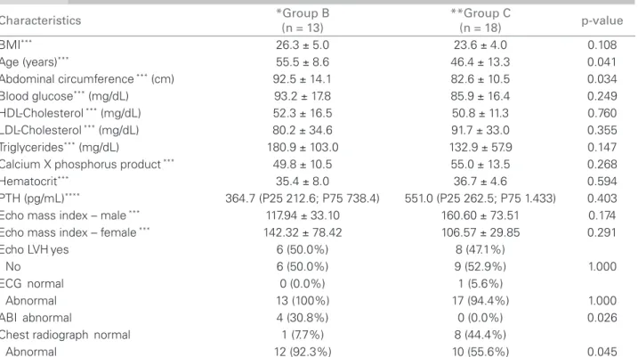
Table 4 CLINICAL, ANTHROPOMETRICANDLABORATORYCHARACTERISTICSOF GROUPS B AND C PATIENTS
Characteristics *Group B
(n = 13)
**Group C
(n = 18) p-value
BMI*** 26.3 ± 5.0 23.6 ± 4.0 0.108
Age (years)*** 55.5 ± 8.6 46.4 ± 13.3 0.041
Abdominal circumference *** (cm) 92.5 ± 14.1 82.6 ± 10.5 0.034
Blood glucose*** (mg/dL) 93.2 ± 17.8 85.9 ± 16.4 0.249
HDL-Cholesterol *** (mg/dL) 52.3 ± 16.5 50.8 ± 11.3 0.760 LDL-Cholesterol *** (mg/dL) 80.2 ± 34.6 91.7 ± 33.0 0.355 Triglycerides*** (mg/dL) 180.9 ± 103.0 132.9 ± 57.9 0.147 Calcium X phosphorus product *** 49.8 ± 10.5 55.0 ± 13.5 0.268
Hematocrit*** 35.4 ± 8.0 36.7 ± 4.6 0.594
PTH (pg/mL)**** 364.7 (P25 212.6; P75 738.4) 551.0 (P25 262.5; P75 1.433) 0.403 Echo mass index – male *** 117.94 ± 33.10 160.60 ± 73.51 0.174 Echo mass index – female *** 142.32 ± 78.42 106.57 ± 29.85 0.291
Echo LVHyes 6 (50.0%) 8 (47.1%)
No 6 (50.0%) 9 (52.9%) 1.000
ECG normal 0 (0.0%) 1 (5.6%)
Abnormal 13 (100%) 17 (94.4%) 1.000
ABI abnormal 4 (30.8%) 0 (0.0%) 0.026
Chest radiograph normal 1 (7.7%) 8 (44.4%)
Abnormal 12 (92.3%) 10 (55.6%) 0.045
BMI: body mass index; HDL: high density lipoprotein; LDL: low density lipoprotein; PTH: parathyroid hormone; Echo: echocardiography; LVH: left ventricular hypertrophy; ECG: electrocardiogram; ABI: ankle-brachial index; CAD: coronary artery disease.
was reported elsewhere.14,15 Furthermore, because
dyspnea, the main symptom reported by our patients, is frequent in patients on dialysis, due to volume over-load, its presence does not always alert the nephrolo-gist to the possibility of CAD. These peculiarities, among others, make the cardiovascular assessment of CKD patients a particularly difficult task.
In spite of the high rate of electrocardiographic abnormalities found, which is in agreement with the literature data,16 there was no significant difference
between Groups A and B. This decreases the impor-tance of electrocardiography for the cardiac assess-ment of CKD patients.
It is noteworthy that liaison between nephrologist and cardiologist is still insufficient in Brazil. This is due, in part, to cardiologists` lack of knowledge about this particular population and the difficulty to find a cardiologist to whom the patient can be referred. In fact, most large cardiac studies exclude CKD patients, further contributing to the paucity of knowledge and strategies targeting this population.
We demonstrated that the cardiac evaluation of our patients was delayed, that is, it was performed in those with PTx indication and mean dialysis time of five years. Besides, the low rate of cardioprotective drug use found indicates the need of greater liaison between cardiologist and nephrologist. Other authors have also shown a low rate of cardioprotective drug use in CKD patients.17 This fact is associated with
high early and late mortality, and inferior therapeutic
response to pharmacological and non-pharmacologi-cal interventions alike.1,2
CKD patients have both classic and uremia-related risk factors. The rate of classic risk factors, such as hy-pertension and smoking, was high, in accordance with what was reported by Gowdak et al.,9 demostrating
that the epidemiologic profile of CKD favors athero-sclerosis. However, the mean 10-year Framingham`s risk of our patients with CAD was low (5.3% for women and 6.3% for men), which does not reflect the high cardiovascular risk seen in CKD, and character-izes this population as an exception, besides confirm-ing the importance of other pro-atherosclerotic factors that are not quantified by this classic score.
The prevalence of CAD in our sample was 32.6%, a high rate, chiefly because it was mainly early CAD. In fact, our patients` mean age was 50 years. This prevalence increased to 53.6%, when those who un-derwent coronary arteriography were analyzed, which demonstrates the high CAD risk in the group selected for coronary angiography.
Because of the higher pre-test likelihood of CAD with non-invasive stratification of the CKD popula-tion, coronary angiography is indicated in high and very high-risk groups, as well as in symptomatic patients, those over the age of 50 years, diabetics, and/or those with structural alterations, such as a re-duced LVEF, even when asymptomatic.16
The high LVH rate we found confirmed the data reported by Kundhal et al.,18 who observed LVH in
Table 5 DISTRIBUTIONOFTHEVARIABLESOFTHEMALNUTRITION, INFLAMMATIONANDATHEROSCLEROSISSYNDROMEIN
GROUPS A AND B PATIENTS
Characteristics *Group A
(n = 15)
**Group B
(n = 13) p-value
Albumin*** (g/dL) (4.5 ± 0.6) (4.5 ± 0.3) 0.819
Normal (n = 13) 86.7% (n = 13) 100.0%
Abnormal (n = 2) 13.3% (n = 0) 0% 0.484
CRP*** (mg/dL) (1.6 ± 1.4) (1.4 ± 1.0) 0.746
Normal (n = 9) 60.0% (n = 9) 69.2%
Abnormal (n = 6) 40.0% (n = 4) 30.8% 0.705
ABI*** (1.1 ± 0.1) (1.0 ± 0.2) 0.053
Normal (n = 14) 100.0% (n = 9) 69.2%
Abnormal (n = 0) 0.0% n = 4 30.8% 0.153
MIA syndrome
No (n = 14) 100.0% (n = 13) 100.0%
Yes (n = 0) 0.0% (n = 0) 0.0%
-CRP: C-reactive protein; ABI: ankle-brachial index; MIA: malnutrition, inlammation and atherosclerosis; CAD: coronary artery disease.
74% of the dialysis population. There are several factors implicated in the development of LVH in the CKD population, such as arterial thickening (due to alterations of the calcium, phosphorus, PTH and vi-tamin D metabolism) and systolic hypertension (due to a chronic state of hypervolemia and anemia-related increased after-load).
Mineral and bone metabolism impairment was evident in this study, as shown by our patients' se-rum PTH levels. In fact, a PTH sese-rum level over 300 pg/mL indicates considerable hyperparathyroid-ism. Furthermore, 53.3% of Group A patients were in the PTx perioperative stage, indicating more severe secondary hyperparathyroidism in these individuals.
Our study should alert nephrologists and cardi-ologists to the fact that CKD patients on the PTx waiting list are at risk of CAD. Hence, should pa-tients being considered for PTx undergo coronary angiography? Because of our small sample size we could not answer this question. It would also be interesting to assess the impact of the PTx waiting time, as this is determinant of all the deleterious ef-fects of the PTH on the heart.
PTH directly affects muscle cells in vessels and the heart, altering the energy metabolism and leading to calcium accumulation. Hyperparathyroidism has been suggested to play a role in the pathogenesis of myo-cardial fibrosis and hypertrophy, vascular calcifica-tion, endothelium-mediated vasodilation dysfunction and alterations in the diastolic function of CKD pa-tients.19 The complexity of the alterations surrounding
the bone metabolism and vascular calcification, both in hyperparathyroidism and adynamic bone disease, in which PTH is reduced, has made it difficult to correctly associate PTH with CAD.18 Decreased and increased
PTH levels are likely to be associated with CVD. As for the MIA syndrome, which is known to be as-sociated with atherosclerosis, it was not diagnosed in any of our patients. Stenvinkel et al.20 reported malnutrition
in 44%, inflammation in 32% and carotid plaques in 72% of their patients, although an association of the three parameters occurred in only 30% of the cases. Inflammation was the parameter most frequently found in our CAD patients. Because of the results reported by Silva Júnior.,3 who demonstrated that inflammation is
the main determinant of the MIA syndrome, we cannot totally exclude the association of this syndrome with the CAD of our patients.
An abnormal ABI indicates peripheral atherosclerosis in the general population, being associated with a higher risk of CAD and carotid disease.21 Gabriel et al.21
ob-served ABI values over 1.3 to be associated with diffuse
atherosclerotic disease, with calcification of the middle layer and vascular rigidity. This phenomenon would be more frequent in high-risk groups, such as diabetics, el-ders and those with CKD. The low rate of abnormal ABI we found could be explained by the high rate of hy-perparathyroidism, once this metabolic disorder is asso-ciated with vascular calcification and increased arterial rigidity, which would then increase the ABI. We might thus have used a parameter that influenced the low diag-nostic rate of atherosclerosis in our population.
We used serum albumin to diagnose the nutritional status of our patients. Although this is not sensitive enough for the diagnosis of malnutrition, albumin mea-surement was the available exam during the study pe-riod. Hypoalbuminemia may be due to a combination of inflammation and low protein-energy intake, contrib-uting to reduced fat stores and muscle mass and being associated with the high mortality seen in patients on hemodialysis.22,23 The combination of a normal serum
albumin level and an increased body mass index (BMI) suggested a good nutritional status.
When the patients who did not undergo coronary arteriography (Group C) were assessed, we observed a younger population, with smaller abdominal cir-cumference, lower rate of chest radiograph alterations and more favorable atherosclerosis parameters (ABI), that is, patients with less severity criteria. Obviously, CAD cannot be safely ruled out in these patients, but this is less likely because of their better clinical profile in comparison with Group B patients, that is, those who underwent coronary arteriography and did not have CAD.
C
ONCLUSIONSWe confirmed the high rate of cardiovascular altera-tions, including CAD in our CKD patients, chiefly those on dialysis.
Although the MIA syndrome was not diagnosed in any patient, we were able to demonstrate the impor-tance of other uremia-related factors, such as mineral and bone metabolism disturbances, in the develop-ment of CAD.
Furthermore, we demonstrated that in spite of the high cardiovascular mortality in the CKD popula-tion, these patients are still assessed and under-treated with cardioprotective drugs, which is against common sense.
R
EFERENCES1. Canziani ME. Doenças cardiovasculares na doença renal crônica. J Bras Nefrol 2004;23:20-1.
2. Neves CL, Custódio MR, Neves KR, et al. O hiperparatireoidismo secundário e a doença cardiovascular na doença renal crônica. J Bras Nefrol 2008;30:18-22. 3. Silva Jr. ACC. Influência do transplante renal e de
polimorfismos genéticos nos níveis de proteína C-reativa em pacientes com doença renal crônica. Universidade de São Paulo [dissertation] [Internet] [cited 2009 May 13]. Available from: http://www.teses.usp.br/2008.
4. Krum H, Iyngkaran P, Lekawanvijit S. Pharmacologic management of the cardiorenal syndrome in heart failure. Current Heart Failure Reports 2009;6:105-11.
5. Gavina C. Doença renal crônica e doença aterosclerótica cardiovascular: o risco cardiorenal. Rev Port Cardiol 2010;29(Suppl):19-25.
6. Suassuna PGA, Bastos MG. Proteína C-reativa, aterosclerose e estatinas na DRCT: novas perspectivas. J Bras Nefrol 2007;29:171-81.
7. Zoccali C. Fatores de risco cardiovascular e renal tradicionais e emergentes: uma perspectiva epidemiológica. Kidney Int 2006;2:89-96.
8. Van der Zee S, Baber U, Elmariah S, et al. Cardiovascular risk factor in patients with chronic disease. Nat Rev Cardiol 2009;6:580-9.
9. Gowdak LHW, Paula FJ, Giorgi DMA, et al. Doença cardiovascular e fatores de risco cardiovasculares em candidatos a transplante renal. Arq Bras Cardiol 2005;84:156-60.
10. Nabais S, Rocha S, Costa J, et al. Prognostic impact of moderate renal dysfunction in acute coronary syndromes. Rev Port Cardiol 2008;27:303-12.
11. Francisco AR, Sousa M, Amador P, et al. Co-morbilidades médicas crônicas em doentes com síndrome coronária aguda. Rev Port Cardiol 2010;29:7-21.
12. Gowdak LHW, Paula FJ, Cesar LA, et al. Screening for significant coronary artery disease in high-risk renal transplant candidates. Coron Artery Dis 2007;18:553-8.
13. Devereux RB. Detection of left ventricular hypertrophy by M-mode echocardiography. Anatomic validation, standardization, and comparison to other methods. Hypertension 1987;9:119-26.
14. Roig EB, Betriu A, Castañer A, et al. Disabling angina pectoris with normal coronary arteries in patients undergoing long-term hemodialysis. Am J Med 1981;71:431-4.
15. Weinrauch L, D’Elia JA, Healy RW, et al. Asymptomatic coronary artery disease: angiographic assessment of diabetic evaluated for renal transplantation. Circulation 1994;58:1184-9.
16. Lima JJG, Sabbaga E, Vieira ML, et al. Coronary angiography is the best predictor of events in renal transplant candidates compared with noninvasive testing. Hypertension 2003;42:263-8.
17. Johnston N, Dargie H, Jardine A. Diagnosis and treatment of coronary artery disease in patients with chronic kidney disease. Heart 2008;94:1080-8.
18. Kundhal K, Lock CE. Clinical Epidemiology of cardiovascular disease in chronic kidney disease. Nephron Clin Pract 2005;101:47-52.
19. United States Renal Data System (URDS). Annual Data Report; 2003. The National Institutes of Health, National Institute of Diabetes and Digestive and Kidney Disease. Bethesda, MD; 2003.
20. Stenvinkel P, Heimbürger O, Paultre F, et al. Strong association between malnutrition, inflamation and atherosclerosis in chronic renal failure. Kidney Int 1999;55:1899-911.
21. Gabriel SA, Serafin PH, Freitas CEM, et al. Doença arterial obstrutiva periférica e índice tornozelo-braço em pacientes submetidos à angiografia coronariana. J Cardiovasc Surg 2007;22:49-59.
22. Freitas ATVS, Vaz IMF, Fornes NS. Estado nutricional de pacientes em hemodiálise no Hospital Universitário de Goiano (GO). J Bras Nefrol 2009;31:125-31.