Rational Structure-Based Rescaffolding
Approach to
De Novo
Design of Interleukin
10 (IL-10) Receptor-1 Mimetics
Gloria Ruiz-Gómez1☯*, John C. Hawkins1☯, Jenny Philipp2, Georg Künze3, Robert Wodtke4, Reik Löser4, Karim Fahmy2, M. Teresa Pisabarro1
*
1Structural Bioinformatics, BIOTEC TU Dresden, Tatzberg, Dresden, Germany,2Helmholtz-Zentrum Dresden Rossendorf, Institute of Resource Ecology, Dresden, Germany,3Institute of Medical Physics and Biophysics, University of Leipzig, Leipzig, Germany,4Helmholtz-Zentrum Dresden Rossendorf, Institute of Radiopharmaceutical Cancer Research, Dresden, Germany
☯These authors contributed equally to this work.
*gloriaruizgomez@biotec.tu-dresden.de(GRG);mayte@biotec.tu-dresden.de(MTB)
Abstract
Tackling protein interfaces with small molecules capable of modulating protein-protein inter-actions remains a challenge in structure-based ligand design. Particularly arduous are cases in which the epitopes involved in molecular recognition have a non-structured and discontinuous nature. Here, the basic strategy of translating continuous binding epitopes into mimetic scaffolds cannot be applied, and other innovative approaches are therefore required. We present a structure-based rational approach involving the use of a regular expression syntax inspired in the well established PROSITE to define minimal descriptors of geometric and functional constraints signifying relevant functionalities for recognition in protein interfaces of non-continuous and unstructured nature. These descriptors feed a search engine that explores the currently available three-dimensional chemical space of the Protein Data Bank (PDB) in order to identify in a straightforward manner regular architec-tures containing the desired functionalities, which could be used as templates to guide the rational design of small natural-like scaffolds mimicking the targeted recognition site. The application of this rescaffolding strategy to the discovery of natural scaffolds incorporating a selection of functionalities of interleukin-10 receptor-1 (IL-10R1), which are relevant for its interaction with interleukin-10 (IL-10) has resulted in thede novodesign of a new class of potent IL-10 peptidomimetic ligands.
Introduction
Protein-protein interactions (PPIs) mediate most biological processes and, therefore, represent relevant avenues as targets for the development of therapeutics. In order to target large pro-tein-protein interfaces with small molecules in a rational fashion, the most relevant molecular interactions in the functional ligand-receptor complex need to be identified and mimicked
a11111
OPEN ACCESS
Citation:Ruiz-Gómez G, Hawkins JC, Philipp J, Künze G, Wodtke R, Löser R, et al. (2016) Rational Structure-Based Rescaffolding Approach toDe Novo
Design of Interleukin 10 (IL-10) Receptor-1 Mimetics. PLoS ONE 11(4): e0154046. doi:10.1371/journal. pone.0154046
Editor:Narayanaswamy Srinivasan, Indian Institute of Science, INDIA
Received:December 8, 2015
Accepted:April 7, 2016
Published:April 28, 2016
Copyright:© 2016 Ruiz-Gómez et al. This is an open access article distributed under the terms of the
Creative Commons Attribution License, which permits unrestricted use, distribution, and reproduction in any medium, provided the original author and source are credited.
Data Availability Statement:All relevant data are within the paper and its Supporting Information file.
Funding:GRG thanks the Alexander von Humboldt
Foundation for a postdoctoral fellowship. The authors acknowledge support by the German Research Foundation and the Open Access Publication Funds of the TU Dresden. The funders had no role in study design, data collection and analysis, decision to publish, or preparation of the manuscript.
Competing Interests:The authors have declared
appropriately. The rational design of molecules that disrupt PPIs is particularly challenging due to the large and structurally complex nature of the interfaces involved. A decisive strategy has been the identification of key binding residues, which contribute to the majority of stabiliz-ing interactions [1–6]. In some cases, the most contributing residues at the binding interface appear organized in a continuous and well-defined manner in regular architectures (i.e. sec-ondary structures:α-helices,β-strands and turns). In these cases, the binding epitope contain-ing such key bindcontain-ing residues can be directly translated into a chemical scaffold consistcontain-ing of the same structural features (for instance, mimetics of proteinα-helices,β-hairpins or turns) [7–11]. However, in most cases, the large size, the discontinuous nature, and even the lack of well-defined secondary structure of protein-protein interfaces [12–14] do not allow a straight-forward transfer of such relevant residues directly into a small scaffold with high affinity to the targeted recognition site [2,4]. Antibody-based strategies have been widely exploited for inter-fering large and structurally complex protein interfaces [15,16]. Nevertheless, antibodies are suboptimal as drugs mainly due to their low oral bioavailability and cell permeability, slow pharmacokinetics and, sometimes, insufficient stability. From this perspective, small mole-cules, which can be engineered to target every potential protein of interest, are advantageous over biomacromolecules. In this regard, natural-like small scaffolds or peptidomimetics bear the potential of combining the benefits of both approaches: favorable pharmacokinetic proper-ties of small molecules and the convenient tailoring of biomacromolecules to disrupt protein-protein interactions. Also extensively used to mimic discontinuous recognition epitopes in pro-teins are phage display techniques, which provide with linear peptides and simple cyclic pep-tides to target protein-protein interfaces [17–19]. Moreover, small chemical scaffolds such as triazacyclophane have been used to covalently attach discontinuous binding epitopes without further spatial arrangement considerations [20,21].
A plausible strategy to successfully transfer a large protein recognition site of non-structured and discontinuous nature into a small scaffold in a straightforward manner consists on first identifying a minimum set of functionalities relevant for the molecular recognition, secondly, having in hand a small scaffold able to present the desired functionalities in a suitable three-dimensional (3D) arrangement and to accommodate all chemical features necessary for stabil-ity and good complementarstabil-ity to the targeted site so that it can achieve potent binding affinstabil-ity. Bringing all these components together constitutes a quite arduous path, and, in order to ease this process, the speedy selection of a suitable scaffold is definitively a determinant step. Once suchideal scaffoldis in place, a right design rationale strategy is crucial to bringing it into the final desired molecule effectively mimicking the targeted recognition site.
Naturally occurring scaffolds such asβ-hairpins,α-helices and structured turns of known
protein structures offer a plethora of combinations of chemical functionalities disposed in a particular manner in 3D space and, therefore, they represent a great source of such ideal molec-ular templates. Thus, protein structure repositories such as the Protein Data Bank (PDB) can be used as a source ofseeding templatesto easede novodesign strategies for tackling challeng-ing protein bindchalleng-ing epitopes.
Here, we report a minimalist computational structured-based approach to ease thede novo
considered as candidateseeding templatesto lead a rational structure-based rescaffolding design strategy to develop molecules that can effectively mimic the targeted recognition site.
We have applied this innovative and simple rescaffolding approach tode novodesign of a new class of potent interleukin 10 (10) ligands that mimic the high affinity receptor IL-10R1. This protein-receptor system constitutes a clear example of a challenging molecular interface in terms of receptor mimicry. IL-10 is a pleiotropic cytokine that plays a crucial role in modulation of immune response and pathological inflammatory processes [24–26]. Struc-turally, IL-10 consists of a symmetric homodimer of two alpha-helical domains [27], and its biological function is modulated by interactions with two cell surface receptors: IL-10R1 (high binding affinity) and IL-10R2 (low binding affinity). The high-resolution 3D crystal structure of IL-10 in complex with IL-10R1 exhibits a quite large binding interface (ca. 800 Å2) in which
the receptor side comprises several discontinuous patches of unstructured nature [28]. To our knowledge, the defiant endeavor of designing small regular architectures as IL-10R1 mimetics has not been previously accomplished. Our work represents a successful example on how to ease the path from discontinuous unstructured protein regions to small regular natural-like architectures inde novorational design of protein mimetics.
Results and Discussion
Downsizing the binding epitope to a minimal functional descriptor in 3D
The recognition of IL10-R1 by IL-10 involves 23 receptor residues (S1A and S1B Fig) [28]. The particularly discontinuous arrangement of these residues in sequence, and their quite scattered disposition in 3D space alongca. 34 Å, makes it quite challenging to assemble all theirfunc-tionalities within a small molecule able to mimic the IL-10R1 interaction to IL-10. Based on available structural data [28], residues IL-10R1Y43, IL-10R1R76, IL-10R1R96, IL-10R1S190and
IL-10R1R191have been proposed as being the most relevant for protein-receptor recognition.
Our energetic calculations (seematerials and methods) confirmed those residues as being the major contributors to the energy of binding of IL-10R1 to IL-10 (S1C Fig).
In our approach, as strategy to ease the definition of key functionalities in the targeted bind-ing site, we downsized the number of residues to be taken into account by a selection procedure based on: i) calculated residue binding energy contribution, ii) interfacial hydrogen bond net-work with IL-10, and iii) proximity in space. Based on these criteria, residues IL-10R1Y43,
IL-10R1R76and IL-10R1R96(S1 Fig), which exhibited top binding energies, an extensive
inter-facial hydrogen bond network with IL-10 and were disposed in close proximity in space (all within 12 Å), were considered as key functionalities for ourseeding templatesearch. Though
downsizing the large binding epitope to three residues would imply at first none or consider-ably low binding affinity of anyseeding templatemolecule containing them, the geometric and
functional constraints demarcated by the side chains of the selected residues would constitute the minimal3D functional descriptorsto generate regular expressions signifying relevant
func-tionalities of the targeted recognition site to be mimicked (Fig 1).
studies such as the template library TESS by using geometric hashing techniques [30] or Jess, which builds on the previous approach by using a back tracking algorithm [31]. Flexibility in the geometric constraints has been previously included as an overall threshold in the accepted RMSD between template and matches structures [32–38]. Our approach to the protein struc-ture search problem has been to develop a procedure that allows us to define structural rela-tionships between flexible sequence patterns. For this, we have taken the commonly used sequence-based PROSITE syntax [22,23] as bases to formulate a3D pattern syntax, which
allows a minimalist representation of relationships in 3D space among functionalities without the necessity of introducing any template coordinates as input file (seematerials and methods for details).
The above-mentioned3D functional descriptorsand the geometric relationships among them has been used to generate the 3D syntax query R-<2,6>-R-<4,5>-Y-, which incorpo-rates the selected key functionalities distributed in 3D as desired. Functionalities (side chains) are specified in our search interface as pseudo-points [39]. In the 3D syntax query, R-<2,6>-R describes a range of distances (2 to 6 Å) covering all possible spatial relationships among the side chains of IL-10R1R96, IL-10R1R76aand IL-10R1R76b(being R76a and R76b the two
con-formers found for residue R76 in equal occupancy in the crystal structure). The relation between the functionalities of the side chains of IL-10R1R96and IL-10R1Y43, or between one
conformer of IL-10R1R76and IL-10R1Y43are described by R-<4,5>-Y. Finally, the hyphen character“-”at the end of the R-<2,6>-R-<4,5>-Y- query specifies a cyclic pattern that rein-forces tyrosine functionalities in a way that should be placed in space between the two arginine functionalities already described by the previous syntax elements (Fig 1).
Fig 1. Template-based rescaffolding strategy.3D functional descriptorsrepresenting the side chain functionalities of selected IL-10R1 binding residues (yellow sticks) are used to define the3D pattern syntax
query R-<2,6>-R-<4,5>-Y- to search forseeding templatesin PDB. One of the best hits, used asseeding template(2ACA149-157, in green), is shown superimposed to the selected IL-10R1 relevant residues for
molecular recognition. Molecular images created with PyMOL [29].
Search and identification of seeding templates
Our approach uses a 3D pattern query and a search algorithm to scrutinize the PDB. The mil-lions of atomic coordinates collected in the PDB (ca. 115.000 experimentally obtained macro-molecular structures) and organized in different 3D topologies accommodating endless combinations of functionalities are exploited to identify small regular architectures matching the selected3D functional descriptors(seematerials and methodsfor details).
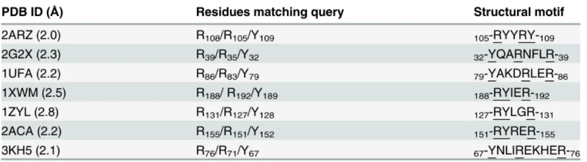
The scrutiny of the PDB by our search engine with the query R-<2,6>-R-<4,5>-Y- threw off a total of 102 matches to our3D functional descriptors(S1 Table). Only those hits contain-ing the desired descriptors in a well-defined or regular secondary structure were considered. Furthermore, we selected the shortest architectures (up to 10 residues long) and, by rejecting those that were redundant, we were left with seven candidate structural motifs (Table 1). Inter-estingly, these contained helical structures, indicating that a helix-like architecture could be considered as a suitable scaffold to achieve an appropriate 3D disposition of the required func-tionalities. Alpha-helical scaffolds are very attractive from the design point of view as they can be easily stabilized by chemical modifications and, furthermore, they offer many possibilities for substitutions [8,10,11]. The selected seven structural motifs were manually superimposed on the X-ray structure of the IL-10/IL-10R1 complex such that a maximal overlap of their func-tionalities with the selected IL-10R1 binding residues was achieved (Fig 1). Those motifs with best atomic overlapping, 2ARZ105-109, 1ZYL127-131and 2ACA151-155, were selected. These
superposition models were used to analyze which motif would provide the possibility of intro-ducing additional functionalities such that they would engage in the maximum number of complementary interactions with IL-10. In this line, we adopted a straightforward strategy con-sidering the possibility to elongate the alpha helix at the N- and C-termini. 1ZYL127-131and
2ACA151-155were disposed longitudinally with respect to the protein-receptor interface,
whereas 2ARZ105-109was transversally oriented (S2 Fig). The longitudinal orientation was able
to provide a larger number of interactions with the protein when considering the N- and C-terminal elongation of the helical scaffold. Therefore, the 1ZYL and 2ACA structural motifs were taken into account with two and five additional residues at the N- and C-termini, respec-tively, which were already in helical conformation in their respective PDB structure. The result-ing dodecameric helical scaffold X1X2R3Y4X5X6R7X8X9X10X11X12(being X any amino acid and
underlined positions those representing the 3D functional descriptors) was considered as seed-ing templatefor further design purposes. Its model in complex with IL-10 was used to investi-gate in atomic detail those positions that would allow structure-stabilizing chemical
modifications excluding positions 3, 4 and 7, which resemble the relevant functionalities of IL-10R1 for IL-10 recognition.
Table 1. Residues in PDB matching the3D pattern syntaxquery R-<2,6>-R-<4,5>-Y-.They are under-lined in their corresponding PDB structural motifs.
PDB ID (Å) Residues matching query Structural motif
2ARZ (2.0) R108/R105/Y109 105-RYYRY-109
2G2X (2.3) R39/R35/Y32 32-YQARNFLR-39
1UFA (2.2) R86/R83/Y79 79-YAKDRLER-86
1XWM (2.5) R188/ R192/Y189 188-RYIER-192
1ZYL (2.8) R131/R127/Y128 127-RYLGR-131
2ACA (2.2) R155/R151/Y152 151-RYRER-155
3KH5 (2.1) R76/R71/Y67 67-YNLIREKHER-76
Rational design strategy and experimental validation
Structure-based design of first generation IL-10R1 mimetics. Peptides of less than 15 residues derived from sequences present in protein helical domains usually lack such confor-mation when they are taken out of the stabilizing environment of the protein. Several approaches have been developed to stabilize short peptides into alpha helical conformations. The formation of covalent linkages between adjacent residues has been one of the preferred methodologies [3–5,8,10,11]. In this regard, lactam bridges between the side chains of lysine and aspartic acid in positionsi,i+ 4 have been shown to stabilize most efficiently short syn-thetic peptides into an alpha-helical structure [40]. We therefore adopted a lactam bridge design strategy. Our 12-merseeding templateX1X2R3Y4X5X6R7X8X9X10X11X12(vide supra)
offers the possibility to accommodate twoi,i+ 4 lactam bridges, which, in order to fix the desired conformation of positions 3, 4 and 7 with respect to IL-10 (i.e. in two consecutive heli-cal turns), could be introduced separately or consecutively: [K1X2R3Y4D5]
X6R7[K8X9X10X11D12] and [K1X2R3Y4D5][K6R7X8X9D10]X11X12, respectively (square brackets
represent lactam bridges between lysine (K) and aspartic acid (D) side chains) (S3A and S3B Fig). In the first scenario, the twelve residues would be constrained in helical conformation. In the second, only residues 1 to 10 would be constrained. Nevertheless, the helical conformation could be extended beyond these constraints at least by one or two more residues [41]. Based on our 3D molecular models, the first option would offer only position 11 for introducing addi-tional H-bond interactions with IL-10, whereas in the second option positions 8 and 11 would be available. In addition, our models suggested that the region of IL-10 interacting with the res-idue in position 11 (loop AB) would require certain flexibility in its counterpart. Therefore, two consecutivei,i + 4lactam bridges were introduced in the scaffold resulting in a bicyclic molecule, which was then subjected to a per residue structure-based mutagenesis process in order to achieve best binding complementarity to IL-10. Here, substitutions at positions 3, 4, 7, 8 and 11 pointed towards IL-10, whereas residues at positions 9 and 12 did not establish con-tacts with the protein, and they were therefore maintained unaltered as in the corresponding PDB template structure (2ACA149-160). Position 12 could be used to enhance helicity in the
C-term. Following this rationale, a lysine residue was introduced at position 8 to favor interac-tions with IL-10D44, and position 11 was used to introduce an arginine side chain for
interac-tions with IL-10D44and IL-10Q42(S3C Fig). In order to analyze the possibility that the four
positive charges so-far introduced in our bicyclic template could engage in non-specific electro-static interactions at other regions of IL-10, we performed energy interaction calculations with a sp2amine NH2cation and a sp3amine NH3cation chemical probes by using GRID [42]. The
most favorable energy was obtained at the predicted binding region for our template (S4A Fig), suggesting that the selected topology of charged residues would confer sufficient binding speci-ficity to the designed molecule.
[KR7K8VD]R11A-NH2(M1,Table 2,Fig 2) was refined in complex with IL-10 using MD
simu-lations (seematerials and methodsfor details). The mimetic-protein binding free energy (ΔGM-P) was estimated with the MM-PBSA method and indicated favorable electrostatic and
van der Waals contributions to the binding [43,44] (Table 2). Hydrogen bonding between both molecules was evaluated (S2 Table). In the refined model, R3participated in a hydrogen
bond with IL-10D144, Y4with IL-10K138and IL-10E142, R7with IL-10Q38and IL-10D41. K8and
R11formed hydrogen bonds with IL-10D44. Furthermore, the aliphatic moieties of the R3, Y4
and R7side chains of the mimetic showed important van der Waals contributions with the side
chains of IL-10I145and IL-10Q38. The relevance of mimetic residues Y4, R5and R7in binding to
IL-10 was further investigated by MM-PBSA alanine mutagenesis [43–45]. The substitution of these residues by alanine revealed a considerable unfavorable effect in binding for all mutants, showing the most dramatic effect for alanine mutation at position 7 (R7A,S3 Table).
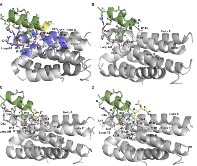
Fig 2. Structure-based design rationale.(A) Atomic representation of the model of the complex between IL-10 (gray, PDB ID 1J7V) and the helical scaffold used in the rationale for the design of IL-10R1 mimetics (green,N-terminal functionalization in yellow). For clarity, no side chains shown for IL-10Q38and
IL-10L48. (B) Snapshot of complex IL-10/M1 (at 2.9 ns from 10 ns MD simulation). (C) Snapshot of complex IL-10/M4 (at 10 ns). (D) Snapshot of complex IL-10/
M6 (at 3.6 ns). Residues involved in recognition are shown in sticks and colored by atom type (IL-10 highlighted in violet in A). Red spheres represent interfacial structural waters observed during MD simulations. Intermolecular H-bonds are depicted by black dashed lines. Figure generated in PyMOL [29].
Experimental validation of first generation IL-10R1 mimetics. M1was chemically syn-thesized, and its experimental binding affinity towards IL-10 was assessed by thermal stability measurements using circular dichroism (CD) [46] and fluorescence [47] (seematerials and methods). For these studies, we used the readily available murine IL-10 mutant mIL-10C149Yin
which Cys149 was replaced by the corresponding Tyr residue of the human protein, as it was previously shown to enhance protein refolding yield [48,49]. Furthermore, this tyrosine would enhance the sensitivity of tryptophan fluorescence measurements(i.e. as stated above, it can act as energy donor for the tryptophan residue introduced as label in the mimetics).
Human and murine IL-10 share high sequence similarity (88%), and the residues forming the mimetic binding site are conserved in both. Furthermore, the selected IL-10R1Y43,
IL-10R1R76and IL-10R1R96binding residues used for our design are fully conserved in both
human and murine receptors. The 3D structure of mouse IL-10 and its receptor are not avail-able. Therefore, in order to investigate the suitability of the use of mIL-10C149Yfor our studies,
and for comparison purposes between the murine and human proteins, atomic models of
mIL-10C149Yand mIL-10R1 were built by applying homology modeling techniques (seematerials
and methodsfor details andS5 Fig). The obtained 3D mouse models were analyzed in detail in context of the crystal structures of human IL-10 and its complex structure with IL-10R1 to investigate any structural differences, in particular at the recognition site (S5 Fig). In
mIL-10C149Y, most residues involved in the mimetic binding interface are conserved in sequence
and 3D space disposition. Residues mIL-10Q32(R in human, helix A), mIL-10T39(M in human,
helix A) and mIL-10T49(K in human, loop AB), although not conserved, were not exposed to
the mimetic and, therefore, were not expected to interfere with its binding. Residue mIL-10N141
(S in human IL-10, helix F’) is located at the recognition site of the conserved receptor residues mIL-10R1R76and mIL-10R1R96, and it could therefore establish similar interactions as in
human IL-10. mIL-10I46(L in human IL-10, loop AB) is semi-conserved and would participate
in van der Waals interactions with mIL-10R1Y43as in the human protein-receptor complex.
mIL-10D50(E in human, helix B) is also semi-conserved and could participate in additional
interactions with the N-terminally elongated IL-10R1 mimetics. Based on the sequence and structural similarity of the mimetic recognition site in the human and mouse proteins and the receptors, we concluded that mIL-10C149Ycould appropriately serve our experimental needs,
and that the designed IL-10R1 mimetics should also bind to it as predicted for the human protein.
Table 2. Mimetic-protein binding free energy obtained by MM-PBSA [43,44] and experimental dissociation constants (Kd).[a],[b]
IL-10R1 Mimetic Sequence ΔGM-P(kcal/mol)[c] K
d(μM)
M1 Ac-[KWRYD][KRKVD]RA-NH2 -27.1±6.6 >30
[a]
M2[d] X-[KWRYD][KRKVD]RA-NH2 -53.3±9.9 5.0
[a]
M3[d][e] X-[KZRYD][KRKVD]RA-NH
2 -59.9±9.2 6.7[a]
M4 Ac-R[KWRYD][KRKVD]RA-NH2 -59.2±7.0 8.4[a]
M5 Ac-EK[KWRYD][KRKVD]RA-NH2 -77.9±9.7 0.07±0.02
[b]
M6 Ac-ER[KWRYD][KRKVD]RA-NH2 -71.8±8.0 0.04±0.02
[b]
[a]K
dvalues calculated from thermal denaturation data. [b]K
dvalues inferred from ITC thermodynamic data. [c]ΔG
M-P= Mimetic-Protein binding free energy. Entropy contribution to binding was not estimated here. [d]X = 4-guanidinobutanoyl.
[e]Z = 5-Hydroxy-L-tryptophan.
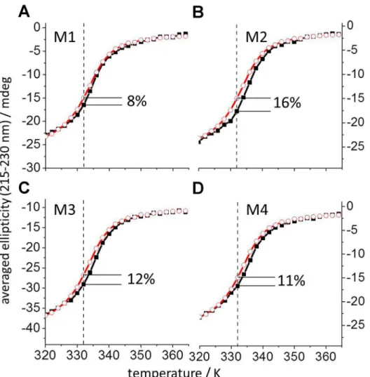
The experimentally estimated binding affinity shows thatM1binds to the protein (Table 2, Figs3and4). Complex formation betweenM1and mIL-10C149Ywas determined by thermal
denaturation using CD and the well-known negative ellipticity of IL-10 between 200 to 300 nm [40]. The average CD signal in this range of wavelengths was measured as a function of temper-ature between 298 and 363 K.Fig 3Ashows a decrease of negative ellipticity, which exhibits a sigmoidal shape typical of thermally induced protein unfolding. The obtained curves shift to higher temperatures in the presence ofM1. The shift is due to a lower degree of unfolded pro-tein at any temperature in the presence of mimeticM1as compared to the denaturation of mIL-10C149Yalone. The largest difference was observed at 332 K and is expressed in percentage
of the total unfolding signal (obtained after subtraction of the initial and final linear segments of the curves). At 332 K, the presence ofM1increased the native protein structure by 8%. Fig 3. Temperature denaturation curves obtained by CD for mIL-10C149Yin complex with IL-10R1 mimetics M1-M4.The average thermal denaturation CD signal in the 215 to 230 nm range is plotted as a function of temperature for mIL-10C149Yalone (2μM, red open circles) and for mIL-10C149Yin the presence of
each mimetic M1-M4 (4.5μM, black filled squares) in panels A-D, respectively. The obtained mIL-10C149YCD
data agree with those published earlier for human and murine IL-10 [50]. The highest amount of native protein structure binding to each of the mimetics M1-M4 was observed at 332 K (dashed lines) and is given in each corresponding panel as percentage of the total change in the CD-signal.
Fig 4. Temperature-dependent excitation spectra of the tryptophan in mimetics M1-M4.Upper plots A-D: Mimetic tryptophan emission (excited between 200 and 290 nm) measured at 360 nm for the temperature range between 298 K and 363 K for each mimetic M1-M4 in the presence of mIL-10C149Y. The
Simultaneously with the thermal stability CD-signals, the excitation spectrum of the trypto-phan residue strategically introduced at position 2 inM1was measured. The results obtained provided an additional assessment of complex formation betweenM1and mIL-10C149Y(Fig
4A). We observed that the emission at 360 nm decreased with increasing temperatures, leading to the decline of the excitation spectra between 200 and 290 nm (Fig 4Aupper plot, excitation spectra at 332 K highlighted in red). The underlying spectral differences are clearly appreciated in the representation of the subtraction of the excitation spectrum at 298 K from that at 332 K in the presence and absence of mIL-10C149Y(Fig 4Alower plot, red and blue line, respectively).
The enhanced temperature sensitivity of tryptophan fluorescence obtained in the presence of mIL-10C149Ysupports a binding interaction. The increased fluorescence emission is explained
by a restriction in movement of the indole ring at the binding interface and the energy transfer from Y149in mIL-10C149Y. Both factors would vanish upon thermal denaturation, leading to
the observed larger drop in fluorecence excitation in the presence of mIL-10C149Y. These data
therefore support the interaction ofM1with mIL-10C149Yas derived from the
CD-denatur-ation curves.
Rational optimization of first generation IL-10R1 mimetics and experimental valida-tion. Two iterative cycles ofin silicooptimization and experimental validation were carried out with the purpose of increasing the interactions betweenM1and IL-10 and, therefore, obtain potent IL-10R1 mimetics.
By comparing the structures of the refined model complex IL-10/M1and the X-ray struc-ture of IL-10/IL-10R1, it was envisaged that N-terminal functionalization ofM1could poten-tially contribute with additional complementary interactions to the protein. Indeed, in the MD simulation of the 10/10R1 complex the following interactions were observed: IL-10R1N73/IL-10E50, IL-10R1N73/IL-10L48, IL-10R1D100/IL-10R27and IL-10R1E101/IL-10K34.
Thus, based on the structure of the refined model of theM1/IL-10 complex (Fig 2A and 2B) and taking into account the interaction energies obtained with the sp3and sp2amine cation chemical probes (vide supra), two new mimetics (M2,M3; differing in tryptophanvs. its 5-hydroxy derivative in position 2) were designed by N-terminal functionalization with 4-gua-nidinobutanoyl, providing a positively charged H-bond donor for potential interaction with the negatively charged side chain of IL-10E50(helix B). The same holds true for the extension of
the N-terminus ofM1by an arginine (M4) or lysine, which could offer stabilizing interactions with the backbone of IL10L48(loop AB) and/or the side chain of IL-10Y149(helix F’). The
bind-ing affinities of IL-10R1 mimeticsM2,M3andM4, determined as described forM1(vide supra), showed a considerable improvement (Table 2,Fig 3).
As forM1, the obtained fluorescence data for mimeticsM2-M4clearly revealed enhance-ment of tryptophan emission when the mimetics were bound to the protein (Fig 4B–4D). As described above, this is explained by the immobilization of the tryptophan of the mimetics and the energy transfer from the protein tyrosine in position 149 upon complex formation, which support the protein-mimetic interaction at the expected binding site. This piece of evidence supported our design rationale.
Further ligand optimization was accomplished by the introduction of an additional N-ter-minal H-bond acceptor such as Gln or Glu (M5,M6), which would allow interactions with the cationic side chains of IL-10R27and IL-10K34(helix A) or with IL-10N148(helix F’) (Fig 2A).
Favourable energies obtained for the interaction of IL-10 with an aliphatic carboxylate chemi-cal probe [42,51] supported this rationale. Furthermore, residues IL-10L46(loop AB) and
IL-10I145(helix F’) would additionally contribute with van der Waals interactions with the
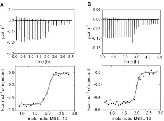
nanomolar affinities forM5andM6as determined in more detail by isothermal titration calo-rimetry (ITC, seematerials and methods) (Fig 5). Furthermore, these measurements supported the expected stoichiometry 2:1 mimetic:dimer-protein binding ratio as previously observed for the IL-10R1/IL-10 complex [28].
All in all, the step-wise increase in binding obtained computationally and experimentally for the designed molecules along our iterative rational strategy support the design rationale.
The analysis of the interfacial H-bond formation observed along the corresponding MD tra-jectories of the protein-mimetic complex series (seematerials and methods) corroborated the envisaged interaction patterns. First, the introduction of new H-bond donors at the N-termi-nus ofM1led to complementary interactions with acceptor groups of protein residues L48, E50, E142 and Y149 (M2-M4,S2 Table,Fig 2C). Second, the N-terminal extension by an addi-tional residue acting as an H-bond acceptor (M5andM6) allowed interactions with the donor groups of protein residues K34, Q38 and N148 (S2 Table,Fig 2D). The data obtained from the MD trajectories were also used to analyze the dynamic behaviour of solvent, in particular those water molecules that were participating in the binding of the mimetics to the protein. In the case of mimeticsM3andM4, a well-defined water molecule was observed bridging by H-bond their guanidinium groups in X and R-1, respectively, with the protein residues L48 and E142
(S2 Table). InM5, a water molecule located in the mimetic-protein interfacial core was observed to bridge residue Y4and protein residues D41 and S141. Bridging water molecules
were also involved in interactions between residue R3inM6and protein residue S141 (S2
Table,Fig 2D). These observations highlight the relevance of interfacial solvent for the protein-mimetic recognition, as demonstrated for other PPIs [52–54], and the importance of the Fig 5. Isothermal titration calorimetry.(A) mIL-10C149Y(43μM) with M5. (B) mIL-10C149Y(25μM) with M6.
Upper and lower panels show the raw data and integrated heat changes with the corresponding fitted binding curves based on a single site model, respectively.
solvation effect for ligand recognition [55]. Furthermore, per residue energetic contributions obtained from MM-PBSA indicated favourable hydrophobic contributions. The most relevant van der Waals interactions were observed between the aliphatic moieties of K-1, R-1, X, R3, Y4,
K8ofM2-M6and the protein residues D44, L46, D144 and I145 (S6 Fig). The aliphatic moiety
of residue R7in the mimetics also contributed to hydrophobic interactions with the protein
res-idues Q38 and Q42. The results obtained from the MM-PBSA alanine scanning carried out on the residues R3, Y4and R7in mimeticsM4andM6were consistent with the decrease in protein
binding observed for these mutations inM1(S3 Table). MimeticsM2,M3andM4 incorporat-ing one H-bond donor at the N-terminus ofM1showedKdvalues below 10μM, reflecting an enhancement of binding higher than 6-fold in the best case with respect to the leadM1. The maximal binding enhancement was obtained forM5andM6, which showed binding affinities values in the nanomolar range, corresponding to a more than 70-fold increase with respect to
M2-M4, and at least more than 400-fold with respect toM1. Thermodynamic parameters determined from ITC measurements for the most potent designed IL-10 ligands (M5andM6) indicated that their free energies of binding were mostly driven by a favourable entropic com-ponent (TΔS 9.5 kcal/Kmol) with a weak favourable enthalpic term (ΔH -0.5 kcal/mol). The observed favourable entropic contribution was attributed to the intrinsic rigid structural nature of the protein, the pre-organized helical nature of the mimetic, as described for other structur-ally constrained systems [56,57], the van der Waals interactions established between several protein and mimetic residues, and the presence of structural bridging waters involved in pro-tein-mimetic binding. The latter was further investigated by analyzing the overlap of the inter-action energy maps obtained for the unbound IL-10 (PDB ID 2ILK (1.6 Å) [27]) with a GRID
“solvent”water and a“solvent exclusion”sp3carbon chemical probes [42,54]. A favourable
energy contribution for solvent was indicative of the presence of three water molecules at the mimetic binding site, which, interestingly, overlapped with three structural waters present in the crystal structure of the free IL-10. Furthermore, the“solvent exclusion”probe provided one of the largest energetic contributions in the same region where these waters are located (S4B Fig). This analysis further indicates an entropic contribution resulting from the release of struc-turally bound water molecules upon mimetic binding.
Conclusions
Materials and Methods
3D Pattern regular expression methodology
Our methodology is inspired on the well-established PROSITE syntax commonly used in pro-tein sequence analysis [22,23], which consists of a collection of pre-defined sequence profiles (defining protein families or functional motifs) that is used to find matches in protein sequence databases. In particular, our methodology involves two technical contributions: a 3D pattern syntax and a structural patternsearch algorithm.
- The3D pattern syntaxdeveloped here uses the PROSITE pattern syntax as starting point. It containssequence patternsdefining functionalities at residue level, and a variety ofstructural connectorsdefining distance and angle ranges and their inter-relationships (Fig 6). This syntax allows multiplesequence patternsto be joined using flexiblestructural connectors. The combi-nation of all these syntax elements confers flexibility in the definition of sequence and struc-tural constraints representing desired functionalities in each individual syntax query.
Asequence patternconsists of a definition of one or a combination of amino acids. The later is represented with square brackets“[]”for allowed residues and curly brackets“{}”for for-bidden residues. The geometric relationship betweensequence patternsin three-dimensional space is established by using geometrical descriptors, which we callstructural connectors. A
structural connectoris defined in terms of a range of values: i) distances (in Angstroms) between the alpha carbons or a functional center of the side chain of amino acids, the latest denoted as pseudo atoms roughly equivalent to the center of mass of the side chain and inspired in previous work of Baharet al[39]; ii) angles (in degrees) formed by the correspond-ing peptide sequence backbones. The use of pseudo-points represents an advantage over calcu-lating all pairwise distances, which would considerably increase the calculation time (about 16 times). Distances between alpha carbons or pseudo-points and angles between alpha carbons
Fig 6.3D pattern syntax: Sequence patterns and structural connectors.
are specified in the search interface by selecting either alpha-carbons or pseudo-points for each search. Astructural connectoris denoted by a pair of angle brackets“< >”. Structural con-straints can be combined by introducing a pipe-line“|”between connectors. The connection between twosequence patternsand astructural connectoris defined by a hyphen character (i.e.
“-“). A cyclic pattern is built by adding this hyphen at the end of the last sequence pattern or structural connector.
A particular3D pattern syntaxquery defining geometric relationships of desired functional-ities is used as input by our structural pattern search algorithm.
- Thestructural patternsearch algorithmoperates across multiple stages designed to mini-mize the computational overhead of querying large protein structure datasets (i.e. the Protein Data Bank PDB) directly.
1. First, and as a pre-filtering step, the search engine utilizes a dataset of sequences (extracted from the 3D structures to be analyzed;i.e. PDB) for candidate hits matching. A candidate hit is any protein sequence that has each of the specified sequence elements or sub-sequences in the pattern. Sub-sub-sequences should not overlap.
2. A data-structure of all available sub-sequences in a given protein (often there are multiple matches for each sub-sequence) is built by considering all hits obtained in the previous step. This data-structure is then used in the next stage.
3. The engine verifies that there is at least one set of sub-sequences that matches the required structural constraints specified in the3D pattern syntaxquery. As the combinatorial space can be extremely large, the engine makes use of a recursive backtracking algorithm to mini-mize redundant computation and permit early termination when one of the required sub-sequences has been found to fail all structural constraints.
As multiples matches could be found for the same structure, and in order to improve compu-tational efficiency and to avoid large numbers of very minor variations, we add the additional constraint that multiple matches in the same structure cannot overlap any components. In addi-tion, we run the structural pattern search algorithm over a redundancy reduced data set. When matches are found, we are able to manually explore similar structures for equivalent matches.
All algorithms developed to interrogate the protein structure space in a systematic manner searching for patterns in databases have to make a trade-off between three key areas: 1) The pattern expressivity (i.e. range of functional motifs that can be captured), 2) The ease of pattern definition, 3) The computational efficiency. In our approach, we scarify some of the first fea-ture in order to achieve better performance on the latter two. Our methodology presents a number of key differences with other comparable computational techniques such as RAMOT-3D [36], ASSAM [37] and IMAAAGINE [38] and overcomes some of their limitations. Typi-cally, in these tools the search syntax does not allow for variations in the identities of residues in particular positions as our does. In our case, the patterns are inspired on the PROSITE syn-tax and therefore defined using amino acid sequence elements. As result easy and flexible pat-terns can be designed instead of using graphs. Furthermore, our patpat-terns can have arbitrary numbers of amino acids containing distance and angle tolerances described for each connec-tion instead of being globally defined. Besides, the chemical properties in our patterns are defined in our case only through sets of amino acids and not via many pre-defined sets.
Computational structure-based design and simulation
molecules. Due to the two-fold axis symmetry in this complex, only one of the two IL-10 domains and a receptor molecule were considered for our studies (S1 Fig).
Analysis of relevant binding residues in the IL-10/IL-10R1 interface. To determine the receptor residues being the major contributors to the protein binding energy, molecular dynamics calculations were carried out with the IL-10/IL-10R1 complex. AMBER11 and AMBER12 packages [58] were used for the simulations, and the MM-PBSA method [43,44] as implemented in AMBER11 was used to obtain per residue energy contributions (S1C Fig). The ff99SB force field [59] and standard protocols as implemented in the AMBER11 and
AMBER12 packages were used (see below) [58]. The IL-10/IL-10R1 complex was solvated in a truncated octahedral box of TIP3P water molecules and neutralized with Cl-counterions. Molecular dynamics simulations were preceded by two energy-minimization steps: i) only the solvent and ions were relaxed; ii) the entire system was minimized. The system was then heated up from 0 K to 300 K in 50 ps. Weak position restraints (10 kcal/molÅ2) were used for the
whole system in the canonical ensemble (NVT). Langevin temperature coupling with a colli-sion frequencyγ= 1 ps−1was used at this step. The system was equilibrated without restraints
at 300 K during 100 ps in the isothermal-isobaric ensemble (NPT) followed of other 100 ps in the canonical ensemble (NVT). The Langevin thermostate and Berendsen barostat under peri-odic boundary conditions were used during the equilibration steps. After this, a total of 20 ns MD production without restraints was carried out at 300 K in the canonical ensemble (NVT) using the Langevin thermostat. The SHAKE algorithm was used to constrain all bonds involv-ing hydrogen atoms. A time step of 2 fs was used. A cutoff of 8 Å was applied to treat the non-bonded interactions, and the Particle Mesh Ewald (PME) method was used to treat long-range electrostatic interactions. MD trajectories were recorded every 2 ps. Trajectories were visual-ized with VMD [60] and evaluated in terms of intermolecular H-bonds using the PTRAJ and CPPTRAJ modules implemented in AMBER. The criterion used to consider a dynamic hydro-gen bond formation was to be found at least 10% of the simulation time using a distance accep-tor-donor cutoff of 3.5 Å and a 120° angle cutoff.
Mimetic refinement and analysis. In each step of the lactam bridge design strategy (S3 Fig), a brief minimization was performed with MOE (Amber12:EHT force field and default parameters were used) [61]. Molecular dynamics simulations of the complex of IL-10 with each of the designed IL-10R1 mimetics were performed using the ff99SB force field [59] and standard protocols as implemented in the AMBER11 and AMBER12 packages (see below) [58]. The mimetic residues Lys and Asp involved in lactam bridges were parametrized accord-ing to the ff99SB force field, and RESP charges were derived at the HF/6-31Gcalculation level
using Gausian09 [62–64]. Same strategy was followed for parameters of the chemical groups introduced in the optimization process that were not included in the ff99SB force field. Each protein-mimetic complex was solvated in a truncated octahedral box of TIP3P water molecules and neutralized with Cl-counterions. Molecular dynamics simulations were preceded by two energy-minimization steps: i) only the solvent and ions were relaxed; ii) the entire system was minimized with low position restraints (10 kcal/molÅ2) for the helical backbone section of the
mimetic. The system was then heated up from 0 K to 300 K in 20 ps. Weak position restraints (10 kcal/molÅ2) were used for the protein and the mimetic in the canonical ensemble (NVT).
Langevin temperature coupling with a collision frequencyγ= 1 ps−1was used at this step.
Along three equilibration steps of 500 ps each, the helical backbone position restraints and also two distance restraints between protein and mimetic (IL-10Q38/MR7and IL-10K138/MY4) were
consecutively decreased (10, 5, and 2 kcal/molÅ2, respectively). This was carried out in the
300 K in the canonical ensemble (NVT) using the Langevin thermostat. The SHAKE algorithm was used to constrain all bonds involving hydrogen atoms. A time step of 2 fs was used. A cut-off of 10 Å was applied to treat the nonbonded interactions, and the Particle Mesh Ewald (PME) method was used to treat long-range electrostatic interactions. MD trajectories were recorded every 2 ps. Trajectories were visualized with VMD [60] and evaluated in terms of intermolecular H-bonds (including bridging water molecules at the interface between the designed mimetics and IL-10) and RMSD using PTRAJ and CPPTRAJ modules implemented in AMBER. The criterion used to consider a dynamic hydrogen bond formation was to be found at least 10% of the total simulation time (S2 Table). Water molecules connecting func-tionalities (bridging waters) were also considered for hydrogen bond analysis using a distance acceptor-donor cutoff of 3.5 Å and angle cutoff of 120°. A structural bridging water was consid-ered to be participating in H-bond in protein-mimetic complex when was found at least 20% of the total simulation time (S2 Table). Energy decomposition per residue and binding free energy post-processing analysis of the trajectories (Table 2) were performed in implicit solvent using the MM-PBSA method [43,44] as implemented in AMBER11. The same method was applied to study the effect of alanine mutation on mimetic residues R3, Y4and R7ofM1,M4andM6in
complex with IL-10 (S3 Table) [43–45]. Data analysis was carried out with the R-package [65]. In order to ensure that main conclusions inferred from the 10 ns MD simulations main-tained valid our strategy design, a further 20 ns extension was carried out for the optimized molecules (i.e. IL-10/M2-M6complexes,S6B Fig).
GRID calculations. GRID [42,51] version 22 was used to predict energetically favorable interactions between IL-10 (PDB ID 1J7V) and the following chemical probes: N2 = (sp2 amine NH2cation), N3+ (sp3amine NH3cation), COO- (aliphatic carboxylate), and between
the unbound IL-10 (PDB ID 2ILK) and the chemical probes: OH2 (water) and C3 (methyl CH3group) (S4 Fig). The GRID box dimensions were set to 51 Å x 58 Å x 46 Å in order to
cover the whole protein for the calculations. A grid spacing of 1 Å was applied. The rest of GRID input parameters values were used as default.
Structure-based modeling of mIL-10C149Y. The crystal structure of human IL-10 at high
resolution (PDB ID 2ILK (1.6 Å)) [27] and its complex structure with IL-10R1 (PDB ID 1J7V (2.9 Å)) [28] were taken as structural templates. Human and mouse proteins and their R1 receptors share high sequence similarities (88% and 73%, respectively). The program Modeller as implemented in Discovery Studio (Accelrys) [66] was used for the modeling. The mouse receptor was modeled following the same procedure.
Peptide synthesis
Designed IL-10R1 mimeticsM1toM4were purchased from GL Biochem (Shanghai, China) with a purity95% by analytical rpHPLC. Their purity was re-checked by rpHPLC and MS
before experimental use. MimeticsM5andM6were synthesized with purity95% by
analyti-cal rpHPLC using similar methods as previously reported [67].
All commercial reagents and solvents were used without further purification unless other-wise specified. Mass spectra (ESI) were obtained on a Micromass Quattro LC or a Waters Xevo TQ-S mass spectrometer each driven by the Mass Lynx software. Preparative HPLC was per-formed on a Varian Prepstar system equipped with UV detector (Prostar, Varian) and auto-matic fraction collector Foxy 200. A Microsorb C18 60–8 column (Varian Dynamax 250 × 21.4 mm) was used as the stationary phase and a binary gradient system of 0.1% TFA/water (solvent A) and 0.1% TFA/CH3CN (solvent B) at a flow rate of 10 mL/min served as the eluent. The
gradient back to 90% A. Analytical HPLC was done in a Merck Hitachi system consisting of a L7100 gradient pump combined with a Jasco DG2080 4-line degasser with UV absorption detection at 220 nm by a Merck Hitachi L7450 diode array detector. The system was operated by the D-700 HSM software using a Merck Hitachi D7000 interface. A Luna C18 5μm column (Phenomenex, 250×4.6 mm) was used as stationary phase and a binary gradient system of 0.1% CF3COOH/water (solvent A) and 0.1% TFA/CH3CN (solvent B) at a flow rate of 1 mL/
min served as the eluent. The programme for elution was as follows: 0–3 min 95% A, 3–25 min gradient to 95% B, 25–30 min 95% B, 30–35 min gradient back to 95% A.
IL-10R1 mimeticsM5andM6were prepared on 0.15 mmol scale by manual stepwise solid phase peptide synthesis using HBTU/DIPEA activation for Fmoc chemistry [68,69] on Fmoc-Ala-Rink Amide MBHA (substitution 0.37 mmolg-1). Fmoc-Ala-Rink Amide MBHA was
pre-pared by Fmoc-deprotection of Fmoc-Rink Amide MBHA (1 g, substitution 0.55 mmolg-1)
resin using the procedure outlined below followed by treating the deprotected Rink Amide MBHA resin with 1.0 equivalent of Fmoc-Ala-OH (0.171 g), 1.25 equivalents of HBTU (0.261 g), and 2.0 equivalents of DIPEA (187μL) in DMF (4 mL) for 2 hours. Subsequently, the resin was filtered off and washed with DMF and CH2Cl2(DCM) (3×5 mL each). Capping of the
unreacted resin-bound amino groups was accomplished by treatment with a 1:1 (v/v) mixture of DCM and acetic anhydride. The resin was shaken for 3 min, and the liquid was removed by filtration. The resin was re-suspended in the capping mixture and shaken again for 7 min. After filtration the resin was washed with DCM (3×5 mL), and the presence of remaining resin-bound amino groups was checked using the ninhydrin assay. In the case of positive result, the capping procedure was repeated [70]. Determination of the loading degree was accom-plished following the protocol described by Gudeet al. [71] using two aliquots of the resin. About 5 mg of the resin (exact value has to be noted) containing the Fmoc-Ala-OH was placed into a PP reaction vessel and swollen in DMF (2 mL) for 30 min. The suspension was filtered, and the resin was treated with 2% DBU in DMF (2 mL) and stirred for another 30 min. The suspension was filtered into a graduated 10 mL flask, and the resin was washed with CH3CN
(3×1 mL). Each filtrate was added to the flask and, the solution was diluted to 10 mL with CH3CN. 2 mL of this solution were transferred to a graduated 25 ml flask and diluted with
CH3CN to 25 mL. A reference solution was prepared the same way but without addition of the
resin. The sample solutions were measured against the reference at 294 nm in the UV/Vis spec-trometer. The loading yield was calculated as follows: Loading (mmol/g)294 nm=
(E×14.214μmol) / mresin(mg). Finally, the average value of the two aliquots was calculated.
Using the procedure outlined above, loading degrees in the range of 0.37–0.44 mmol/g were obtained.
Fmoc deprotections were achieved with 2 × 5 min treatments with DMF/Piperidine (1:1). For each coupling, 4.0 equivalents of amino acid and 4.2 equivalents of diisopropylethylamine (DIPEA) were employed in each coupling step (20–25 min), except for Fmoc-Asp(OAll)-OH and Fmoc-Lys(Alloc)-OH, where only 2 equivalents were coupled using HATU and DIPEA activation. Coupling yields were monitored by quantitative ninhydrin assay [72]. After the first Fmoc-Lys(Alloc)-OH residue was coupled, the resin was washed with DMF/DCM, and swollen in DCM. Then, the Ally and Alloc orthogonal protecting groups were removed by treating the peptide resin with PhSiH3in DCM (20 equiv) and Pd(PPh3)4(0.15 equiv.) (2 × 20 min) under
peptide resin was then washed with DMF, MeOH/DCM, DCM, and driedin vacuofor 1 hour. Peptide was cleaved by using 95% TFA, 2.5% TES, 2.5% H2O (6 mL) for 2 hours. The resin was
then filtered off and the solvent evaporated under a stream of nitrogen. The peptide was precip-itated with cold diethyl ether. The ether was decanted, and the precipprecip-itated peptide was re-dis-solved in 1:1 acetonitrile/water and lyophilized. Crude peptide was purified by reverse-phase HPLC and assessed for purity by using the conditions specified above. IL-10R1 mimeticM5:Rt
13.0 min, ESMS observed (calculated): [M+2H]2+= 942.18 (942.03), [M+3H]3+= 628.29
(628.36), [M+4H]4+= 471.49 (471.52); IL-10R1 mimeticM6:Rt11.9 min, ESMS observed
(cal-culated): [M+2H]2+= 956.25 (956.04), [M+3H]3+= 637.83 (637.69), [M+4H]4+= 478.67
(478.52).
Protein expression and purification
Recombinant murine IL-10 (rmIL-10) was expressed inEscherichia coli(E.coli) Rosetta(DE3) (Merck) using a pET41b(+) vector (Novagen). For purification purposes, the C-terminal histi-dine tag (LEHHHHHHHH) of the vector was used. The rmIL-10 coding region was further modified by replacement of cysteine 149, which is not involved in disulfide bond formation, by a tyrosine,i.e., the corresponding amino acid in the human IL-10 sequence. That modification improved protein refolding yield by a factor of 20 as previously shown for rat IL-10 but without any loss in activity [48]. The C149Y mutation was introduced according to the instructions of the QuikChange mutagenesis kit (Stratagene). The primers for the introduction of the C149Y mutation were: forward5’-CTTCATCAACTACATAGAAGC-3’and reverse complementary 5’-GCTTCTATGTAGTTGATGAAG-3’. Correctness of the rmIL-10 coding region and intro-duction of the point mutation were confirmed by DNA sequencing.
E.colicells were grown at 37°C in a minimal salt medium containing 9.29 g/L sodium sul-fate, 29.2 g/L di-potassium hydrogenphosphate, 8.14 g/L sodium dihydrogenphosphate dihy-drate, 1.2 g/L magnesium sulfate, 5 g/L glucose, 1 g/L ammonium chloride, 4.92 g/L
ammonium sulfate, 100μg/mL thiamin, 100μg/mL kanamycin, and 34μg/mL chlorampheni-col. Protein expression was induced with 1 mM isopropyl-β-D-thiogalactopyranoside (IPTG)
and was conducted for 4 hours. The cell pellet was resuspended in 50 mM Tris-HCl (pH 7.4), 100 mM NaCl, 1 mM EDTA, 0.1 mM PMSF, and cells were lysed by using a french press and by adding 0.3 mg/mL lysozyme. rmIL-10 was obtained inE.coliinclusion bodies which were washed three times in 50 mM Tris-HCl (pH 7.4), 1 M NaCl, 5% (v/v) Triton X-100, 1 mM EDTA and finally dissolved in 50 M Tris-HCl (pH 7.4), 100 mM NaCl, 6 M guanidine hydro-chloride, 200 mM dithiothreitol, 1 mM EDTA. Prior to refolding of rmIL-10, dithiothreitol was removed by dialysis against 100 mM Tris-HCl (pH 9.0), 100 mM NaCl, 6 M guanidine hydrochloride. Protein refolding was initiated by rapid dilution into 100 mM Tris-HCl (pH 9.0), 100 mM NaCl, 1 M L-arginine, 5 mM reduced glutathione, 1 mM oxidized glutathione until a final protein concentration of 0.1 mg/mL was reached. Protein aggregates were immedi-ately removed by filtration of the protein solution through a 0.45μm nitrocellulose membrane. Refolding was allowed to further proceed for two days.
rmIL-10 was confirmed by NMR spectroscopy including the resonance assignments of the pro-tein backbone and Cβside chain atoms [49].
Circular Dichroism (CD) and Fluorescence Spectroscopy
CD spectra were recorded with a J-815 instrument (Jasco, Gross-Umstadt, Germany) from 195 to 300 nm in 1-cm cuvettes at concentrations of 2μM of mIL-10C149Y(dimer) and 4.5μM of
mimeticsM1-M4. Ellipticityθwas recorded in millidegrees. Samples were heated from 298 to 364 K at a rate of 2 K/min under constant stirring to determine stabilization of IL-10/mimetic complexes. Estimates of binding affinities were derived with the assumption of a two state unfolding of mIL-10C149Y. The mimetics were assumed to stabilize exclusively the folded state.
These simplifications allow applying equilibrium thermodynamics, such that the fraction Snof
the native state of mIL-10C149Ycan be approximated: Sn= (1+ [M]Kd-1+Ku)-1.Kdis the
esti-mated dissociation constant of the mimetic, [M] its concentration.Kuis the temperature-dependent unfolding equilibrium constant (Ku= [denatured]/[native]) as measured by the
unfolding curve of mIL-10C149Yin the absence of mimetics. This model provides a relative
ranking of mimetic affinities but does not allow for a more detailed analysis with respect to absolute binding enthalpy and entropy for which partially unfolded states need to be included. In this case, the measured CD curves would be actually compatible with much higher mimetic affinities, because a local stabilization of the mimetic binding epitope on mIL-10C149Yis not
counted as binding in the applied two state model but it is very likely to occur. For consistency, we have not explored the higher affinity limit that would still be compatible with the data. Instead, we report only the lower affinity range that complies with the assumptions, in order to prevent bias for overestimated mimetic affinities. Due to the complicated underlying processes of thermally induced complex dissociation and unfolding of both, mIL-10C149Yand designed
mimetics, we cannot relate the shift of the temperature mid point of unfolding (Tm) to more specific thermodynamic parameters by a simple two state unfolding model. Absolute thermo-dynamic parameters could only be obtained by isothermal titration calorimetry with the most promising mimetics. This of course required the initial confirmation of the binding of the pre-cursor molecules which was accomplished by CD and Fluorescence Spectroscopy. Fluorescence emission at 360 nm was monitored simultaneously with the CD scan (the measuring beam served as the excitation light) using a second monochromator and photomultiplier in 90° geometry with respect to the CD absorption path. Thereby, temperature-dependent excitation spectra of the tryptophan at position two in the synthetic IL-10R1 mimetics were additionally obtained with the CD data acquisition. The thermally induced spectral differences between 334 K and 298 K were calculated for IL-10R1 mimetics in the absence and presence of
mIL-10C149Y.
Isothermal titration calorimetry (ITC)
ITC was performed with a Microcal VP-ITC calorimeter (GE Healthcare, Buckinghamshire, U. K.) at 298 K. Aliquots (5–10μL) of synthetic IL-10R1 mimeticsM5andM6(300–500 mM) dissolved in the original dialysis buffer of the mIL-10C149Ypreparation were injected into
mIL-10C149Ysolutions (19–50μM) until the molar ratio of mimetic: mIL-10C149Yexceeded 3 and
Supporting Information
S1 Fig. Cartoon representation of the crystal structure of the complex of human IL-10 (in gray/violet) and IL-10R1 (in brown/yellow) (PDB ID 1J7V, 2.9 Å) and energetic details of IL-10R1 residues involved in IL-10 binding.(A) Two molecules of IL-10R1 bind to two iden-tical two-fold related surface areas of the protein composed of helix A, loop AB and helix F’of one IL-10 domain. (B) Close-up of the protein-receptor interface. The recognition site involves 27 protein residues (in violet) and 23 receptor residues (in yellow). Selected key binding resi-dues of IL-10R1 (Tyr43, Arg76 and Arg96) and their binding counterparts in IL-10 are shown in sticks, labeled and colored by atom type. The two red spheres represent interfacial crystallo-graphic water molecules interacting with the key binding residues. Intermolecular H-bonds are depicted by black dashed lines. Panels A and B were generated with PyMOL. (C) MM-PBSA IL-10R1 residue binding energy contribution calculated from MD simulation.
(PDF)
S2 Fig. Cartoon and surface representation of an IL-10 domain (in gray) in complex with the manually docked selected structural motifs that best matched the defined 3D functional descriptors (Table 1in main text).Motifs 1ZYL, 2ACA (A), and 2ARZ (B) are represented by a green ribbon, and their helical elongation is depicted in yellow. Those motif residues overlap-ping with the selected IL-10R1 key binding residues (i.e. IL-10R1 residues Tyr43, Arg76 and Arg96 in orange) are shown in sticks. The helix axis (represented by a black line) is oriented longitudinally (A) or transversally (B) with respect to the helices A, B, F’of IL-10.
Figure generated with PyMOL. (PDF)
S3 Fig. Lactam bridge design strategy to stabilize theseeding templatein an alpha-helical scaffold.IL-10 and the scaffolds are represented as gray and green cartoons, respectively. Rele-vant residues in each of the protein/scaffold complex are labeled and shown in sticks. The scaf-folds Ac-[K1X2R3Y4D5]X6R7[K8X9X10X11D12]-NH2(A), Ac-[K1X2R3Y4D5][K6R7X8X9D10]
X11X12-NH2(B) and Ac-[K1X2R3Y4D5][K6R7K8X9D10]R11X12-NH2(C) and their respective
180° rotation view are shown. Lactam bridges between the side chains of Lys and Asp are repre-sented by brackets in sequence and in sticks in the structure models. Residue positions available for substitutions in the scaffolds are highlighted with spheres at their Cα. Figure generated with
PyMOL. (PDF)
S4 Fig. Interaction energy maps obtained for IL-10 with a series of GRID chemical probes.
(A) Interaction energies calculated for one IL-10 domain from the structure in complex with the receptor-1 (in gray cartoon, PDB ID 1J7V (2.9 Å)) with an N3+ probe (sp3amine NH
3
cat-ion). Two contour energy levels are shown at cutoffs -7.5 kcal/mol and -13.5 kcal/mol (cyan and blue, respectively). The most favorable interactions with IL-10 involve residues from helix A (Gln38) and loop AB (Asp41, Gln42) (highlighted in pink). Further favorable interactions in the same region comprise residues of the loop AB (Asp44) and helix F’(Ser141, Glu142, Asp144) (highlighted in violet). (B) Interaction energies calculated for the unbound IL-10 (PDB ID 2ILK (1.6 Å)) with a water molecule or“solvent”chemical probe (contour energy level at cutoff -7.5 kcal/mol shown in red) and for a carbon sp3or“solvent exclusion”chemical probe (contour energy level at cutoff -2.8 kcal/mol shown in green). Favorable interactions of both,solventandsolvent exclusionprobes, overlap with the mimetic binding region indicating solvent exclusion. Three crystallographic waters found in the free IL-10 in this region are shown as yellow spheres. Figure generated in VMD.
S5 Fig. Molecular modeling of murine IL-10 mutant mIL-10C149Y.Top: sequence alignment
of murine and human proteins used for the homology modeling (sequence numbering as in PDB ID 2ILK). Bottom: detail of one domain of the obtained 3D molecular model of
mIL-10C149Y(beige cartoon) superimposed with one domain of the human IL-10 used as template
(PDB ID 2ILK, gray cartoon) (RMSDCα: 0.8 Å). The protein residues involved in mimetic
rec-ognition are shown in sticks (pink for murine and violet for human. Figure generated with PyMOL.
(PDF)
S6 Fig. Surface representation of a domain of IL-10 (in gray) in complex with mimetic M5 (in green, N-terminal functionalization in yellow) and RMSD along MD simulation.(A) Hydrophobic interactions between mimetic residues K8, Y4, K-1and protein residues D44, L46
and I145 are highlighted with labels. Structure taken from a snapshot of the MD simulation. Figure generated with PyMOL. (B) Relative movement of mimetic M5 (given as backbone RMSD) with respect to IL-10 along a 30 ns MD simulation.
(PDF)
S1 Table. List of motifs in the PDB database that match the3D pattern syntaxquery R-<2,6>-R-<4,5>-Y-.Matches corresponding to short regular secondary structures (maximum 10 residues long in the same chain) are highlighted in bold.
(PDF)
S2 Table. Summary of dynamic H-bond formation between IL-10 and IL-10R1 mimetics M1-M6 along MD simulations.M1: Ac-[KW2R3Y4D][KR7K8VD]R11A-NH2, M2:
X-[KWR3Y4D][KR7K8VD]R11A-NH2(X = 4-guanidinobutanoyl), M3: X-[KZR3Y4D]
[KR7K8VD]R11A-NH2(X = 4-guanidinobutanoyl, Z = 5-Hydroxy-L-tryptophan), M4: Ac-R -1[KWR3Y4D][KR7K8VD]R11A-NH2, M5: Ac-E-2K-1[KWR3Y4D][KR7K8VD]R11A-NH2, M6:
Ac-E-2R-1[KWR3Y4D][KR7K8VD]R11A-NH2.#H-bonds observed along MD simulations of 10
ns for IL-10/M1 and 30 ns for IL-10/M2-M6 are shown in black. Additional H-bonds observed during last 10 ns MD production for IL-10/M2-M6 are shown in gray.Interactions via
struc-tural bridging water. S: side chain. M: main chain.aNot observed during the last 10 ns MD pro-duction.
(PDF)
S3 Table. Binding free energies for alanine mutants of M1, M4 and M6 calculated with the MM-PBSA alanine scanning method.
(PDF)
Acknowledgments
We are most indebted to Axel Roers, Sebastian Lanvermann, Stephan Theisgen, Daniel Huster and Sandra Franz for materials, invaluable support and scientific discussions. We would also like to thank Jose C. Martinez Herrerías for his valuable suggestions during the reviewing pro-cess of this manuscript.
Author Contributions
References
1. Clackson T, Wells JA. A hot-spot of binding-energy in a hormone-receptor interface. Science. 1995; 267(5196):383–6. PMID:7529940
2. Wells JA, McClendon CL. Reaching for high-hanging fruit in drug discovery at protein-protein inter-faces. Nature. 2007; 450(7172):1001–9. PMID:18075579
3. Wilson AJ. Inhibition of protein-protein interactions using designed molecules. Chem Soc Rev. 2009; 38(12):3289–300. doi:10.1039/b807197gPMID:20449049
4. Milroy L-G, Grossmann TN, Hennig S, Brunsveld L, Ottmann C. Modulators of Protein-Protein Interac-tions. Chem Rev. 2014; 114(9):4695–748. doi:10.1021/cr400698cPMID:24735440
5. Nevola L, Giralt E. Modulating protein-protein interactions: the potential of peptides. Chem Commun. 2015; 51(16):3302–15.
6. Pelay-Gimeno M, Glas A, Koch O, Grossmann TN. Structure-Based Design of Inhibitors of Protein-Pro-tein Interactions: Mimicking Peptide Binding Epitopes. Angew Chem Int Ed. 2015; 54(31):8896–927; Angew Chem 127(31), 9022–54.
7. Robinson JA. beta-Hairpin Peptidomimetics: Design, Structures and Biological Activities. Acc Chem Res. 2008; 41(10):1278–88. doi:10.1021/ar700259kPMID:18412373
8. Ross NT, Katt WP, Hamilton AD. Synthetic mimetics of protein secondary structure domains. Philos Trans R Soc A-Math Phys Eng Sci. 2010; 368(1914):989–1008.
9. Robinson JA. Protein epitope mimetics as anti-infectives. Curr Opin Chem Biol. 2011; 15:379–86. doi: 10.1016/j.cbpa.2011.02.015PMID:21419690
10. Azzarito V, Long K, Murphy NS, Wilson AJ. Inhibition of alpha-helix-mediated protein-protein interac-tions using designed molecules. Nat Chem. 2013; 5(3):161–73. doi:10.1038/nchem.1568PMID: 23422557
11. Hill TA, Shepherd NE, Diness F, Fairlie DP. Constraining cyclic peptides to mimic protein structure motifs. Angew Chem Int Ed. 2014; 53(48):13020–41; Angew Chem 126(48): 13234–257.
12. Janin J, Bahadur RP, Chakrabarti P. Protein-protein interaction and quaternary structure. Q Rev Bio-phys. 2008; 41(2):133–80. doi:10.1017/S0033583508004708PMID:18812015
13. Teyra J, Paszkowski-Rogacz M, Anders G, Pisabarro MT. SCOWLP classification: Structural compari-son and analysis of protein binding regions. BMC Bioinformatics. 2008; 9.
14. Teyra J, Hawkins J, Zhu H, Pisabarro MT. Studies on the inference of protein binding regions across fold space based on structural similarities. Proteins: Struct Funct Bioinf. 2011; 79(2):499–508. 15. Chon JH, Zarbis-Papastoitsis G. Advances in the production and downstream processing of antibodies.
N Biotechnol. 2011; 28(5):458–63. doi:10.1016/j.nbt.2011.03.015PMID:21515428
16. Putnam WS, Prabhu S, Zheng Y, Subramanyam M, Wang Y-MC. Pharmacokinetic, pharmacodynamic and immunogenicity comparability assessment strategies for monoclonal antibodies. Trends Biotech-nol. 2010; 28(10):509–16. doi:10.1016/j.tibtech.2010.07.001PMID:20691488
17. Sidhu SS, Lowman HB, Cunningham BC, Wells JA. Phage display for selection of novel binding pep-tides. Method Enzymol. 2000; 328:333–63.
18. Sidhu SS, Koide S. Phage display for engineering and analyzing protein interaction interfaces. Curr Opin Struc Biol. 2007; 17(4):481–7.
19. Omidfar K, Daneshpour M. Advances in phage display technology for drug discovery. Expert Opin Drug Discov. 2015; 10(6):651–69. doi:10.1517/17460441.2015.1037738PMID:25910798
20. Hijnen M, van Zoelen DJ, Chamorro C, van Gageldonk P, Mooi FR, Berbers G, et al. A novel strategy to mimic discontinuous protective epitopes using a synthetic scaffold. Vaccine. 2007; 25(37–38):6807– 17. PMID:17689841
21. Mulder GE, Kruijtzer JAW, Liskamp RMJ. A combinatorial approach toward smart libraries of discontin-uous epitopes of HIV gp120 on a TAC synthetic scaffold. Chem Commun. 2012; 48(80):10007–9. 22. Sigrist CJA, Cerutti L, Hulo N, Gattiker A, Falquet L, Pagni M, et al. PROSITE: a documented database
using patterns and profiles as motif descriptors. Brief Bioinform. 2002; 3(3):265–74. PMID:12230035 23. Sigrist CJA, de Castro E, Cerutti L, Cuche BA, Hulo N, Bridge A, et al. New and continuing
develop-ments at PROSITE. Nucleic Acids Res. 2013; 41(D1):E344–E7.
24. Sabat R, Gruetz G, Warszawska K, Kirsch S, Witte E, Wolk K, et al. Biology of interleukin-10. Cytokine Growth F R. 2010; 21(5):331–44.

![Table 2. Mimetic-protein binding free energy obtained by MM-PBSA [43, 44] and experimental dissociation constants (K d )](https://thumb-eu.123doks.com/thumbv2/123dok_br/18289471.346441/8.918.57.866.133.271/table-mimetic-protein-binding-obtained-experimental-dissociation-constants.webp)