ssDNA Viruses from Insectivorous Bats in
Southern Brazil
Francisco Esmaile de Sales Lima1*, Samuel Paulo Cibulski1,2, Helton Fernandes dos Santos2, Thais Fumaco Teixeira2, Ana Paula Muterle Varela2, Paulo Michel Roehe1,2, Eric Delwart3, Ana Cláudia Franco1
1Virology Laboratory, Department of Microbiology, Immunology and Parasitology, Institute of Basic Health Sciences, Federal University of Rio Grande do Sul (UFRGS), Porto Alegre, Rio Grande do Sul (RS), Brazil,2
FEPAGRO Animal Health, Institute of Veterinary Research "Desidério Finamor" (IPVDF), Eldorado do Sul, Rio Grande do Sul (RS), Brazil,3Blood Systems Research Institute, San Francisco, California, United States of America; University of California, Department of Laboratory Medicine, San Francisco, California, United States of America
Abstract
Circovirusesare highly prevalent porcine and avian pathogens. In recent years, novel circu-lar ssDNA genomes have recently been detected in a variety of fecal and environmental samples using deep sequencing approaches. In this study the identification of genomes of novel circoviruses and cycloviruses in feces of insectivorous bats is reported. Pan-reactive primers were used targeting the conservedrepregion of circoviruses and cycloviruses to screen DNA bat fecal samples. Using this approach, partialrepsequences were detected which formed five phylogenetic groups distributed among theCircovirusand the recently proposedCyclovirusgenera of theCircoviridae. Further analysis using inverse PCR and Sanger sequencing led to the characterization of four new putative members of the family Circoviridaewith genome size ranging from 1,608 to 1,790 nt, two inversely arranged ORFs, and canonical nonamer sequences atop a stem loop.
Introduction
Viruses of theCircoviridaefamily are known to infect a wide range of vertebrates. The virions consist of naked nucleocapsids of about 20 nm in diameter, with a circular single stranded DNA (ssDNA) genome of approximately 2.0 kb [1]. They have an ambisense genome organi-zation containing two major open reading frames (ORFs) inversely arranged, responsible for encoding the replicase (Rep) and capsid (Cap) proteins, and are separated by a 3’intergenic re-gion (IGR) between the stop codons and a 5’IGR between the start codons [2]. Some circo-viruses are major pathogens of pigs [3–5], e.g. porcine circovirus 2 (PCV2) which causes either asymptomatic infections or clearly apparent disease which may be responsible for significant economic losses [6–10]. In birds, avian circoviruses, within the genusGyrovirus, have been
OPEN ACCESS
Citation:Lima FEdS, Cibulski SP, dos Santos HF, Teixeira TF, Varela APM, Roehe PM, et al. (2015) Genomic Characterization of Novel Circular ssDNA Viruses from Insectivorous Bats in Southern Brazil. PLoS ONE 10(2): e0118070. doi:10.1371/journal. pone.0118070
Academic Editor:Jean-Pierre Vartanian, Institut Pasteur, FRANCE
Received:September 11, 2014
Accepted:January 4, 2015
Published:February 17, 2015
Copyright:© 2015 Lima et al. This is an open access article distributed under the terms of the
Creative Commons Attribution License, which permits unrestricted use, distribution, and reproduction in any medium, provided the original author and source are credited.
Data Availability Statement:All relevant data are within the paper.
identified in a broad range of avian species; one of them, chicken anemia virus (CAV), is a major cause of disease, associated to lymphoid depletion, immunosuppression and develop-mental abnormalities [11–15]. According to the document 2014.006a-gV from ICTV, there is a proposal of theGyrovirusgenus removal fromCircoviridaetoAnelloviridaefamily due to re-cent metagenomic studies on gyroviruses showing a very high sequence divergence when com-pared to other circoviruses members.
Recent metagenomic approaches, high-throughput sequencing techniques and degenerate PCRs have led to the identification of small circular DNA genomes in fecal samples of wild mammals, in insects as well as from environmental samples [2,16–18]. Some of the newly de-scribed circular genomes are similar to those of circoviruses, but phylogenetically different from the previously known avian and porcine circoviruses [18]. Their distinct nucleotide/ amino acid composition and genome organization allowed authors to propose the creation of a new genus within theCircoviridae, which was namedCyclovirus. In comparison to members of the genusCircovirus, bothrepandcapcycloviruses genes are smaller, with shorter or no 3’IGR between the stop codons of the two major ORFs and a longer 5’IGR between the start codons of the two major ORFs [2].
Sequences related to circoviruses have been identified based on the detection of the con-served Rep region involved in rolling circle replication (RCR) [19].Cyclovirusgenomes were detected in wild animal’s samples, human feces and cerebrospinal fluids; muscular tissues of farm animals such as chickens, cows, sheep, goats, and camels [20,21]. Currently, eight differ-ent species of cycloviruses have been detected in winged-insect populations highlighting they circulate in a wide host range possessing a high genetic diversity, as well [18–20,22–24].
So far, classification for the genusCircovirusconsiders circoviruses sharing>75%
genome-wide nucleotide identity and>70% amino acid identity in the capsid (Cap) protein to the same
species. Although, there are no species demarcation criteria for the genusCyclovirus, the taxo-nomic classification for the familyCircoviridaeconsiders viruses sharing>60% in their Cap
amino acid identity level as belonging to distinct genera [19].
In the present article, the detection of ssDNA genomes from bat fecal samples is reported. Genome segments were amplified by consensus/degenerate PCR. Whole genome sequencing and phylogenetic analyses of the sequences obtained revealed that four of the sequences repre-sent viral genomes of new members of the familyCircoviridae.
Materials and Methods
Ethics Statement
Permission for this work on protected bats was granted by Health Monitoring (CEVS—Centro Estadual de Vigilância em Saúde) of the Brazilian federal state of Rio Grande do Sul. The study did not involve any direct manipulations of bats and relied entirely on collection of fecal sam-ples from the attic floor. All experiments were performed in compliance with the European Convention for the Protection of Vertebrate Animals Used for Experimental and Other Scien-tific Purposes (European Treaty Series—No. 170 revised 2005) and the procedures of the Bra-zilian College of Animal Experimentation (COBEA). It must be highlighted that we had the owner’s permission to access the attic for the purposes of this study. In case of future surveys in Porto Alegre, the Health Monitoring (CEVS) will be contacted to obtain the permissions.
Sample collection and preparation
A maternity roost of bats was identified in the summer of 2012 in the attic of a private resi-dence in the central area of the municipality of Porto Alegre, Rio Grande do Sul, Southern Bra-zil. The colony was estimated to harbor about 500 bat specimens of insectivorous bats of two
species, velvety free tailed bats (Molossus molossus) and brazilian free tailed bats (Tadarida bra-siliensis) [25]. Speciation was confirmed by DNA extraction from fecal pellets, amplification and sequencing of the mitochondrial cytochrome b (cytb) gene as described [26].
One hundred fecal samples were collected from the attic floor as follows: a plastic film was spread on the ground of the attic compartment and fresh droppings were collected with clean disposable forks in the following night. Each sample consisted of pool of 5 fecal droppings, which were immediately sent to the laboratory and stored at -80°C. The samples were then thawed, resuspended and in 1 mL of Hank’s balanced salt solution (HBSS), vortexed and cen-trifuged at 10.000 xgfor 5 min. The supernatants were then transferred to fresh tubes, filtered through 0.45μm pore-size syringe filters (Fisher Scientific, Pittsburgh, PA) and submitted to
DNA extraction.
DNA extraction, PCR and sequencing
Total fecal DNA was extracted from 400μL of the supernatants (above) with
phenol-chloro-form (Invitrogen) [27]. The extracted DNA was eluted in 50μL of TE (Tris-hydrochloride
buffer, pH 8.0, containing 1.0 mM EDTA), treated with 20μg/mL of RNase A (Invitrogen) and
stored at -80°C. Subsequently, samples were submitted to amplification in a nested-PCR target-ing therepgene of circoviruses/cycloviruses with the following degenerate primers: CV-F1 (5´-GGIAYICCICAYYTICARGG-3´), R1 (5`-AWCCAICCRTARAARTCRTC-3`), CV-F2 (5´-GGIAYICCICAYYTICARGGITT-3´), and CV-R2 (5´-TGYTGYTCRTAICCRTCCC ACCA-3´) [2]. Briefly, the nested PCR was performed as follows: the first reaction was per-formed in a 25μL volume containing 20 to 50 ng of sample DNA 1 mM MgCl2, 0.2μM of each
primer (CV-F1 and CV-R1), 1.5 U Taq DNA polymerase (Invitrogen), 10% PCR buffer and 0.6 mM dNTPs. The cycling conditions were: 5 min at 95°C; 40 cycles of 1 min at 95°C, 1 min at 52°C, 1 min at 72°C and a final incubation at 72°C for 10 min. For the second (nested) reac-tion, the 25μL mix components were: 1μL of the 1streaction product, 1 mM MgCl2, 0.2μM of
each primer (CV-F2 and CV-R2), 1.5 U Taq DNA polymerase (Invitrogen), 10% PCR buffer and 0.6 mM dNTPs. The cycling conditions were: 5 min at 95°C; 40 cycles of 1 min at 95°C, 1 min at 56°C, 1 min at 72°C, and a final incubation at 72°C for 10 min. Products with a size of approximately 400 bp were purified and directly sequenced using primer CV-R2. To confirm the sequences, each product was sequenced three times. Samples were sequenced with the Big Dye Terminator Cycle Sequencing Ready Reaction (Applied Biosystems, UK) in an ABI-PRISM 3100 Genetic Analyzer (ABI, Foster City, CA), according to the protocol of the manu-facturer. Sequences similar to therepgene sequences of circovirus-like-genomes were aligned for designing of new sets of primers to perform the inverse PCR (iPCR). The iPCR were carried out in a 25μL reaction mixture optimized with Platinum Taq Hi-Fi (Invitrogen™) (cycling
primers as described above. The full-length sequence of genomes was constructed by“genome walking”using the Geneious software (version 7.1.3).
Gene identification and phylogenetic analysis
Identification of putative ORFs was made with aid of ORF Finder (NCBI;http://www.ncbi. nlm.nih.gov/gorf/gorf.html). Sequence analyses were performed with the BLASTX software (http://www.ncbi.nlm.nih.gov/blast/). Nucleotide sequences were aligned and compared to se-quences of human, animal and sewage-associated members of theCircoviridaeavailable at GenBank database using ClustalW [28]. The alignments were optimized with the BioEdit Se-quence Alignment Editor Program version 7.0.9 [29]. The hairpin and stem-loop structures were identified in Mfold [30]. Phylogenetic analysis was carried out in MEGA5 [31]. The confi-dence of each branch in the phylogeny was estimated with bootstrap values calculated from 2000 replicates. For the purpose of this work, the samples were named Bat Circovirus Porto Alegre (BatCV POA), followed by the cluster number to which each one was assigned.
Results
Molecular detection and genetic diversity of circovirus-like rep
sequences in feces of insectivorous bats
Amplicons with the expected size (about 400 bp) were obtained from 24 out of the 100 (24%) fecal samples screened. The amplified DNA was direct sequenced. The nucleotide sequences corresponding to part of therepgene were determined and submitted to GenBank
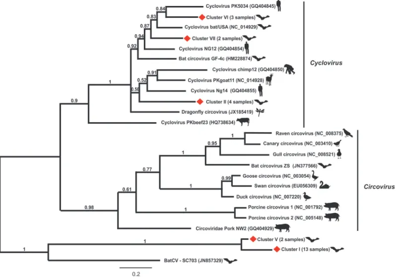
(KM401658-KM401681). BLASTX analysis showed that these partialrepsequences have an amino acid identity of 10–76% with those of known circoviruses and 87–100% among them-selves. A phylogenetic tree was constructed based on the alignment of the deduced amino acid sequences herein detected with those of the representativeCircovirusandCyclovirussequences (Fig. 1). As shown in the tree, it was observed the arrangement of five main groups with clusters II (4 sequences), VI (3 sequences) and VII (2 sequences) falling into the clade of cycloviruses, in contrast to clusters I (13 sequences) and V (2 sequences) that formed distinct and distant groups from those formed by circoviruses and cycloviruses. The arbitrary division of these se-quences in clusters was carried out to analyze their genomic features, assuming that according to the criteria used forCircovirusdiversity analysis, distinct species comprising more than>20%
sequence divergence are considered to be classified as an individual viral [32]. According to this, we could infer the detection of five potential new species from bat samples (3 cycloviruses and 2 circoviruses).
Genomic characterization of the new putative circovirus-like sequences
in insectivorous bats
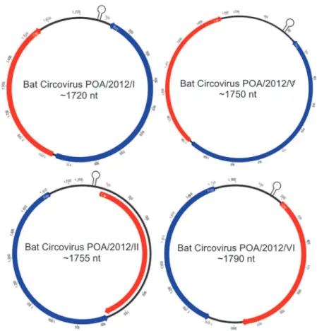
Two full-length circular ssDNA genomes of 1,755 and 1,790 nt (named BatCV POA/2012/II and BatCV POA/2012/VI, respectively) and two nearly complete circular DNA genomes of 1,720 and 1,750 nt (named BatCV POA/2012/I and BatCV POA/2012/V) were identified (GenBank accession numbers: KM382269-KM382272). It was not possible to amplify the ge-nome comprising those of cluster VII.
The predicted two ORFs,repandcap, are present and inversely arranged in all sequences as shown inFig. 2. The predicted CAP protein sequences consist of 197–231 amino acids and share an amino acid identity of 24–76% with the known cycloviruses/circoviruses and 15.5– 88.8% among themselves (Tables1and2). The predicted REP protein sequences ranged from 232 to 280 amino acid and have an amino acid identity ranging from 9.2–44.4% among them-selves (Tables1and2).
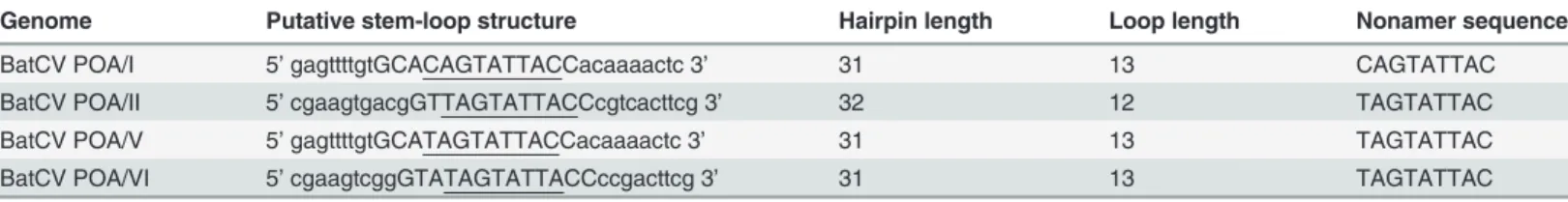
Stem-loop structures were found in all 4 bat circular genomes. They have a conserved nona-nucleotide motif located at the 5’IGR (NANTATTAC) and are considered to be responsible for initiating the rolling-cycle replication of circoviruses [18,34]. As shown inTable 3, all four BatCV POA also contain a conserved nonamer sequence in the loop region of the 5’IGR, dif-ferent from the conservedCyclovirusandCircovirusnonanucleotide motif sequence, but simi-lar to the loop motif of cycloviruses found on bat, human and chimpanzee feces (BatCV POA II, V, VI) and slightly modified from those ofCyclovirusandCircovirus(BatCV POA I) [2,17,18,20].
Fig 1. Phylogenetic analysis of partial REP protein sequences obtained from pooled bat fecal samples compared to representative members of Circoviridaefamily.The twenty-four REP sequences of bat-sourced circoviruses formed five clusters, labelled by red diamonds from I–VII. The evolutionary history was inferred by using the maximum likelihood method based on the Poisson correction model. Evolutionary analyses were conducted in MEGA5 [31].
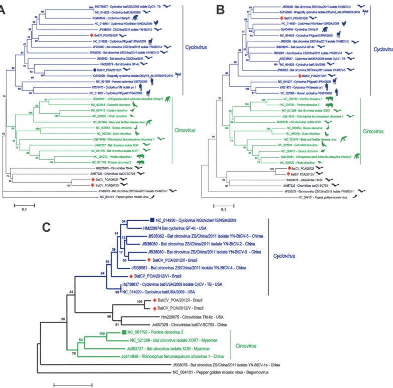
The predicted protein sequences encoded by ORF2 (cap) and ORF1 (rep) of BatCV I-VI ge-nomes were used for phylogenetic analysis with representative and recently discovered circo-viruses/cycloviruses; Pepper golden mosaic virus was used as outgroup, as they are somewhat related to other members in theCircoviridaefamily (Fig. 3A,3Band3C). As shown in the trees, BatCV POA/2012/II and VI fell into the cyclovirus clade already identified in chickens, chimps, bats, goats, humans and dragonflies [2,17,18,20,22]. When analyzing thecap-encoding region (Fig. 3A), BatCV POA/2012/II was related to aCyclovirusdetected in muscle tissues of a
Fig 2. Predicted genome organization of the four circular ssDNA sequences with the locations of the potential stem-loop structures recovered from bat feces in Southern Brazil.The two inversely arranged ORFs responsible for encoding the putatives replication associated protein (REP) and capsid protein (CAP) are shown in blue and red boxes, respectively.
doi:10.1371/journal.pone.0118070.g002
Table 1. Main features of BatCV POA genomes.
Genome (nt*) Cap Rep
Position (nt) Strand Size (nt) Size (aa) Position (nt) Strand Size (nt) Size (aa)
BatCV POA/I 1720 967–1560 + 594 197 953–120 - 834 277
BatCV POA/II 1755 1676–981 - 696 231 142–984 + 843 280
BatCV POA/V 1750 643–47 + 597 198 657–1490 - 834 277
BatCV POA/VII 1790 1583–924 - 660 219 88–786 + 699 232
*The nucleotide sizes of partial genomes I and V (GenBank accession numbers KM382269 and KM382271, respectively) were estimated by ImageJ. Genomes II (KM382270) and VI (KM38272) were completely sequenced.
goat from Pakistan through degenerate/consensus PCR [20], and BatCV POA/2012/VI was more related to dragonflyCyclovirusdetected through viral metagenomics [22]. However, when analyzing both genomes according to the conservedrep-encoding region, it was observed that they formed a monophyletic clade (Fig. 3B). On the other hand, BatCV POA/2012/I and V fell outside theCircovirusandCyclovirusclades, not yet related to any genus ofCircoviridae
family along with Bat circovirus-like virus TM6 and batCV-SC703 [17,18]. This situation was confirmed based on the alignments of the whole genomes, producing a similar tree topology (seeFig. 3C). These sequences are closer to sequences detected in guano and fecal samples col-lected from bats in the United States and China through metagenomic approaches, suggesting that these viruses have the same host origin, likely from bats [17,18]. However, currently, no classification has been fully considered to these sequences.
Discussion
In this work we report the discovery of 4 novel circular ssDNA genomic sequences from insec-tivorous bats feces from Brazil. In the recent years, many genomes of circoviruses, cycloviruses andrep-containing circular DNA viruses have been characterized in mammals, birds, insects and environmental samples [19] bringing to light a high level of genetic diversity among these viruses [19,35]. According to our results, two genomes belong to genusCyclovirus(BatCV POA II and VI). These genomes are organized and contain two major ORFs in opposite direc-tions, presenting in their 5’IGR of therepORF the cyclovirus-conserved nonanucleotide motif (5’-TAATACTAT-3’) in their loop region (Table 3). BatCV POA I and V present theircap lo-cated in the positive strand and the largerreplocated on the minus strand, as expected for cir-coviruses, but this pattern was not present in BatCV POA II and VI, as shown inTable 1. The phylogenetic analysis constructed based on the alignments of the complete REP and CAP pro-tein confirms that BatCV POA/II and VI cluster into the genusCyclovirusalong with the Chi-nese cycloviruses sequences clade detected in bat feces [18] and sharing less than 65% of identity at the CAP/REP amino acid level. BatCV POA I and V had a low amino acid identity with CAP (<20%) and REP (<10%) sequences of two other sequences detected in bat feces in
this study with known circoviruses/cycloviruses (Table 2). Consequently, they formed a
Table 2. Pairwise comparison of the BatCVs POA I, II, V and VI based on amino acid identities (%) shared by CAP and REP proteins.
Cap protein Rep protein
Genomes BatCV POA/I BatCV POA/II BatCV POA/V BatCV POA/VI BatCV POA/I BatCV POA/II BatCV POA/V BatCV POA/VI
BatCV POA/I - 15.5 88.8 17.5 - 30.4 9.2 21.4
BatCV POA/II 15.5 - 15.9 33.9 30.4 - 31.8 44.4
BatCV POA/V 88.8 15.9 - 17.5 9.2 31.8 - 20.7
BatCV POA/VI 17.5 33.9 17.5 - 21.4 44.4 20.7
-doi:10.1371/journal.pone.0118070.t002
Table 3. Organization and genomic features of the potential stem-loop structures found in the four BatCV/POA genomes.
Genome Putative stem-loop structure Hairpin length Loop length Nonamer sequence
BatCV POA/I 5’gagttttgtGCACAGTATTACCacaaaactc 3’ 31 13 CAGTATTAC
BatCV POA/II 5’cgaagtgacgGTTAGTATTACCcgtcacttcg 3’ 32 12 TAGTATTAC
BatCV POA/V 5’gagttttgtGCATAGTATTACCacaaaactc 3’ 31 13 TAGTATTAC
BatCV POA/VI 5’cgaagtcggGTATAGTATTACCccgacttcg 3’ 31 13 TAGTATTAC
distinct clade along with other bat-sourced sequences, expanding the view of diversity in these new ssDNA viruses that are divergent enough at the sequence level that they could very likely be part of a different genus.
Fig 3. Phylogenetic analysis of the complete CAP (A), REP (B) and complete genomes (C) fromCircovirusandCircovirus-like genomes identified in mammals, birds and insects.Host denomination is demonstrated after each retrieved sequence from GenBank with their accession numbers and clustered along, according to genus classification. The sequences identified in bat feces from Southern Brazil are labeled by red diamonds. Evolutionary analyses were conducted in MEGA5 [31].
In our study, we detectedCyclovirusandCircovirusrelated sequences at a frequency of 24% in the examined samples. However, due to methodological limitations, restriction in location and variety of bat species, we were not able to extrapolate our results to epidemiological data (such as incidence and prevalence) or to which bat species the ssDNA positive samples be-longed. As performed by Ge et al. in China [36], further investigation is needed to determine the prevalence of circoviruses in other Brazilian bat species. Nevertheless, it becomes clear that such study is worthy to understand the great diversity of circoviruses found worldwide.
Our study was based on the phylogenetic analysis and comparison to the sequences recov-ered. The finding of known insect viruses in bat feces simply reflects the diet of these insectivo-rous bats, which play an important role on predating insects. Viral DNA detection in bat feces does not allow one to differentiate between viral replication in bats or simple passage through the digestive track from ingested food [20,35].
To date, few members of theCircovirusgenera can be related to severe clinical conditions in animals, with the exception of PCV2 and some of the avian circoviruses [5]. Even with the re-cent discovery of many cycloviruses, circoviruses-like or rep-like sequences in a variety of mammals tissues and feces, including humans fecal samples [20,36,37], there is no syndrome yet associated with these viruses. Nevertheless, a recent identification of a newCyclovirusfrom Vietnamese and Malawi patients with acute central nervous system infection of unknown etiol-ogy raises the possibility of disease association, yet to be proven [38,39], although possibly with limited geographic distribution [38].
In this work, two more circular DNA genomes were characterized which did not fall within the circo/cycloviruses clade grouping instead distantly with TM6 and batCV-SC703 [17,18] both also from bat feces. These new genomes have in common the presence in the Rep N-ter-minus of the same motifs associated with rolling circle replication (FTLNN, TPHLQGY) and dNTP-binding (GXGKS), as well as the conserved identified in the carboxy half of Rep amino acid motifs associated with 2C helicase function (WWDGY and DRYP) [19]. The N-terminal regions related to Cap proteins of BatCV POA I and V are highly basic and arginine-rich, as is typical for circoviruses capsid proteins with arginine residues ranging from 36%-42% (Genome I and V, respectively) along the first 50 aa, in contrast to TM6 (28%) and SC703 (26%). They are also distinguishable based on their CAP and REP sizes (data not shown), as well as on the low amino acid level for both proteins, as the percentage of amino acid identity of BatCV POA I and V shows a REP identity<45% and<35% for CAP identity in relation to TM6 and
SC703. Based on these genomes characteristics, even though they are clustered in a separate clade, not yet characterized, they are new viral species. Upon the discovery of other sequences grouping along with these genomes, it will be of interest to propose the creation of a new genus withinCircoviridaeby the International Committee on Taxonomy of Viruses (ICTV).
Here we report the detection of four novel circular ssDNAs from bat feces after whole-ge-nome characterization within the familyCircoviridae. So far, it is not clear if these new ssDNA detected have some important role on pathogenesis. In addition to bioinformatics analysis, fu-ture investigations must include attempts in virus isolation to confirm host origin, which will give some light to better understand the relationships between these circular DNA viruses and bats.
Author Contributions
References
1. King AMQ, Adams MJ, Carstens EB, Lefkowitz EJ (2012) Virus Taxonomy: Classification and Nomen-clature of Viruses. USA: Elsevier. doi:10.1016/j.mjafi.2012.08.017PMID:25609874
2. Li L, Kapoor A, Slikas B, Bamidele OS, Wang C, et al. (2010) Multiple diverse circoviruses infect farm animals and are commonly found in human and chimpanzee feces. J Virol 84: 1674–1682. doi:10. 1128/JVI.02109-09PMID:20007276
3. Darwich L, Segales J, Mateu E (2004) Pathogenesis of postweaning multisystemic wasting syndrome caused by Porcine circovirus 2: An immune riddle. Arch Virol 149: 857–874. PMID:15098103
4. Ellis J, Hassard L, Clark E, Harding J, Allan G, et al. (1998) Isolation of circovirus from lesions of pigs with postweaning multisystemic wasting syndrome. Can Vet J 39: 44–51. PMID:9442952
5. Firth C, Charleston MA, Duffy S, Shapiro B, Holmes EC (2009) Insights into the evolutionary history of an emerging livestock pathogen: porcine circovirus 2. J Virol 83: 12813–12821. doi:10.1128/JVI. 01719-09PMID:19812157
6. Allan GM, Ellis JA (2000) Porcine circoviruses: a review. J Vet Diagn Invest 12: 3–14. PMID:10690769
7. Segales J, Calsamiglia M, Olvera A, Sibila M, Badiella L, et al. (2005) Quantification of porcine circo-virus type 2 (PCV2) DNA in serum and tonsillar, nasal, tracheo-bronchial, urinary and faecal swabs of pigs with and without postweaning multisystemic wasting syndrome (PMWS). Vet Microbiol 111: 223– 229. PMID:16289542
8. Chae C (2005) A review of porcine circovirus 2-associated syndromes and diseases. Vet J 169: 326– 336. PMID:15848776
9. Opriessnig T, Meng XJ, Halbur PG (2007) Porcine circovirus type 2 associated disease: update on cur-rent terminology, clinical manifestations, pathogenesis, diagnosis, and intervention strategies. J Vet Diagn Invest 19: 591–615. PMID:17998548
10. Grau-Roma L, Fraile L, Segales J (2011) Recent advances in the epidemiology, diagnosis and control of diseases caused by porcine circovirus type 2. Vet J 187: 23–32. doi:10.1016/j.tvjl.2010.01.018 PMID:20211570
11. Bassami MR, Berryman D, Wilcox GE, Raidal SR (1998) Psittacine beak and feather disease virus nu-cleotide sequence analysis and its relationship to porcine circovirus, plant circoviruses, and chicken anaemia virus. Virology 249: 453–459. PMID:9791035
12. Hattermann K, Schmitt C, Soike D, Mankertz A (2003) Cloning and sequencing of Duck circovirus (DuCV). Arch Virol 148: 2471–2480. PMID:14648300
13. Todd D, Weston JH, Soike D, Smyth JA (2001) Genome sequence determinations and analyses of novel circoviruses from goose and pigeon. Virology 286: 354–362. PMID:11485403
14. Todd D (2000) Circoviruses: immunosuppressive threats to avian species: a review. Avian Pathol 29: 373–394. doi:10.1080/030794500750047126PMID:19184829
15. Stewart ME, Perry R, Raidal SR (2006) Identification of a novel circovirus in Australian ravens (Corvus coronoides) with feather disease. Avian Pathol 35: 86–92. PMID:16595298
16. Blinkova O, Rosario K, Li L, Kapoor A, Slikas B, et al. (2009) Frequent detection of highly diverse vari-ants of cardiovirus, cosavirus, bocavirus, and circovirus in sewage samples collected in the United States. J Clin Microbiol 47: 3507–3513. doi:10.1128/JCM.01062-09PMID:19794058
17. Li L, Victoria JG, Wang C, Jones M, Fellers GM, et al. (2010) Bat guano virome: predominance of die-tary viruses from insects and plants plus novel mammalian viruses. J Virol 84: 6955–6965. doi:10. 1128/JVI.00501-10PMID:20463061
18. Ge X, Li J, Peng C, Wu L, Yang X, et al. (2011) Genetic diversity of novel circular ssDNA viruses in bats in China. J Gen Virol 92: 2646–2653. doi:10.1099/vir.0.034108-0PMID:21795473
19. Rosario K, Duffy S, Breitbart M (2012) A field guide to eukaryotic circular single-stranded DNA viruses: insights gained from metagenomics. Arch Virol 157: 1851–1871. PMID:22760663
20. Li L, Shan T, Soji OB, Alam MM, Kunz TH, et al. (2011) Possible cross-species transmission of circo-viruses and cyclocirco-viruses among farm animals. J Gen Virol 92: 768–772. doi:10.1099/vir.0.028704-0 PMID:21177928
21. Tan le V, van Doorn HR, Nghia HD, Chau TT, Tu le TP, et al. (2013) Identification of a new cyclovirus in cerebrospinal fluid of patients with acute central nervous system infections. MBio 4: e00231–00213. doi:10.1128/mBio.00231-13PMID:23781068
23. Padilla-Rodriguez M, Rosario K, Breitbart M (2013) Novel cyclovirus discovered in the Florida woods cockroach Eurycotis floridana (Walker). Arch Virol 158: 1389–1392. doi:10.1007/s00705-013-1606-x PMID:23358613
24. Dayaram A, Potter KA, Moline AB, Rosenstein DD, Marinov M, et al. (2013) High global diversity of cycloviruses amongst dragonflies. J Gen Virol 94: 1827–1840. doi:10.1099/vir.0.052654-0PMID: 23596268
25. Lima FE, Campos FS, Kunert Filho HC, Batista HB, Carnielli P Jr., et al. (2013) Detection of Alphacoro-navirus in velvety free-tailed bats (Molossus molossus) and Brazilian free-tailed bats (Tadarida brasi-liensis) from urban area of Southern Brazil. Virus Genes 47: 164–167. doi: 10.1007/s11262-013-0899-xPMID:23504146
26. Drexler JF, Gloza-Rausch F, Glende J, Corman VM, Muth D, et al. (2010) Genomic characterization of severe acute respiratory syndrome-related coronavirus in European bats and classification of coronavi-ruses based on partial RNA-dependent RNA polymerase gene sequences. J Virol 84: 11336–11349. doi:10.1128/JVI.00650-10PMID:20686038
27. Sambrook J, Russell DW (2001) Molecular Cloning: A Laboratory Manual: Cold Spring Harbor Labora-tory Press. PMID:25506954
28. Larkin MA, Blackshields G, Brown NP, Chenna R, McGettigan PA, et al. (2007) Clustal W and Clustal X version 2.0. Bioinformatics 23: 2947–2948. PMID:17846036
29. Hall TA. BioEdit: a user-friendly biological sequence alignment editor and analysis program for Win-dows 95/98/NT; 1999. pp. 95–98.
30. Zuker M, Jacobson AB (1995) "Well-determined" regions in RNA secondary structure prediction: analy-sis of small subunit ribosomal RNA. Nucleic Acids Res 23: 2791–2798. PMID:7544463
31. Tamura K, Peterson D, Peterson N, Stecher G, Nei M, et al. (2011) MEGA5: molecular evolutionary ge-netics analysis using maximum likelihood, evolutionary distance, and maximum parsimony methods. Mol Biol Evol 28: 2731–2739. doi:10.1093/molbev/msr121PMID:21546353
32. Biagini P, Bendinelli M, Hino S, Kakkola L, Mankertz A, et al. (2012) Circoviridae. In: King AMQ, Adams MJ, Cartens E.B., Lefkowitz EJ, editors. Virus Taxonomy, IXth Report of the International Committee for the Taxonomy of Viruses. UK: Elsevier/Academic Press,. pp. 343–349.
33. Rijsewijk FA, Dos Santos HF, Teixeira TF, Cibulski SP, Varela AP, et al. (2011) Discovery of a genome of a distant relative of chicken anemia virus reveals a new member of the genus Gyrovirus. Arch Virol 156: 1097–1100. doi:10.1007/s00705-011-0971-6PMID:21442232
34. Cheung AK (2006) Rolling-circle replication of an animal circovirus genome in a theta-replicating bacte-rial plasmid in Escherichia coli. J Virol 80: 8686–8694. PMID:16912316
35. Delwart E, Li L (2012) Rapidly expanding genetic diversity and host range of the Circoviridae viral family and other Rep encoding small circular ssDNA genomes. Virus Res 164: 114–121. doi:10.1016/j. virusres.2011.11.021PMID:22155583
36. Abe C, Uto Y, Nakae T, Shinmoto Y, Sano K, et al. (2011) Evaluation of the in vivo radiosensitizing ac-tivity of etanidazole using tumor-bearing chick embryo. J Radiat Res (Tokyo) 52: 208–214. PMID: 21436611
37. Acosta-Leal R, Bryan BK, Rush CM (2010) Host effect on the genetic diversification of beet necrotic yel-low vein virus single-plant populations. Phytopathology 100: 1204–1212. doi: 10.1094/PHYTO-04-10-0103PMID:20649415
38. Le VT, de Jong MD, Nguyen VK, Nguyen VT, Taylor W, et al. (2014) Limited geographic distribution of the novel cyclovirus CyCV-VN. Sci Rep 4: 3967. doi:10.1038/srep03967PMID:24495921