O
h
r
c
i
r
g
a
in
e
a
s
l
R
e
Olcay Ünver, Büşra Kutlubay, Gülten Thomas, Nilüfer Eldeş Hacifazlioğlu, Güneş Sağer, Dilşad Türkdoğan Çocuk Nöroloji BD, Çocuk Sağlığı ve Hastalıkları ABD, Marmara Üniversitesi, İstanbul, Türkiye
Acute Cerebellar Ataxia
Postinfectious Acute Cerebellar Ataxia in Childhood
Çocukluk Çağında Postenfeksiyöz Akut Serebellar Ataksi
DOI: 10.4328/JCAM.4337 Received: 24.01.2016 Accepted: 24.02.2016 Printed: 01.09.2016 J Clin Anal Med 2016;7(5): 652-5
Corresponding Author: Olcay Ünver, Fevzi Çakmak Mah. Muhsin Yazıcıoğlu Cad. No: 10 Üst Kaynarca, Pendik, İstanbul, Türkiye. T.: +90 2166254545 F.: +90 2166254580 E-Mail: [email protected]
Özet
Amaç: Postenfeksiyöz akut serebellar ataksi çocukluk çağı ataksilerinin en sık sebebidir. Olgular çocuk acil servis veya çocuk nöroloji polikliniklerine ani başlangıçlı ataksi ile başvururlar. Varisella virüsü en sık ilişkilendiren virüs-tür. Bu çalışmanın amacı postenfeksiyöz akut serebellar ataksi nedeniyle iz-lenen çocuklarda klinik özellikleri, etiyoloji ve prognozu belirlemek ve çocukluk çağında akut ataksiye genel bir yaklaşım önermektir. Gereç ve Yöntem: Ocak 2011-Haziran 2015 tarihleri arasında başvurmuş 16 çocuğun dosyaları geri-ye dönük olarak incelendi. Bulgular: Dokuz olgu erkekti (%56,2). Olguların ço-ğunluğunun yaşı 2-5 yaş aralığındaydı (%62,5). Olguların %87,5’inde bulgula-rın ortaya çıkmasından önce viral enfeksiyon öyküsü mevcuttu: Çocuklabulgula-rın iki-sinde suçiçeği enfeksiyonu, birinde Epstein Barr enfeksiyonu geri kalanlarda nonspesifik ateşli hastalık saptandı. Ateşli hastalık ve bulguların ortaya çıkışı arasındaki ortalama süre 7,4 (±5) gündü. Ortalama hastane yatış süresi 4,37 (± 1,4) gündü. Ortanca iyileşme süresi 7 gündü. En uzun iyileşme süresi 4 aydı. Tartışma: Çocukluk çağında postenfeksiyöz akut serebellar ataksi, ani başla-yan aylar içinde iyileşme gösteren benign bir durumdur. Ancak akut serebel-lar ataksinin bir dışlama tanısı olduğu unutulmamalı; bu çocukserebel-larda labora-tuar testleri ve görüntüleme incelemelerinin uygun kullanımı ile santral sinir sistemi enfeksiyonu ve kitle lezyonu gibi daha ciddi medikal durumların ayı-rıcı tanısı mutlaka yapılmalıdır.
Anahtar Kelimeler
Postenfeksiyöz Akut Serebellar Ataksi; Çocuk; Serebellit; Varisella Enfeksiyo-nu; Serebellar Ataksi
Abstract
Aim: Postinfectious acute cerebellar ataxia is the most common cause of childhood ataxia. Cases present with acute onset of ataxia to pediatric emer-gency and pediatric neurology clinics. Varicella zoster is the most commonly associated virus. The aim of this study is to assess the clinical features, eti-ology, and prognosis of children with postinfectious acute cerebellar ataxia and to propose a diagnostic approach to acute cerebellar ataxia in children. Material and Method: Files of 16 children admitted between January 2011 and June 2015 were retrospectively evaluated. Results: Nine patients were male (56.2%). The majority of the cases were in the 2-5 years age group (62.5%). A history of a preceding febrile infection was noted in 87.5% of the cases: Two children had varicella infection, one Epstein Barr infection, and the rest nonspeciic febrile illness. The mean time interval between the prodromal febrile illness and the onset of the symptoms was 7.4 (±5) days. The mean time of hospitalization was 4.37 (± 1.4) days. The median time for recovery was 7 days. The longest time for recovery was 4 months. Discus-sion: Postinfectious acute cerebellar ataxia in childhood is the most common cause of childhood ataxia, which presents abruptly and requires recovery over weeks. However, it should be kept in mind that it is a diagnosis of exclusion. Appropriate utilization of laboratory tests and imaging studies is necessary for the diferential diagnosis from other serious causes of acute cerebellar ataxia including central nervous system infections and mass lesions.
Keywords
Postinfectious Acute Cerebellar Ataxia; Children; Cerebellitis; Varicella Infec-tion; Cerebellar Ataxia
Acute Cerebellar Ataxia
2
Introduction
Acute ataxia is a relatively common presentation to pediatric emergency departments or pediatric neurology clinics. It is characterized by motor incoordination of fewer than 72-hours duration in a previously healthy child and is usually most prominently seen in the child’s movements, such as walking and picking up objects. [1]. Acute cerebellar ataxia is the most common cause of acute ataxia in childhood, accounting for 30-50% of all cases [2]. It is characterized by the sudden onset of ataxia following a viral infection, usually varicella [2,3]. How-ever, other infectious agents including Epstein Barr virus (EBV), mumps, Legionella pneumophila, hepatitis A, inluenza, herpes simplex, enterovirus, parvovirus B19, rubeola, and mycoplasma pneumoniae are also associated with acute cerebellar ataxia [1,4]. Acute cerebellar ataxia usually results from postinfectious cerebellar demyelination; it less commonly occurs as a result of direct infection of the cerebellum. Postinfectious cerebellar demyelination is thought to be an autoimmune phenomenon in-cited by infection or immunization [5]. The diferential diagnosis of acute cerebellar ataxia is broad. It is a diagnosis of exclusion ater other serious conditions including posterior fossa tumors, neuroblastoma (opsoclonus-myoclonus syndrome), acute hem-orrhage, drug intoxications, acute labyrinthitis, and metabolic diseases (Hartnup disease, Maple syrup urine disease) have been ruled out [2]. In this study, we aim to analyze all cases of acute cerebellar ataxia presented to our pediatric neurology clinic in order to characterize the clinical features, etiology, and prognosis of the disease and to propose a diagnostic approach to acute ataxia in children.
Material and Method
In this descriptive study, the iles of the children diagnosed with acute cerebellar ataxia admitted between January 2011 and June 2015 to our pediatric neurology clinic were examined for age, sex, etiology, accompanying neurological indings, labora-tory and imaging indings, hospitalization time, healing time and follow-up time ater hospital discharge. A total of 16
pa-tients were included in the study. The diagnosis of acute cer-ebellar ataxia was based on the following criteria: acute-onset loss of coordination or gait diiculties with or without nystag-mus, lasting fewer than 72 hours in a previously healthy child, and the absence of known genetic disorders presenting with ataxia, drug intoxication, bacterial meningitis, episodic ataxia syndromes, and metabolic disorders. The local ethics commit-tee approved the study.
Results
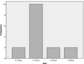
The study included 9 males (56.25%). Mean age at presentation was 4.5 years (±3.06). The youngest patient was 1 year of age and the oldest patient was 13. The demographic and clinical characteristics of the patients are presented in Table 1. Ten of the included cases were in the 2-5 year age group (Figure 1). A febrile illness preceded the onset of symptoms in 14 of the cases. This illness was a nonspeciic febrile illness in 11 of the cases. Two patients had a preceding varicella infection whereas one patient had a preceding EBV infection. The mean time in-terval between the prodromal febrile illness and the onset of
Figure 1. The distribution of cases according to age
Tablo 1. Demographic and clinical characteristics of the patients Patient
number
Gender Age Etiology Prodrome
(days)
Associated neurological Symptoms
Lumbar Puncture Protein levels (mg/dl)
Cranial Imaging
Treatment Recovery
(days)
1 male 2.5 Nonspeciic febrile illness 5 - 15,50 MRI - 7
2 male 3,5 Nonspeciic febrile illness 7 dysarthria 14.20 MRI - 5
3 female 4 Nonspeciic febrile illness 2 nystagmus - MRI - 4
4 male 2.5 Nonspeciic febrile illness 5 dysarthria, dysmetria 22 MRI - 7
5 male 1.75 Nonspeciic febrile illness 10 - - MRI - 8
6 female 9.5 Nonspeciic febrile illness 7 dysarthria - MRI - 21
7 male 5.5 Nonspeciic febrile illness 2 nystagmus - MRI - 7
8 female 2 - - nystagmus - MRI - 10
9 female 4 EBV 14 - 19 MRI - 10
10 female 4.5 Nonspeciic febrile illness 21 dysmetria - MRI - 7
11 male 2.5 Nonspeciic febrile illness 7 dysmetria 24 MRI IVIG 11
12 male 5.5 Varisella 7 - 21 MRI - 5
13 female 6 Varisella 7 dysmetria - MRI - 5
14 male 1 Nonspeciic febrile illness 3 - - CT - 2
15 female 4.5 Nonspeciic febrile illness 7 dysmetria 28 MRI IVIG 120
16 male 13 - - dysmetria - MRI Pulse steroid 10
Acute Cerebellar Ataxia
3
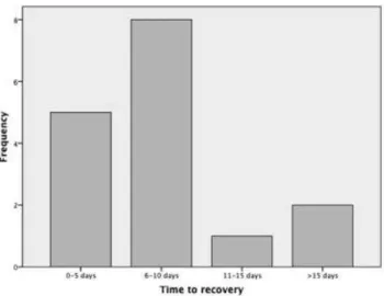
the symptoms was 7.4 (±5) days. Dysmetria and dysarthria were the most common accompanying neurological symptoms. The median time for recovery was 7 days. Eight patients healed during the 6-10 day interval (Figure 2). The longest time for
recovery was 4 months in a 4½-year-old girl. Lumbar puncture was performed in 7 of the cases. Cerebrospinal luid protein lev-els were normal in all of the cases, with a mean of 20,8 mg/dl (±4,9). All patients underwent a cranial imaging, 15 underwent brain magnetic resonance imaging (MRI), and one underwent brain computerized tomography (CT). The results were normal in all of the cases. Electromyography (EMG) was performed in 4 of the cases to exclude Guillain Barre Syndrome (GBS). The results did not reveal any pathology. Two patients were treated with intravenous immunoglobulin (IVIG) and one patient was treated with intravenous methylprednisolon because of the se-verity of the symptoms and the lack of clinical improvement during the hospitalization period. The mean time for hospital-ization was 4.4 (±1.4) days. Three patients were lost to follow-up; the mean follow-up time for the rest of the cases was 2.2 (±1,4) months. Full recovery was observed in all of the cases. One case still had minor degrees of gait ataxia and abnormal cerebellar examination in the 2-month follow-up visit; however, these signs had subsided by the 4-month follow-up visit.
Discussion
There is a small number of case series involving acute cerebel-lar ataxia in the published literature. In 1959, Weiss and Carter [6] described 18 patients with acute cerebellar ataxia. Six of these children had permanent neurological sequelae including gait disturbances and delayed speech development.
Connolly et al [7] reported the evaluation of 73 patients in 1994. Thirty-six of the cases were related to nonspeciic viral infec-tions. Varicella infection was responsible for 19 of the cases, EBV for 2. Fourteen cases were found to be idiopathic and 2 secondary to immunization. Cerebrospinal luid examination re-vealed pleocytosis and CSF protein levels ranged between 7-99 mg/dl. Nine patients underwent cranial MRI, of which only 1 examination revealed pathology. Ataxia resolved in 91% of the cases ater 4 months of follow-up; however 20% of the cases exhibited behavioral problems and learning diiculties, which
subsided during follow-up. Recurrence of symptoms was ob-served in 4 patients.
Nussinovich et al [2] reported a prodromal illness in 29 of the 39 cases in 2003. About one-third of these cases had varicella infection. Mumps, EBV, mycoplasma, and nonspeciic infections were noted in the remainder of the cases. Full recovery without any neurological sequelae was achieved within 24 days in all of the cases. A lumbar puncture was performed in all cases. Pleo-cytosis was present in 48% of the cases and abnormal CSF pro-tein levels (>40 mg/dl) were observed in 23.5%. Twelve patients underwent CT imaging that revealed normal results. The most commonly-associated neurological indings were dysmetria and dysarthria; nystagmus was noted in only 3 of the cases. Electro-encephalography (EEG) was performed in 12 cases. Background slowing was observed in 4 of the cases and epileptic discharges without clinical seizures were observed in 2 cases. The authors stated that acute cerebellar ataxia is a self-limiting disease. In 2006, Martinez-Gonzalez et al. [8] reported a favorable prog-nosis in 20 cases with acute cerebellar ataxia attributed to varicella, mycoplasma, enterovirus, EBV, and nonspeciic viral infections.
Acute cerebellar ataxia is common in children between 2 and 4 years of age, but also may be seen in older children and ado-lescents (3,7). Boys are more commonly afected [7]. A history of a febrile illness 5-21 days before the irst appearance of the symptoms is evident in about 70% of patients. In our study group, there was a slight male preponderance, the incidence of acute cerebellar ataxia was higher in the 2-5 years age group, and a preceding illness accompanied most of the cases. All of these indings were consistent with the literature.
Varicella may be responsible in as many as 26% of the cases; rarely does ataxia occur before the eruptions [1]. Compared with the literature, in our study group, the rate of acute cer-ebellar ataxia following varicella infection is lower, which may be attributed to the routine use of varicella vaccine ater 2012 in our country.
In our series, all of the cases recovered without any neurologi-cal sequelae. Only one case had slight gait ataxia and abnormal cerebellar examination ater 2 months of discharge; they even-tually subsided completely. This case was a 4 1/2 year-old-girl with a severe gait ataxia at onset causing an inability to walk. She was treated with IVIG.
Acute postinfectious cerebellar ataxia is a diagnosis of exclu-sion. Detailed evaluation with history and physical examination is more valuable than laboratory tests and imaging techniques in the diferential diagnosis. An altered state of consciousness, presence of hallucinations, behavioral changes, and a sleepy state should raise the suspicion of drug intoxication. Acute disseminated encephalitis (ADEM) and meningoencephalitis should be on the diferential diagnosis list when fever is add-ed to these symptoms [9]. Drug intoxication constitutes about 32.5% of all childhood acute ataxia cases; therefore children and parents should be questioned about drug intake [3]. Anti-convulsant drugs, benzodiazepines, alcohol, and antihistaminic drug intoxication may cause ataxia. Motor examination should be carefully performed because ataxia may be the irst sign of hemiparesis or paraparesis in younger children. Posterior circu-lation infarcts are relatively rare in young children but should Figure 2. The distribution of cases according to time to recovery
| Journal of Clinical and Analytical Medicine 654
Acute Cerebellar Ataxia
4
be considered ater neck trauma (causing vertebral artery dis-section) or in those predisposed to thromboembolic disease [10,11]. Focal cerebral (usually parietal or frontal) and pyra-midal tract lesions manifest with a positive Babinski sign and increased deep tendon relexes [12]. Vomiting and nystagmus should raise the suspicion of labyrinthitis and in the presence of chaotic eye movements, opsoclonus myoclonus syndrome should be considered. Guillain Barre Syndrome should be on the diferential diagnosis list when weakness and absence of deep tendon relexes predominate [9].
Posterior fossa tumors usually present with slowly progressive ataxia and symptoms of increased intracranial pressure such as papilledema and sixth nerve palsy [13]. Brain imaging should be performed in suspected cases. In our study group, all children underwent brain imaging. The results were normal. Although cranial imaging was performed in selected cases in the liter-ature [2,6,7], in countries where patient follow-up is a major problem, it might be more appropriate to evaluate all acute-onset ataxia cases with brain imaging upon admission for the early diagnosis and intervention of brain tumors.
Cerebrospinal luid examination usually reveals normal results or mild pleocytosis and slight elevation of protein levels. Lum-bar puncture should only be performed when central nervous system (CNS) infection is suspected [3]. Rarely, an LP may be helpful in diferential diagnosis from GBS, when EMG is not available. However it should always be kept in mind that the CSF protein levels may be normal during the irst week of GBS [14]. Therefore an LP is generally not indicated at the initial presentation of acute ataxia [1].
Electromyography is indicated when GBS is considered in the diferential diagnosis. We performed EMG on only 4 patients. None of the patients underwent EEG because an altered state of consciousness was not observed in any of them.
Opsoclonus-myoclonus syndrome is a rare autoimmune condi-tion mostly associated with neuroblastoma in childhood [15]. Thorax and abdomen CT or MRI or I123 metaiodobenzilguanidin (MIBG) scanning might be helpful for diagnosis of neuroblas-toma in these patients because the levels of cathecolamines in the urine are increased in only 47-60% of the cases [16,17,18]. The treatment of acute cerebellar ataxia is usually watchful waiting; physiotherapy may be helpful in selected cases [15]. Since an autoimmune process has been suggested in the etiol-ogy, high dose corticosteroids and IVIG has been used in treat-ment with favorable results [19,20]. However, the results are mostly limited to case reports and the yield of corticosteroid and IVIG therapy is uncertain (1). We tried IVIG in 2 patients and high dose corticosteroids in 1 patient because of a severe and resistant course without an evident clinical response. Recovery usually begins within the irst week ater the onset of symp-toms [1]. The average duration of sympsymp-toms is about 2 months; ataxia remains persistent in a small group of patients [7]. In conclusion, postinfectious acute cerebellar ataxia is the most common cause of acute ataxia in childhood. The prognosis is excellent; however, it causes great anxiety to the parents. Af-ter the exclusion of more serious medical conditions presenting with acute ataxia, informing the parents about the course of the disease and close follow-up during recovery is necessary.
Competing interests
The authors declare that they have no competing interests.
References
1. Ryan MM, Engie EC. Acute ataxia in childhood. J Child Neurol 2003;18:309-16. 2. Nussinovich M, Prais D, Volowitz B, Shapiro R, Amir J. Postinfectious acute cer-ebellar ataxia in children. Clin Pediatr (Phila) 2003;42:581-4.
3. Gieron-Korthals MA, Westberry KR, Emmanuel PJ. Acute childhood ataxia: 10 year experience. J Child Neurol 1994;9:381-4.
4. Harai T, Kanegane H, Ito Y, Saito M, Hongo K, Miyawaki T. Case of acute cer-ebellar ataxia associated with primary Epstein-Barr virus infection. Pediatr Int 2010;52(4):178-80.
5. Adams C, Diadori P, Schoenroth L, Fritzler M. Autoantibodies in childhood post-varicella acute cerebellar ataxia. Can J Neurol Sci 2000;27:316-20.
6. Weiss S, Carter S: Course and prognosis of acute cerebellar ataxia in children. Neurology 1959;9:711–21.
7. Connolly AM, Dodson WE, Prensky AL, Rust RS. Course and outcome of acute cerebellar ataxia. Ann Neurol 1994;35:673-9.
8. Martinez-Gonzalez MJ, Martinez-Gonzalez S, Garcia-Ribes A, Mintegi-Raso S, Benito-Fernandez J, Prats-Viñas JM. Acute ataxia in infancy: its etiology, treatment and follow-up. Rev Neurol 2006;42(6):321-4.
9. Sivaswamy L. Approach to acute ataxia in childhood: diagnosis and evaluation. Pediatric Annals 2014;43(4):153-9.
10. Echenne B, Gras M, Astruc J, Castan P, Brunel D. Vertebro-basilar arterial occlu-sion in childhood—report of a case and review of the literature. Brain Dev1983; 5:577–81.
11. Garg BP, Ottinger CJ, Smith RR, Fishman MA. Strokes in children due to verte-bral artery trauma. Neurology 1993;43:2555–8.
12. Freund H-J. Diferential efects of cortical lesions in humans. Ciba Foundation Symposium 1987;132:269–81.
13. Pollack IF. Brain tumors in children. N Engl J Med 1994;331:1500-7. 14. Hadden RDM, Hughes RAC. Management of inlammatory neuropathies. J Neurol Neurosurg Psychiatry 2003;74:9-14.
15. Desai J, Mitchell MG. Acute cerebellar ataxia, acute cerebellitis, and opsoclo-nus-myoclonus syndrome. J Child Neurol 2012;27(11):1482-8.
16. Whealan HT, Verma S, Gun Y, Thabet Farouq, Bozarth Xiuhua, Nwosu Michelle et al. Evaluation of the child with acute ataxia: a systematic review. Ped Neurol 2013;49:15-24.
17. Koh PS, Rafensperger JG, Berry S, Larsen MB, Johnstone HS, Chou P, et al. Long-term outcome in children with opsoclonus-myoclonus and ataxia and coinci-dent neuroblastoma. J Pediatr 1994;125:712–6.
18. Laug WE, Siegel SE, Shaw KN, Landing B, Baptista J, Gutenstein M. Initial uri-nary catecholamine metabolite concentrations and prognosis in neuroblastoma. Pediatrics 1978;62:77–83.
19. Kato Z, Shimozawa N, Kokuzawa J, Iwamura M, Hirata T, Yamasishi A, et al. Magnetic resonance imaging of acute cerebellar ataxia: report of a case with gadolinium enhancement and review of the literature. Acta Paediatr Jpn 1998;40:138-42.
20. Go T. Intravenous immunoglobulin therapy for acute cerebellar ataxia. Acta Paediatrica 2003;92(4):504-6.
How to cite this article:
Ünver O, Kutlubay B, Thomas G, Hacifazlioğlu NE, Sağer G, Türkdoğan D. Postin-fectious Acute Cerebellar Ataxia in Childhood. J Clin Anal Med 2016;7(5): 652-5.