Mobilization-with-movement and Exercises with EMG
Biofeedback in Subjects with Subacromial
Impingement Syndrome
Projeto elaborado com vista à obtenção
do grau de Mestre na Escola Superior de Saúde do Alcoitão, na Especialidade de Fisioterapia em Condições Músculo-Esqueléticas
Orientador: Professor Doutor Hugo Gamboa Coorientador: Professor Doutor José Esteves
João Pinto
Setembro,2014
Júri:
Presidente: Professor Doutor João Manuel Cunha da Silva Abrantes
Professor Catedrático e Presidente do Conselho Técnico-Científico da Escola Superior de Saúde do Alcoitão
Vogais: Professor Doutor Hugo Filipa Silveira Gamboa
Professor Auxiliar da Faculdade de Ciências e Tecnologia da Universidade Nova de Lisboa
Abstract
Objective: The purpose of this study was to investigate the effects of Mobilization-with-movement (MWM), Exercises with EMG Biofeedback (BFB) and Conventional physiotherapy (CP) in subjects with Subacromial Impingement Syndrome (SAIS). Methods: Nineteen subjects with shoulder pain and restricted ROM referred by physicians were included. Participants were randomly assigned to 1 of 4 groups: CP group, MWM group, BFB group and MWM+BFB group. The main outcome measures were active ROM, speed of movement, strength, pain and functionality. Active ROM and speed of movement were measured for Abduction, Flexion and Scaption movements. Pain was measured with VAS and functionality with Constant-Murley score. Measurements were performed at baseline and at the end of a three weeks intervention period.
Results: Intra-group repeated-measurement analysis indicated that subjects in the three intervention groups showed significant improvements on pain, Constant-Murley score, strength, speed of movement and Abduction and Scaption active ROM, pre- to post-intervention period (P<0,05). On Flexion active ROM variable no differences were found in MWM and BFB groups. Improvements in active ROM, Flexion and Scaption speed, strength, functionality were significantly higher in MWM+BFB group (P<0,05). Pain was significantly lower and Abduction speed was significantly and higher in MWM group.
Conclusion: This study suggests that combining MWM with exercises using EMG Biofeedback can result in greater improvements from pre- to post-treatment in subjects with SAIS.
Keywords: MWM, EMG Biofeedback, Exercise, Subacromial Impingement Syndrome
Resumo
Objectivo: O objetivo deste trabalho foi investigar os efeitos da Mobilização-com-movimento (MWM), Exercícios com Biofeedback EMG (BFB) e Fisioterapia convencional (CP) em sujeitos com Síndrome de Conflito Subacromial (SCS).
Métodos: Foram incluídos dezanove sujeitos com dor e restrição da ROM no ombro, referidos por Fisiatria. Os participantes foram distribuídos aleatoriamente em 1 de 4 grupos: grupo CP, grupo MWM, grupo BFB e grupo MWM+BFB. As principais medidas de resultados foram ROM ativa, velocidade de movimento, força, dor e funcionalidade. A dor foi medida com a EVA e a funcionalidade com a escala de Constant-Murley. As medições foram realizadas no momento inicial e no final do período de três semanas de intervenção.
Resultados: A análise das medições repetidas intra-grupo indicaram que os sujeitos nos três grupos de intervenção mostraram melhorias significativas na dor, Constant-Murley, força, velocidade de movimento e na ROM ativa de abdução e elevação no plano da omoplata, entre o período pré e pós intervenção (P<0,05). Na variável ROM ativa de flexão não foram encontradas diferenças nos grupos MWM e BFB.
Melhorias na ROM ativa, velocidade de flexão e elevação no plano da omoplata, força, funcionalidade foram significativamente maiores no grupo MWM+BFB (P<0,05). A dor foi significativamente mais baixa e a velocidade de abdução significativamente mais alta no grupo MWM.
Conclusão: Este estudo sugere que combinando MWM com exercícios usando o Biofeedback EMG pode resultar em maiores melhorias do início para o final do tratamento em sujeitos com SCS.
1. Introduction
Subacromial Impingement Syndrome (SAIS) is defined as the abrasion caused from
mechanical compression of the structures of the rotator cuff, subacromial bursa, and/or tendon of
the long head of the biceps brachii when they pass under the coracoacromial arch during the
elevation of the upper limb (Michener, McClure e Karduna, 2003; Neer, 1983).
SAIS represents about 44-65% of all shoulder pain and shoulder movement disorders
during a medical consultation (Michener et al., 2003), and it is the most common cause of
shoulder pain in athletes practicing sports that recruit mostly the upper limb (Jobe, Coen e
Screnar, 2000).
Shoulder pain with subsequent restriction of movement is a common problem in both
athletes and workers. Approximately 1% of adults consult health professionals with an episode
of shoulder pain annually (Monu, Pruett, Vanarthos e Pope, 1994).
These conditions are arrise from multifactorial causes. Some authors refer the kinematic
changes, including the scapular-humeral rhythm and changes in the patterns of motor control
(Chester, Smith, Hooper e Dixon, 2010; Cools, Geerooms, Van den Berghe, Cambier e
Witvrouw, 2007a; Cools, Declercq, Cagnie, Cambier e Witvrouw, 2008; Fayad, Hoffmann,
Hanneton, Yazbeck, Lefevre-Colau, Poiraudeau, Revel e Roby-Brami, 2006; Ludewig & Cook,
2000).
Imbalance of the muscles of the shoulder girdle has been found in patients with shoulder
disorders (Cools, Witvrouw, Declercq, Danneels e Cambier, 2003). These muscles may result in
the scapula mobility change, contributing to worse states of the impingement (Kibler &
McMullen, 2003; Ludewig & Cook, 2000). Specifically, over-activity of the upper trapezius
(UT) combined with reduced activity of the lower trapezius (LT) and the serratus anterior (SA),
have been observed in patients with SAIS (Cools et al., 2003; Ludewig & Cook, 2000).
Exercise training should be prescribed according to the kinematic mechanism related to
the impingement syndrome. In general, exercises for the rehabilitation of scapular-humeral
rhythm should promote lower trapezius, middle trapezius (MT) and serratus anterior activity and
reduce the activity of the upper trapezius (Cools et al., 2003; Kibler & McMullen, 2003; Ludewig & Cook, 2000).
In electromyographic (EMG) biofeedback training an electronic device is used to
instantly provide information about the occurrence of muscle activation events and intensity. By
manipulating the signals shown, the subject can be taught to control these events that otherwise
could be involuntary or uncontroled (Basmajian, 1981). According to various studies, EMG
UT (Holtermann, Mork, Andersen, Olsen e Sogaard, 2010; Holtermann, Roeleveld, Mork,
Gronlund, Karlsson, Andersen, Olsen, Zebis, Sjogaard e Sogaard, 2009). In a study by Levargie
& Humphrey (2000) and Paterson & Sparks (2006), the authors verified that using the EMG
biofeedback can reduce shoulder rehabilitation time by near 50% .
The Mulligan concept is a manual therapy approach which involves a set of joint
mobilization techniques that generate changes in neuro-muscular activity, being based on ideas
and methods of Kaltenborn (Wilson & Hazen, 1995). Mobilization-with-movement (MWM) is a
passive mobilization technic, which promotes a painless correction of a "positional fault", with
active movement. This combination of joint mobilization with active motion may be responsible
for the rapid return to pain-free movement (Exelby, 1995). Wright (1995) defined that the
mechanisms responsible for the effects of manual therapy were due to changes in the joint,
muscle, pain and motor control. There is already relevant literature supporting the benefits of
MWM in the treatment of shoulder disorders (Exelby, 1996; Ho, Sole e Munn, 2009;
Kachingwe, Phillips, Sletten e Plunkett, 2008; Teys, Bisset e Vicenzino, 2008; Yang, Chang,
Chen, Wang e Lin, 2007).
The aim of this work is to investigate the effects of exercise with EMG biofeedback
and/or the use of Mulligan Concept, on pain, range of motion, muscle recruitment patterns,
strength and speed of movement, comparatively with Conventional Physiotherapy, in individuals
with Subacromial Impingement Syndrome.
2. Methods
2.1 Subjects
In this study was followed the rehabilitation process of 19 participants (6 men and 13
women), aged between 22 and 68 years (mean 47,3 years SD+13,4), who were diagnosed with
rotator cuff injury and/or shoulder impingement syndrome by their referring physician. All the
participants were treated between April and July 2013 at Health Care Services of the biggest
Portuguese bank, held in Lisbon. Informed consent for participation in this study was obtained
from subjects.
Their main complaints were shoulder pain and restricted ROM that compromised the
activities of daily living. The inclusion criteria were: presence of superior-lateral shoulder pain
combined with three or more from the five following signs and symptoms: Positive Neer
impingement test; positive Hawkins-Kennedy impingement test; positive Empty can test; painful
Shoulder girdle fractures and/or dislocation; shoulder surgery; rotator cuff lesion (grade III);
medical diagnosis of adhesive capsulitis; cervicobrachial pain due to cervical spine pathology;
neuromuscular disorders in upper extremities; use of corticosteroid therapy in the last month and
traumatic event that provoked pain and/or restricted shoulder motion.
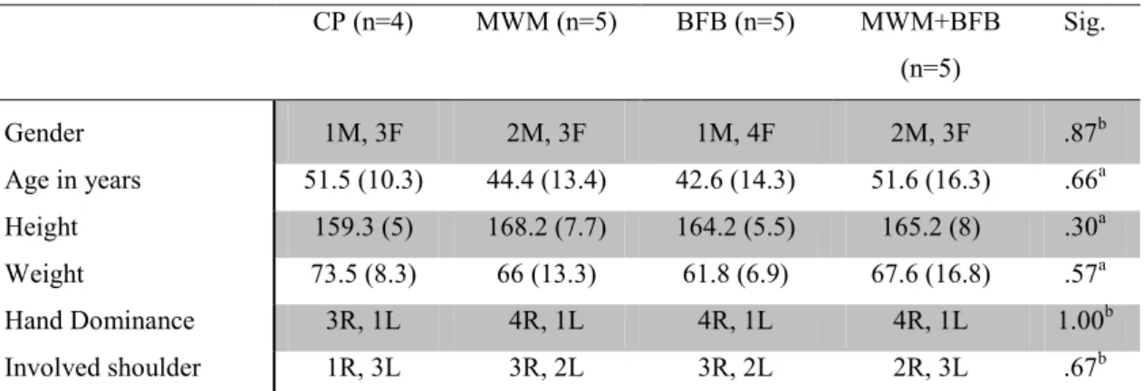
Subjects were randomly allocated to four groups and their baselines demographic are
described on table 1.
Table 1 Baseline demographics means (sd) for the dependent variable for each group
CP (n=4) MWM (n=5) BFB (n=5) MWM+BFB (n=5)
Sig.
Gender 1M, 3F 2M, 3F 1M, 4F 2M, 3F .87b Age in years 51.5 (10.3) 44.4 (13.4) 42.6 (14.3) 51.6 (16.3) .66a Height 159.3 (5) 168.2 (7.7) 164.2 (5.5) 165.2 (8) .30a Weight 73.5 (8.3) 66 (13.3) 61.8 (6.9) 67.6 (16.8) .57a Hand Dominance 3R, 1L 4R, 1L 4R, 1L 4R, 1L 1.00b Involved shoulder 1R, 3L 3R, 2L 3R, 2L 2R, 3L .67b
Abbreviations: CP = conventional physiotherapy group; MWM = mobilization-with-movement group; BFB = EMG Biofeedback group; MWM+BFB = mobilization-with-movement combined with EMG Biofeedback group; M = males; F = females; R = right; L = left; aOne-way ANOVA; bChi square tests.
2.2 Procedure
Participants were initially assessed for their suitability for inclusion in the study. All
subjects were recruited after medical consultation, diagnosed with rotator cuff injury and/or
shoulder impingement syndrome.
Than the participants were randomly allocated to one of the following four groups:
Conventional Physiotherapy (CP group); Mobilization-with-movement (MWM group); EMG
Biofeedback (BFB group); Mobilization-with-movement combined with EMG Biofeedback
(MWM+BFB group).
In the first session were performed speed, strength, active ROM, pain at the maximum
active ROM and Constant-Murley Score variables and were applied the physical therapy
intervention according group allocation. All subjects were asked to decline other forms of
treatment for their shoulder as well as analgesics and anti-inflammatory medication, during the
course of the study.
Participants attended three sessions per week with at least a 48h interval to reduce fatigue
Revaluations of all variables on study were performed at the end of the intervention
period.
A physiotherapist with clinical experience and Mulligan techniques skills performed all
measurements and interventions.
2.3 Instruments and Outcome measures
Speed of movement, range of movement and pain at the maximum active ROM data
were recorded synchronously. These measurements were obtained from three repetitions of each
movement. In order to evaluate the dependent variables in study were performed the following
procedures:
2.3.1 Active range of movement
To evaluate shoulder range of motion, a Triaxial Accelerometer (Plux Wireless
Biosignals S.A., Lisbon, Portugal) was connected by cable to Biosignalsplux pro® (Plux Wireless Biosignals S.A.), a wireless analog/digital signal converter with a resolution of 16 bits. A
Butterworth 2nd order filter were used, with precision sensitivity levels between 270-330 mV/G (±1.7°). The accelerometer was placed on the outer face of the arm 10 cm above the lateral
epicondyle of the humerus in study, fixed with tape.
2.3.2 Speed of movement
The speed of movement was obtained from the Triaxial Accelerometer (Plux Wireless
Biosignals S.A.). Maximum range was maintained for 3 seconds to increase the signal accuracy.
Subjects were asked to perform movement at a comfortable speed to the maximum active active
ROM of flexion, scaption and abduction.
2.3.3 Pain at the maximum active ROM
To measure pain, subjects were asked to complete 10-point visual analog scale (VAS) to
describe pain intensity at the end of each movement. This scale obtained a intra- and inter-rater
reliability range from 0.69 to 0.91 and 0.55 to 0.97 respectively, assessed using the intra-class
correlation coefficient (Taddio, O'Brien, Ipp, Stephens, Goldbach e Koren, 2009).
2.3.4 Pain-free isometric contraction at 90° of abduction and internal rotation of the
For the evaluation of strength a MicroFET3® (HOGGAN Health Industries Inc., Salt
Lake City, UT, USA) hand held dynamometer was used. This instrument showed a moderate to
excellent intra-test reliability, with associations ranging from ICC 0:56-092 and high
concordance for the knee and hip measurements. The inter-test reliability was weak, associations
with hip and knee ICC ranging from 0.60-0.66 (Clarke, Ni Mhuircheartaigh, Walsh, Walsh e
Meldrum, 2011).
For strength assessment, subjects kept the arm at 90° of abduction and internal rotation
and performed isometric contraction against resistance with a MicroFET3® dynamometer placed
next to their wrist, until the moment of pain.
2.3.5 Functionality
The Constant-Murley Score, developed by Christopher Constant and Murley (1987), was
used in order to measure shoulder functionality. This scale aims to measure and assess the
shoulder functional status and was translated and adapted for the Portuguese population by Leal
(2001).
The Constant-Murley Score is a 100-point scoring system that is divided into 4 subscales:
pain, 15 points; activities of daily living, 20; range of motion, 40; and strength, 25.The pain and
activities of daily living subscales are self-reported by the patient. The total score is displayed in
a positive direction scale of 0 (low functionality) to 100 (high functionality).
The intra-tester reliability of the Constant-Murley Score was evaluated by Livain et al
(2007) and Rocourt et al (2008). They observed reliability coefficients varying from P = 0.94 to
0.96 in subjects with different shoulder pathologies with 13% to 19% of patients exhibiting an
exact match in terms of total score. For inter-tester reliability, Livain et al (2007) and Rocourt et
al (2008) observed correlations varying from P = 0.89 to 0.91.
2.4 Experimental conditions
Subjects in CP group underwent Conventional Physiotherapy treatment consisting of
TENS, Ultrasound and Laser for 3 weeks with a frequency of 3 times per week.
MWM Group received Conventional Physiotherapy and Mobilization-with-movement
according to the principles described by Mulligan (1999). The technique consisted in performing
the symptomatic movement for 3 sets of 10 repetitions, with 30 second rest periods between sets,
with a frequency of 3 times per week for three weeks. During the MWM treatment, the
participant was seated and the therapist was positioned on the opposite side of participant’s
participant’s humeral head and the other hand on his/her scapula. The hand placed on the humeral head performed a posterolateral glide, while the other stabilized the scapula. During this
maneuver, the participant was encouraged to perform active shoulder movement to the point of
the first onset of pain (Djordjevic, Vukicevic, Katunac e Jovic, 2012).
In BFB group, Conventional Physiotherapy treatment and an Exercise Protocol with
PhysioPlux® EMG Biofeedback system (Plux Wireless Biosignals S.A.) were applied with a frequency of 3 times per week for three weeks.
In order to monitorize muscle activity during Exercise protocol a Biosignalsplux pro® (Plux Wireless Biosignals S.A.) device was used. Reusable circular electrodes of Ag/AgCl with
self-adhesive silicon were used, placed in a bipolar configuration and a inter-electrode distance
of about 20 mm (De Luca, 1997). These electrodes were connected by cable to a wireless
analog/digital signal converter with a resolution of 16 bits (Biosignalsplux pro®, Plux Wireless Biosignals S.A.). The detection of the differential mode signal occurred with an input impedance
of 100 G and 100 CMRR; a Butterworth filter 5th order (10-300 Hz, 40 dB/dec) was used for these procedures. The electromyographic signal was amplified (overall gain=1000) and then
captured at a sampling frequency of 1000 Hz, properly identified and stored in a computer file
for further "off-line" processing.
To ensure the best electromyographic signal quality, procedures included shaving and
cleansing (alcohol) of the skin. The electrodes were placed in the middle of the muscle,
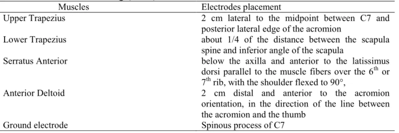
following to the fiber orientation of each muscle (Ludewig & Cook, 2000), according to Table 2.
Each EMG signal was visually monitored during resisted contractions of each of the 4
muscles (using manual muscle testing positions) to ensure an adequate signal-noise ratio and to
minimize crosstalk from adjacent or underlying muscles.
Muscles Electrodes placement
Upper Trapezius 2 cm lateral to the midpoint between C7 and posterior lateral edge of the acromion
Lower Trapezius about 1/4 of the distance between the scapula spine and inferior angle of the scapula
Serratus Anterior below the axilla and anterior to the latissimus dorsi parallel to the muscle fibers over the 6th or 7th rib, with the shoulder flexed to 90°,
Anterior Deltoid 2 cm distal and anterior to the acromion orientation, in the direction of the line between the acromion and the thumb
Ground electrode Spinous process of C7
The exercise protocol was composed by the following exercises:
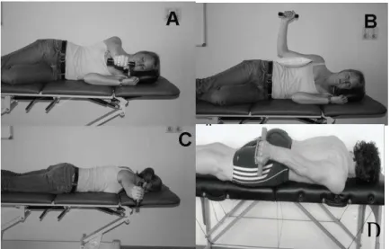
- Exercises directed to Lower Trapezius (Fig 1)
- Shoulder flexion in lateral decubitus (Cools, Dewitte, Lanszweert, Notebaert,
Roets, Soetens, Cagnie e Witvrouw, 2007b)
- External rotation in lateral decubitus (Cools et al., 2007b; De Mey, Danneels,
Cagnie, Huyghe, Seyns e Cools, 2013)
- Horizontal abduction with external rotation in prone position (Cools et al.,
2007b)
- Shoulder extension in prone position (De Mey et al., 2013)
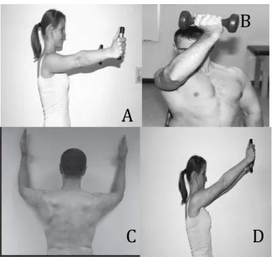
- Exercises directed to Serratus Anterior (Fig 2)
- Scaption in standing position (Cools et al., 2007b)
- Diagonal movement combining flexion, horizontal adduction and external
rotation (Ekstrom, Donatelli e Soderberg, 2003)
- Wall-slide (Hardwick et al., 2006)
- Shoulder flexion (Cools et al., 2007b)
Figure 1 Exercises directed to Lower Trapezius: A - Shoulder flexion in lateral decubitus (Cools et al., 2007) B -External rotation in lateral decubitus (Cools et al., 2007; De Mey et al., 2013) C - Horizontal abduction with external rotation in prone position (Cools et al., 2007) D - Shoulder extension in prone position (De Mey et al., 2013)
Figure 2 Exercises directed to Serratus Anterior: A - Scaption in standing position (Cools et al., 2007) B - Diagonal movement combining flexion, horizontal adduction and external rotation (Ekstrom et al., 2003) C - Wall-slide (Hardwick et al., 2006) D - Shoulder flexion (Cools et al., 2007)
The described exercises protocol was created according to the literature(Cools et al.,
2007b; De Mey et al., 2013; Ekstrom et al., 2003; Hardwick et al., 2006). According to Huang
and colleagues (2013) and Cools (2007b) the stability exercises should be performed in three
phases: concentric, isometric and eccentric with a period of 3 seconds each, measured with a
metronome at 60 bps in order to guarantee a constant speed of movement. Fleck & Kraemer
(1987) recommended three sets of 15–20 repetitions to create a fatigue response and target the
development of local muscular endurance. Descriptive studies of electromyographic activity of
rotator cuff and scapular muscles of Ellenbecker and Cools (2010) have shown favorable
activation of these muscles using free weights 0.5 and 1kg.
In a study by Cools et al (2007b) three exercises were identified with lower ratio UT/LT:
shoulder flexion (Figure 1A) and external rotation (Figure 1B) in lateral decubitus position and
horizontal abduction with external rotation in prone position (Figure 1C). In the same work were
not identified exercises for UT/SA ratio lower than 60% MVIC. The high row (60-80% MVIC)
and shoulder scaption (Figure 2A) and flexion (Figure 2D) (> 80% MVIC) were the highest
ranked. Due to material limitations only the last 2 exercises were incorporated. In another study
by (De Mey et al., 2013) it was concluded that autocorrection of scapular orientation increases
the three sections of Trapezius muscle during Extension exercise in prone position (Figure 1D)
and external rotation in lateral decubitus position. The diagonal movement exercise combining
B
A
flexion, horizontal adduction and external rotation (Figure 2B) generated high levels of
electromyographic activity in SA muscle (100% MVIC) (Ekstrom et al., 2003). Wall slide
exercise (Figure 2C) proved to be a proper exercise to activate SA in positions up to and beyond
90° of shoulder elevation. This exercise seems to be indicated for SIS patients with altered
scapular kinematics and decreased activity of SA (Hardwick et al., 2006).
MWM+BFB group received, along with Conventional Physiotherapy treatment, Exercise
Protocol shoulder stability exercises as described in BFB group with Biofeedback and
Mobilization-with-movement in the symptomatic limb with a frequency of 3 times per week for
3 weeks.
2.6 Signal Processing
The signal accelerometry was filtered with a cut-off frequency of 1Hz to remove linear
acceleration. To measure the angles made during the motion, a vector of the initial acceleration
was recorded and used as a reference for the calculation of the angles with the following vectors.
After conversion to angles, the maximum amplitude of movement and the speed at which it was
done were calculated.
2.7 Statistical Analyses
SPSS® Statistics 20 (IBM Corp., Armonk, NY, USA) for Mac was used for the statistical
analysis. Descriptive statistics were analysed in order to obtain values of mean and standard
deviation.
ANOVA was conducted to determine whether the four groups differed on day 0 and at
the end of treatment in the following variables: pain; strength; active pain-free shoulder flexion;
abduction and scaption of the upper limb; muscle activation moments; speed of movement;
Constant Score. On the variables that showed significant differences, a post hoc test was
conducted to determine differences between groups. Paired Samples t Test was used to obtain
intra-group differences between initial and final observations.
Differences were considered statistically significant when p< 0.05.
3. Results
All subjects completed the intervention program study. By Chi-square analysis no
statistically significant differences were found between the four groups on gender, hand
dominance and involved shoulder. One-way ANOVA analysis indicated no statistically
No significant differences were found between the 4 groups at the first measurement
regarding One-Way ANOVA analysis on pain, Constant-Murley Score, active ROM and Speed
of movement. A significant difference between CP and MWM+BFB groups (P = 0,03) was
obtained on the Strength variable using the parametric analysis however, with Kruskal Wallis
test this difference was not observed (P = 0,054).
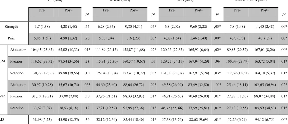
Paired Samples t Tests were used to determine intra-group differences between the first
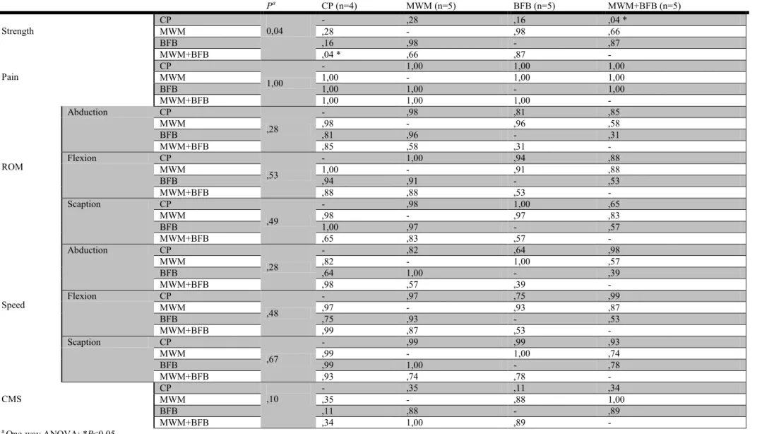
and last measurements (Table 3). To obtain inter-group differences at first and last
measurements, One-way ANOVA with post-hoc Scheffe test was used (Tables 4 and 5).
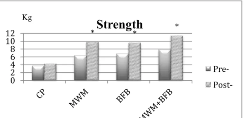
There were intra-group significant differences on Strength variable in MWM, BFB and
MWM+BFB groups (P<0,05). MWM+BFB group showed the highest mean increase (3.6 kg, P
= 0,00) (Fig. 3). Inter-group differences were found between CP and MWM+BFB groups at last
measurement (P = 0,02).
Intra-group significant differences were obtained in MWM, BFB and MWM+BFB
groups on Pain variable. MWM group had the highest mean decrease registered (4.9/10 VAS, P
= 0,00) (Fig.4). Inter-group differences were found between CP and other groups at last
measurement (P = 0,00) (Table 5).
*
*
* 0
2 4 6 8 10
CP MWM BFB MWM+BFB
Pain
Pre-
Post-
/10
* * *
0 2 4 6 8 10
12
Strength
Pre-
Post-
Kg
Figure 3 Strength outcomes between initial and final moment.
The Constant-Murley Score showed intra-group significant differences in MWM, BFB
and MWM+BFB groups (P<0,05). MWM+BFB group had the highest score change (41.9
points, P = 0,00) (Fig. 5). Inter-group differences were found between CP and other groups at
last measurement (P = 0,00).
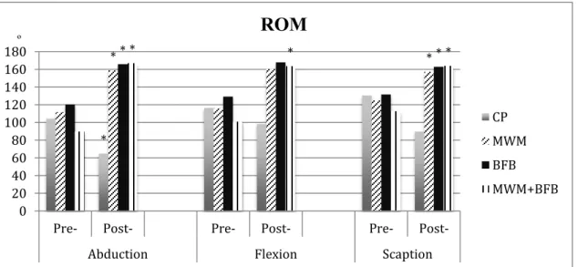
In intra-group analysis, on Abduction ROM outcome, a significant decrease was
observed in CP group (-39,4º; P = 0,01), while significant increases were found in the other
groups (P<0,05). MWM+BFB group had the highest increase (77,2º; P = 0,00). A significant
difference was found in MWM+BFB group on Flexion movement (62,7º; P = 0,01). On Scaption
ROM, significant differences were found in MWM, BFB and MWM+BFB groups. MWM+BFB
group had the highest mean increase (51,4º; P = 0,01) (Fig. 6). Inter-group differences were
found between CP and other groups at last measurement on Abduction, Flexion and Scaption
movements (P = 0,00).
Figure 5 Constant-Murley outcomes between initial and final moment. * P<0,05
* * *
0 20 40 60 80 100
Constant-Murley
Pre-
Post-
%
Figure 6 ROM outcomes between initial and final moment. * P< 0,05
*
* * * * * * *
0 20 40 60 80 100 120 140 160 180
Pre- Post- Pre- Post- Pre- Post-
Abduction Flexion Scaption
ROM
CP
MWM
BFB
MWM+BFB
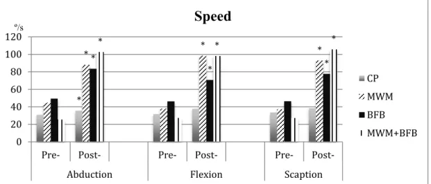
Intra-group significant differences were found on all groups in Abduction speed
(P<0,05). MWM group had the highest improvement in Abduction speed (43,4º/s, P = 0,00).
Inter-group difference was found between CP and MWM+BFB groups at last measurement (P =
0,03). On Flexion and Scaption Speed, intra-group significant differences were obtained in
MWM, BFB and MWM+BFB groups. MWM+BFB group obtained the highest improvement in
Flexion and Scaption speed (70,8º/s; P = 0,01 and 78,5º/s; P = 0,01 respectively) (Fig. 7).
Inter-group differences were found on Scaption speed between CP and MWM+BFB Inter-groups at last
measurement (P = 0,02), and on Flexion speed between CP group and MWM and MWM+BFB
groups (P = 0,05).
*
* * * *
* *
* * *
0 20 40 60 80 100 120
Pre- Post- Pre- Post- Pre- Post-
Abduction Flexion Scaption
Speed
CP
MWM
BFB
MWM+BFB
º/s
Table 3 Descriptive statistics andintra-group differences between observations
CP (n=4)
Pa
MWM (n=5)
Pa
BFB (n=5)
Pa
MWM + BFB (n=5)
Pa
Pre- Post- Pre- Post- Pre- Post- Pre- Post-
Strength 3,7 (1,38) 4,28 (1,48) ,44 6,28 (2,35) 9,80 (4,31) ,05* 6,8 (2,02) 9,60 (2,22) ,05* 7,8 (1,48) 11,40 (2,48) ,00*
Pain 5,05 (1,69) 4,98 (1,32) ,76 5,08 (,84) ,16 (,23) ,00* 4,88 (1,54) 1,46 (1,40) ,00* 4,98 (,90) ,40 (,89) ,00*
ROM
Abduction 104,45 (25,83) 65,02 (15,33) ,01* 111,89 (23,13) 158,87 (11,68) ,02* 120,33 (27,63) 165,93 (6,64) ,02* 89,85 (20,52) 167,01 (8,26) ,00*
Flexion 116,62 (33,72) 98,54 (34,56) ,23 115,91 (35,30) 160,37 (10,67) ,06 129,25 (24,16) 167,94 (4,29) ,06 100,99 (23,49) 163,72 (5,04) ,01*
Scaption 130,77 (19,06) 89,98 (29,56) ,10 125,04 (17,04) 157,41 (10,72) ,03* 131,70 (27,07) 162,91 (5,24) ,03* 112,69 (18,61) 164,10 (5,37) ,01*
Speed
Abduction 30,97 (10,78) 35,67 (10,74) ,05* 44,60 (23,60) 88,04 (26,72) ,00* 49,38 (26,09) 83,49 (32,80) ,00* 25,46 (18,11) 102,65 (36,94) ,02*
Flexion 31,70 (13,21) 37,88 (7,80) ,50 37,86 (21,51) 98,33 (32,93) ,01* 46,21 (26,60) 70,69 (26,80) ,01* 27,32 (11,50) 98,07 (34,44) ,01*
Scaption 33,62 (3,07) 38,53 (6,18) ,12 37,21 (19,57) 92,95 (27,36) ,01* 46,32 (22, 66) 77,59 (25,81) ,01* 27,13 (10,55) 105,59 (34,53) ,01*
CMS 38,98 (5,23) 43,90 (12,55) ,36 52,12 (12,34) 85,44 (18,40) ,01* 57,38 (13,76) 88,62 (9,69) ,01* 52,26 (6,29) 94,12 (6,75) ,00*
Table 4 Multiple Comparasions using One-way ANOVA with post-hoc Scheffe test at first measurement
Pa CP (n=4) MWM (n=5) BFB (n=5) MWM+BFB (n=5)
Strength
CP
0,04
- ,28 ,16 ,04 *
MWM ,28 - ,98 ,66
BFB ,16 ,98 - ,87
MWM+BFB ,04 * ,66 ,87 -
Pain
CP
1,00
- 1,00 1,00 1,00
MWM 1,00 - 1,00 1,00
BFB 1,00 1,00 - 1,00
MWM+BFB 1,00 1,00 1,00 -
ROM
Abduction CP
,28
- ,98 ,81 ,85
MWM ,98 - ,96 ,58
BFB ,81 ,96 - ,31
MWM+BFB ,85 ,58 ,31 -
Flexion CP
,53
- 1,00 ,94 ,88
MWM 1,00 - ,91 ,88
BFB ,94 ,91 - ,53
MWM+BFB ,88 ,88 ,53 -
Scaption CP
,49
- ,98 1,00 ,65
MWM ,98 - ,97 ,83
BFB 1,00 ,97 - ,57
MWM+BFB ,65 ,83 ,57 -
Speed
Abduction CP
,28
- ,82 ,64 ,98
MWM ,82 - 1,00 ,57
BFB ,64 1,00 - ,39
MWM+BFB ,98 ,57 ,39 -
Flexion CP
,48
- ,97 ,75 ,99
MWM ,97 - ,93 ,87
BFB ,75 ,93 - ,53
MWM+BFB ,99 ,87 ,53 -
Scaption CP
,67
- ,99 ,99 ,93
MWM ,99 - 1,00 ,74
BFB ,99 1,00 - ,78
MWM+BFB ,93 ,74 ,78 -
CMS
CP
,10
- ,35 ,11 ,34
MWM ,35 - ,88 1,00
BFB ,11 ,88 - ,89
MWM+BFB ,34 1,00 ,89 -
a
Table 5 Multiple Comparasions using One-way ANOVA with post-hoc Scheffe test at last measurement
Pa CP (n=4) MWM (n=5) BFB (n=5) MWM+BFB (n=5)
Strength CP 0,01 - ,08 ,10 ,02 *
MWM ,08 - 1,00 ,86
BFB ,10 1,00 - ,81
MWM+BFB ,02 * ,86 ,81 -
Pain CP
0,00
- ,00 * ,00 * ,00 *
MWM ,00 * - ,32 ,99
BFB ,00 * ,32 - ,49
MWM+BFB ,00 * ,99 ,49 -
ROM
Abduction CP
,00
- ,00 * ,00* ,00 *
MWM ,00 * - ,78 ,70
BFB ,00 * ,78 - 1,00
MWM+BFB ,00 * ,70 1,00 -
Flexion CP
,00
- ,00 * ,00 * ,00 *
MWM ,00 * - ,92 ,99
BFB ,00 * ,92 - ,98
MWM+BFB ,00 * ,99 ,98 -
Scaption CP
,00
- ,00 * ,00 * ,00 *
MWM ,00 * - ,95 ,92
BFB ,00 * ,95 - 1,00
MWM+BFB ,00 * ,92 1,00 -
Speed
Abduction CP
,02
- ,11 ,16 ,03 *
MWM ,11 - 1,00 ,89
BFB ,16 1,00 - ,79
MWM+BFB ,03 * ,89 ,79 -
Flexion CP
,02
- ,05 * ,43 ,05 *
MWM ,05 * - ,52 1,00
BFB ,43 ,52 - ,53
MWM+BFB ,05 * 1,00 ,53 -
Scaption CP
,01
- ,06 ,22 ,02 *
MWM ,06 - ,85 ,90
BFB ,22 ,85 - ,46
MWM+BFB ,02 * ,90 ,46 -
CMS CP ,00 - ,00 * ,00 * ,00 *
MWM ,00 * - ,98 ,76
BFB ,00 * ,98 - ,92
MWM+BFB ,00 * ,76 ,92 -
a
4. Discussion
Intra-group repeated-measurement analysis indicated that subjects in the three
intervention groups had significant improvements on pain, Constant-Murley score, strength,
speed of movement and active ROM of Abduction and Scaption, pre- to post-intervention
period. On Flexion active ROM variable, no differences pre- to post-intervention were found
in MWM and BFB groups. In the CP group, significant differences were found on abduction
ROM and abduction speed: The first variable showed a decrease on active ROM, while
improvements were found on speed.
Inter-group analysis showed significant differences between BFB and MWM+BFB
groups on flexion speed. No differences were found in the remaining variables, between
MWM, BFB and MWM+BFB groups. Additioning CP group to the others, significant
differences were found at the last measurement moment.
Results suggest that the group receiving MWM in combination with EMG
biofeedback had the higher percentage of change in most measurements.
4.1 Effects on Pain
Results suggest that the three intervention groups had a significant change in pain
intensity. However, groups receiving MWM (MWM and MWM+BFB groups) had a higher
percentage of change, compared to conventional physiotherapy and EMG biofeedback
applied separately. These results are consistent with the assumption that the Mulligan
technique promotes a pain-free movement and with the study of Djordjevic and colleagues
(2012), which refer higher improvement in active pain-free shoulder range of motion in the
group treated with MWM and Kinesiotaping. Also Kachingwe and colleagues (2008) - the
two groups receiving manual therapy (Glenohumeral mobilizations and MWM) in
combination with supervised exercise had a higher percentage of change from pre- to
post-treatment in pain.
Some studies have identified that altered shoulder kinematics are associated with
shoulder pain (Halder, Zhao, Odriscoll, Morrey e An, 2001; Howell, Galinat, Renzi e Marone,
1988; Ludewig & Cook, 2000). It would appear that excessive translation of the humeral head
along the glenoid results in pain and functional impairment (Matsen, Fu e Hawkins, 1993),
and the application of MWM can correct this “positional fault” and promote a pain-free
movement. Other explanation is that MWM technique causes a capsular stretching and
Paungmali et al (2003) found that hypoalgesic effects after MWM for chronic
epicondylalgia concurred with signs of sympatoexcitation and these effects seem to be
nonopioid origin.
As in the studies by Djordjevic et al (2012) and Kachingwe et al (2008), it would be
interesting to verify the different effects between MWM with BFB and MWM with tape or
stability exercises without EMG biofeedback. The combination of techniques seems to
suggest an optimal outcome compared with the techniques applied separately. Since MWM is
a technique of instant results, unlike exercices which involve slower results, it would be
interesting to investigate the effects of these combined techniques at medium/long term.
Another interesting aspect would be to assess other dimensions of pain, using scales or
other assessment measures both for short and long term evaluation. Outcomes like the impact
of pain on function, activities of daily life, at work or in how it is perceived by others, should
be considered in future works.
Since the sample selected for this study did not specify the duration of symptoms, it
would be interesting to compare the results of applying these techniques in contexts of acute
and chronic pain.
4.2 Effects on Strength
In this study a significant strength increase was observed in the three intervention
groups, of which MWM+BFB group had the higher percentage of change.
These results are consistent with studies by Bang & Deyle (2000) that verified that
patients who received manual therapy and exercise demonstrated greater short-term
improvements in muscle strength. Senbursa and collaborators (2011) verified that groups
receiving exercise, exercise with joint and soft tissue mobilization, and home-based rehabilitation program, experienced significant decrease in pain and an increase in shoulder muscle strength and function by both 4th and 12th weeks of treatment.
In this work, isometric contraction was measured in empty can position at which the
onset of pain is related to the clamping of the supraspinatus tendon in the subacromial space.
It is assumed that an increase in the isometric contraction is related to the decrease of
supraspinatus tendon clamping in this space. Strength evaluation was conducted from the
perspective of kinematic evaluation rather than a direct assessment of muscle strength output.
Another reason for choosing this test position was because it was part of the Constant-Murley
capacity, peak torque, quality of movement or patterns of activaction of both stabilizing and
mobilizing muscles of the shoulder girdle after applying MWM and/or exercises with BFB.
Since MWM + BFB group obtained a greater difference than the application of the
techniques separately, this seems to indicate that the increase of the subacromial space was
due both to joint (MWM) and muscle interventions (BFB). On one hand, Mulligan technique
can increase the subacromial space and thereby reduce the clamping; on the other, motor
control improves dynamic stability of the scapula which allows a reduced impingement.
4.3 Effects on Active ROM
Results revealed significant changes on active Abduction and Scaption ROM between
all the three intervention groups, and on Flexion ROM in MWM+BFB group. MWM with
EMG Biofeedback had a higher percentage of change in active ROM than the other two
intervention groups. MWM technique applied alone had better outcomes than the group that
received exercises alone. The abduction movement showed the greatest differences pre-to
post-treatement, in all three groups.
It has been suggested that the application of a anterior glide MWM to the shoulder
may correct this positional fault and allow optimal pain-free motion to occur (Mulligan,
1999). This hypothesis was reinforced in a study by Hsu and collaborators (2000) who found
that the application of an anterior–posterior glide towards the end of range of abduction was
effective in improving the range of glenohumeral abduction.
If pain is the primary factor limiting glenohumeral active ROM in individuals with
SAIS, the MWM technique may be more effective at decreasing pain, resulting in better ROM
outcomes (Kachingwe et al., 2008). Conroy & Hayes (1998) found significant improvements
in pain when exercise was combined with manual therapy but not for exercise alone. They
documented significant statistical and clinical improvements in range of motion for both
groups, with a 3-week follow up.
These results suggest that gains in range of motion are more associated with pain
relief than motor control increase, because the group that received MWM showed better
results than the group who performed exercises with EMG Biofeedback. When the MWM
was performed in combination with exercise, these results seemed to be enhanced.
Our results are in agreement with the study of Teys and colleagues (2008), who
demonstrated that the application of the MWM technique produced an immediate and
significant improvement in ROM (16º, p=0.000) pre-to post-intervention. In another study by
was sustained over one week follow-up. MWM demonstrated an improvement in ROM but
only up to 30-min follow-up. Djordjevic and colleagues (2012) verified in subjects with
SAIS, an improvement in active pain-free shoulder ROM in the group treated with MWM and
KT.
Again, this seems to indicate that combining techniques in a well-structured clinical
reasoning results in better reduction of pain and increase of pain-free ROM. It would be
interesting to verify the effectiveness of MWM in combination with various types of tape.
Performing follow-ups at medium and long term would be interesting for the assessment of
the ability to maintain the gains after the application of the techniques.
4.4 Effects on Speed of movement
Results revealed significant changes in Abduction speed in all groups, and in Scaption
and Flexion speed in MWM, BFB and MWM+BFB groups. No statistically significant
differences between intervention groups were found.
MWM+BFB group had a higher percentage of change on Abduction and Scaption
speed than the other two intervention groups. BFB group had the higher improvement in
Flexion speed. Groups on which EMG Biofeedback was performed obtained the highest
percentage differences.
In order to control the bias that involves carrying out motion at various speeds, speed
was established as a dependent variable, so as to understand how speed varies with the
learning of the movement or the confidence with which it is performed. Gains in speed cannot
be related with functionality or motor control improvement; other, more specific instruments
such as video kinematic evaluation of functional activities could have been considered. In this
study, speed was controlled with a metronome so as to standardize the performance of the
exercice protocol.
Only one study was found in which speed was measured as a dependent variable. In
this study by Roy and colleagues (2009), maximal reaching speeds were significantly reduced
during and following training in subjects with SAIS. These results differs considerably from
the present study, but they can’t be compared because on the study by Roy et al (2009), the
intervention was carried out only once and measurements were carried out before, during and
after this intervention moment.
The higher percentage changes on movement speed in the groups who performed
exercises with EMG Biofeedback seemed to indicate better motor control and greater
4.5 Effects on Functionality
Results revealed significant changes in the Constant-Murley score between all three
intervention groups and conventional physiotherapy group. No statistically significant
differences were found between MWM, BFB and MWM+BFB groups on all three
measurements. MWM with EMG Biofeedback had the highest percentage of change on the
Constant Murley Score.
These results are in agreement with Senbursa and collaborators (2011) who verified an
improvement on functionality after exercise with joint and soft tissue mobilization in subjects with SAIS and with Kachingwe and colleagues (2008) who concluded that the two groups receiving manual therapy (Glenohumeral mobilizations and MWM) in combination with
supervised exercise had a higher percentage of change from pre- to post-treatment on
function, compared to the supervised exercise group and the control group.
In a study that used EMG biofeedback in subjects with glenohumeral instability,
Gisbon and colleagues (2004) found that a functional endurance program twice a week using
EMG biofeedback is more effective than an isokinetic exercise endurance program with the
same frequency, to improve functionality and reduce pain (Gibson et al., 2004).
It would be interesting to investigate the influence of several protocols with or without
EMG biofeedback combined with other techniques. The fact that various methodologies used
in the creation of protocols showed good results up-to-date, this may indicate that activating
certain muscles regardless of speed, duration or type of contraction will have positive effects.
Protocols using different speed criteria should be compared to further investigate the
kinematic and functional aspects of speed.
The therapeutic dosage of the exercise protocol could have been regulated for all
subjects according to their personal levels of strenght and anthropometry, using a fatigue
index instead of a fixed number of sets and repetitions. This would have promoted a better
and more homogenous muscle endurance training.
Both MWM and BFB have shown to be effective in decreasing pain and increasing
ROM, speed, strength and functionality for these subjects. The exercises effectively improved
these outcomes after a treatment period of only 3 weeks. When applied with MWM, EMG
biofeedback can increase its effectiveness, since it transmits an instant response of the muscle
contracted and intensity of the contraction. The use of this approach allows for reduction of
MWM demonstrate an assumptions defended by Mulligan: immediate results in
pain-free movement. This technique comes easy to apply with immediate and effective results. It
would be interesting to see if these results are due to the joint action - the correction of a
“positional fault” - or to changes in the pattern of shoulder muscle activation, allowing for a
pain-free movement.
A generalization of the results found in this study can’t be made since it presents a
small sample and thereby jeopardize both these and the results of other authors. It would be
important to apply the same interventions with the same conditions in a larger sample to see if
the trend of results here obtained is confirmed.
It would be important to perform follow-ups at 6 and 12 months to check the
maintenance of gains.
In agreement with other studies conducted on the application of MWM in subjects
with SAIS, this study appears to strenghten MWM as the technique of choice for immediate
and consistent pain relief. MWM in combination with EMG biofeedback suggest an even
faster pain relief.
5. Conclusion
The physical therapy intervention consisting of combined MWM with exercises using
EMG Biofeedback resulted in the greatest improvements (statistically significant) from pre-
to post-treatment on active ROM, strength, functionality and Flexion and Scaption speed,
compared to exercises with EMG Biofeedback, MWM and conventional physiotherapy
separately in subjects with SAIS. MWM group had the higher percentage of change on pain
and Abduction speed.
This study, despite having a small sample size, seems to suggest that the use of
manual techniques, such as MWM, and stability exercises of the shoulder girdle performed
with EMG Biofeedback, when used in combination, show the best results.
However, further studies are needed to confirm the trend of the results obtained in this
study, using larger samples and longer intervention times. It is also important to conduct
follow-ups at 6 and 12 months to verify the maintenance of gains during the intervention
period, as well as monitorizing other dimensions of pain and functionality.
References
Basmajian, J.V. (1981). Biofeedback in rehabilitation: a review of principles and practices.
Arch Phys Med Rehabil, 62 (10), 469-75.
Chester, R., Smith, T.O., Hooper, L.,Dixon, J. (2010). The impact of subacromial
impingement syndrome on muscle activity patterns of the shoulder complex: a systematic review of electromyographic studies. BMC Musculoskelet Disord, 11 45.
Clarke, M.N., Ni Mhuircheartaigh, D.A., Walsh, G.M., Walsh, J.M.,Meldrum, D. (2011). Intra-tester and inter-tester reliability of the MicroFET 3 hand-held dynamometer.
Physiotherapy Ireland, 32 (1), 6.
Conroy, D.E. & Hayes, K.W. (1998). The effect of joint mobilization as a component of comprehensive treatment for primary shoulder impingement syndrome. J Orthop Sports Phys Ther, 28 (1), 3-14.
Constant, C.R. & Murley, A.H. (1987). A clinical method of functional assessment of the shoulder. Clin Orthop Relat Res, (214), 160-4.
Cools, A.M., Witvrouw, E.E., Declercq, G.A., Danneels, L.A.,Cambier, D.C. (2003).
Scapular muscle recruitment patterns: trapezius muscle latency with and without impingement symptoms. Am J Sports Med, 31 (4), 542-9.
Cools, A.M., Geerooms, E., Van den Berghe, D.F., Cambier, D.C.,Witvrouw, E.E. (2007a). Isokinetic scapular muscle performance in young elite gymnasts. J Athl Train, 42 (4), 458-63.
Cools, A.M., Declercq, G., Cagnie, B., Cambier, D.,Witvrouw, E. (2008). Internal
impingement in the tennis player: rehabilitation guidelines. Br J Sports Med, 42 (3), 165-71.
Cools, A.M., Dewitte, V., Lanszweert, F., Notebaert, D., Roets, A., Soetens, B., Cagnie, B.,Witvrouw, E.E. (2007b). Rehabilitation of scapular muscle balance: which exercises to prescribe? Am J Sports Med, 35 (10), 1744-51.
De Luca, C.J. (1997). The use of surface electromyography in biomechanics. Journal of Applies Biomechanics, 13 135-163.
De Mey, K., Danneels, L.A., Cagnie, B., Huyghe, L., Seyns, E.,Cools, A.M. (2013). Conscious correction of scapular orientation in overhead athletes performing selected shoulder rehabilitation exercises: the effect on trapezius muscle activation measured by surface electromyography. J Orthop Sports Phys Ther, 43 (1), 3-10.
Djordjevic, O.C., Vukicevic, D., Katunac, L.,Jovic, S. (2012). Mobilization with movement and kinesiotaping compared with a supervised exercise program for painful shoulder: results of a clinical trial. J Manipulative Physiol Ther, 35 (6), 454-63.
Ekstrom, R.A., Donatelli, R.A.,Soderberg, G.L. (2003). Surface electromyographic analysis of exercises for the trapezius and serratus anterior muscles. J Orthop Sports Phys Ther, 33 (5), 247-58.
Exelby, L. (1995). Mobilisation with movement: a personal view. Physiotherapy, 81 (12), 724-729.
Exelby, L. (1996). Peripheral mobilisations with movement. Man Ther, 1 (3), 118-126.
Fayad, F., Hoffmann, G., Hanneton, S., Yazbeck, C., Lefevre-Colau, M.M., Poiraudeau, S., Revel, M.,Roby-Brami, A. (2006). 3-D scapular kinematics during arm elevation: effect of motion velocity. Clin Biomech (Bristol, Avon), 21 (9), 932-41.
Fleck, S.J. & Kraemer, W.J., (1987). Designing resistance training programs Human Kinetics Books.
Gibson, K., Growse, A., Korda, L., Wray, E.,MacDermid, J.C. (2004). The effectiveness of rehabilitation for nonoperative management of shoulder instability: a systematic review. J Hand Ther, 17 (2), 229-42.
Halder, A.M., Zhao, K.D., Odriscoll, S.W., Morrey, B.F.,An, K.N. (2001). Dynamic contributions to superior shoulder stability. J Orthop Res, 19 (2), 206-12.
Hardwick, D.H., Beebe, J.A., McDonnell, M.K.,Lang, C.E. (2006). A comparison of serratus anterior muscle activation during a wall slide exercise and other traditional exercises. J Orthop Sports Phys Ther, 36 (12), 903-10.
Hermens, H., Merletti,Freriks, B., (1996). European Activities on Surface Electromyography - Proceedings of the first general SENIAM workshop Torino, Italy: Roessingh Research and Development.
Ho, C.Y., Sole, G.,Munn, J. (2009). The effectiveness of manual therapy in the management of musculoskeletal disorders of the shoulder: a systematic review. Man Ther, 14 (5), 463-74.
Holtermann, A., Mork, P.J., Andersen, L.L., Olsen, H.B.,Sogaard, K. (2010). The use of EMG biofeedback for learning of selective activation of intra-muscular parts within the serratus anterior muscle: a novel approach for rehabilitation of scapular muscle imbalance. J Electromyogr Kinesiol, 20 (2), 359-65.
Holtermann, A., Roeleveld, K., Mork, P.J., Gronlund, C., Karlsson, J.S., Andersen, L.L., Olsen, H.B., Zebis, M.K., Sjogaard, G.,Sogaard, K. (2009). Selective activation of
neuromuscular compartments within the human trapezius muscle. J Electromyogr Kinesiol, 19 (5), 896-902.
Howell, S.M., Galinat, B.J., Renzi, A.J.,Marone, P.J. (1988). Normal and abnormal
mechanics of the glenohumeral joint in the horizontal plane. J Bone Joint Surg Am, 70 (2), 227-32.
Hsu, A.T., Ho, L., Ho, S.,Hedman, T. (2000). Joint position during anterior-posterior glide mobilization: its effect on glenohumeral abduction range of motion. Arch Phys Med Rehabil, 81 (2), 210-4.
Jobe, C.M., Coen, M.J.,Screnar, P. (2000). Evaluation of impingement syndromes in the overhead-throwing athlete. J Athl Train, 35 (3), 293-9.
Kachingwe, A.F., Phillips, B., Sletten, E.,Plunkett, S.W. (2008). Comparison of manual therapy techniques with therapeutic exercise in the treatment of shoulder impingement: a randomized controlled pilot clinical trial. J Man Manip Ther, 16 (4), 238-47.
Kibler, W.B. & McMullen, J. (2003). Scapular dyskinesis and its relation to shoulder pain. J Am Acad Orthop Surg, 11 (2), 142-51.
Leal, S., (2001). Constant Score e Shoulder Pain and Disability Index (SPADI) - Adaptação cultural e linguística. Physiotherapy. Escola Superior de Tecnologia da Saúde de Coimbra. Coimbra
Levargie, P. & Humphrey, E. (2000). The shoulder Girgle: Kinesiologie Rewiew. Physical Therapy, 20
Livain, T., Pichon, H., Vermeulen, J., Vaillant, J., Saragaglia, D., Poisson, M.F.,Monnet, S. (2007). [Intra- and interobserver reproducibility of the French version of the Constant-Murley shoulder assessment during rehabilitation after rotator cuff surgery]. Rev Chir Orthop
Reparatrice Appar Mot, 93 (2), 142-9.
Ludewig, P.M. & Cook, T.M. (2000). Alterations in shoulder kinematics and associated muscle activity in people with symptoms of shoulder impingement. Phys Ther, 80 (3), 276-91.
Matsen, F., Fu, F.,Hawkins, R. (1993). The shoulder: a balance of mobility and stability.
American Academy of Orthopaedic Surgeons,
Michener, L.A., McClure, P.W.,Karduna, A.R. (2003). Anatomical and biomechanical mechanisms of subacromial impingement syndrome. Clin Biomech (Bristol, Avon), 18 (5), 369-79.
Monu, J.U., Pruett, S., Vanarthos, W.J.,Pope, T.L., Jr. (1994). Isolated subacromial bursal fluid on MRI of the shoulder in symptomatic patients: correlation with arthroscopic findings.
Skeletal Radiol, 23 (7), 529-33.
Mulligan, B., (1999). Manual Therapy "NAGS", "SNAGS", "MWM" etc Plane view Services Ltd.
Neer, C.S., 2nd (1983). Impingement lesions. Clin Orthop Relat Res, (173), 70-7.
Paterson, C. & Sparks, V. (2006). The effects of a six week scapular muscle exercise programme on the muscle activity of the scapular rotators in tennis players with shoulder impingement - a pilot study. Physical Therapy in Sport, 7 (4),
Paungmali, A., O'Leary, S., Souvlis, T.,Vicenzino, B. (2003). Hypoalgesic and
Rocourt, M.H., Radlinger, L., Kalberer, F., Sanavi, S., Schmid, N.S., Leunig, M.,Hertel, R. (2008). Evaluation of intratester and intertester reliability of the Constant-Murley shoulder assessment. J Shoulder Elbow Surg, 17 (2), 364-9.
Roy, J.S., Moffet, H., McFadyen, B.J.,Lirette, R. (2009). Impact of movement training on upper limb motor strategies in persons with shoulder impingement syndrome. Sports Med Arthrosc Rehabil Ther Technol, 1 (1), 8.
Senbursa, G., Baltaci, G.,Atay, O.A. (2011). The effectiveness of manual therapy in supraspinatus tendinopathy. Acta Orthop Traumatol Turc, 45 (3), 162-7.
Taddio, A., O'Brien, L., Ipp, M., Stephens, D., Goldbach, M.,Koren, G. (2009). Reliability and validity of observer ratings of pain using the visual analog scale (VAS) in infants undergoing immunization injections. Pain, 147 (1-3), 141-6.
Teys, P., Bisset, L.,Vicenzino, B. (2008). The initial effects of a Mulligan's mobilization with movement technique on range of movement and pressure pain threshold in pain-limited shoulders. Man Ther, 13 (1), 37-42.
Teys, P., Bisset, L., Collins, N., Coombes, B.,Vicenzino, B. (2013). One-week time course of the effects of Mulligan's Mobilisation with Movement and taping in painful shoulders. Man Ther, 18 (5), 372-7.
Wilson, R. & Hazen, J. (1995). Rehabilitation of the Hand. Surgery and Therapy,
Wright, A. (1995). Hypoalgesia post-manipulative therapy: a review of a potential neurophysiological mechanism. Man Ther, 1 (1), 11-6.
