Maria G. GuzmBn,2 Gustav0 Kouri,2 Luis Morier,2 Maritza Soler,2 and
Armando FernBndez2
As a result
of its
1981 dengue-2 epidemic, Cuba suffered over300,OOO dengue cases and 159 recorded deaths. This article reports the resultsof
a postmortem study and virus isolation effort performed with blood and tissue specimensfrom 13 children who died in that epidemic.Introduction
In 1954 the first known epidemic of dengue hemorrhagic fever (DHF) struck the Philippines; depending upon the promptness with which cases were hospitalized, between 5% and 15% of the patients died. Since then DHF has consti- tuted a significant health problem for the coun- tries of Southeast Asia and the Western Pacific
(1, 2).
In 1977 dengue- 1 virus, which had not previ- ously been reported in the Caribbean area, en- tered that area and Cuba (3). giving rise to the first cases of classic dengue fever reported in Cuba since 1945 (4) and causing a massive den- gue epidemic throughout the island. The &us, identified both serologically and by direct isola- tion, circulated widely among the Cuban popu- lation during 1977 and 1978 (5), and until 1981 maintained a more limited circulation-produc- ing a small number of serologically confirmed cases (6). During this period only sporadic cases of hemorrhagic dengue were reported, either in Cuba or elsewhere in the Caribbean area, despite the combined presence of dengue serotypes 1, 2, and 3 within the area (3, 7, 8).
Toward the end of May 198 1, cases of illness were reported in the city of Havana involving a febrile syndrome accompanied in some cases by hemorrhage (of various types and degrees of
‘Also appearing in Spanish in the Boletin de la Oficino Sanitaria Panamericana, 93(5):414-420, 1984.
‘lnstituto de MedicmaTropical “Pedro Kouri,” Apartado 601, Zona Postal Mmanao 13, Havana, Cuba.
severity) and occasionally by shock and death. Serologic and viral isolation studies showed that the causal agent of the disease was dengue-2 virus, even though there was not yet any evi- dence of an epidemic caused by that serotype- either in the Caribbean region or in the countries engaged in extensive interchanges of travelers to and from Cuba (9).
This dengue-2 epidemic began simultane- ously in three parts of the country far removed from one another; and owing to high densities of the Aedes aegypti mosquito vector, it spread rapidly throughout the country. By the end of the epidemic a total of 344,203 cases, including 9,203 described as serious and 1,109 as very serious, had been reported. The total number of deaths reported (including both adults and chil- dren) was 159 (10).
It took slightly over four months to bring the epidemic under control, the last case being re- ported on 10 October 198 1. Effective epidem- iologic interventions, including hospital isola- tion of dengue-2 patients and extensive vector control operations, were instrumental in break- ing the chain of transmission. No cases have been reported since October 198 1; and since then vector indices have been kept very low by means of an Aedes aegypti eradication campaign that has made any transmission of the virus ex- tremely unlikely.
This article reports the results of a postmortem study performed with specimens obtained from 13 children who had been clinically diagnosed as having dengue hemorrhagic feverldengue shock syndrome (DHF/DSS); these specimens
214 PAHO BULLETIN l vol. 18, no. 3, 1984
had been sent to our laboratory for virus isola- tion. It also describes the clinical course of the disease in these children, relevant anatomapath- ologic findings, and the measures taken to suc- cessfully isolate the dengue-2 virus from the remains (a liver specimen) of one subject.
Materials and Methods
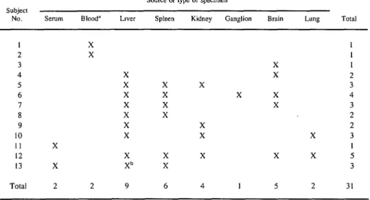
The postmortem specimens, obtained from 13 subjects under 13 years of age, were sent to our laboratory during the epidemic by various pedia- tric hospitals. The samples were transported to the laboratory at -20°C and were processed im- mediately upon arrival. Table 1 identifies the specific types of specimens received in each case.
Each specimen was processed as follows: A 10% suspension was prepared in medium 199 containing 10% calf serum, 500 units of penicil- lin per ml, and 500 micrograms of streptomycin per ml, The suspension was then diluted 1 :5,
1: 10, and I:50 using the same medium, and 0.02 ml of each dilution was inoculated intra-
cerebrally and subcutaneously into unweaned mice 24-48 hours old. The mice were then ob- served 21 days for any signs of disease, and successive passages were made in doubtful cases.
Virus identification was accomplished by means of the indirect immunofluorescence technique described in an earlier report (II).
In two cases where serum from the patient was available (see Table 1), the hemagglutina- tion-inhibition procedure described by Clarke and Casals, modified for microtechnique, was used to determine the subject’s antibody titer (12). Eight units of antigen of the four dengue serotypes were used; sera with antibody titers of l/20 or more were considered positive.
Clinical Data
Two of the study subjects were less than three months old, eight were four to six years old, and three were eight to 12 years old. Nine were girls and four were boys. Racially, 10 were whites and three were mulattoes. The illnesses
Table 1. Postmortem specimens processed for virus isolation, showing the subject from whom each specimen was obtained.
Subject
NO. Serum Blood*
Source or type of spec*men
LlVW Spleen Kidney Ganglion Brain Lung Total
I 2 3 4 5 6 7 8 9 IO
II X
12
13 X
Total 2 2 9 6
X X
X
X X
X X
X X
X X
X X
X X
Xb X
X X X
X X
X
X
X X
X X X
4 I 5 2
1 1 1 2 3 4 3 2 2 3 I 5 3
31
‘Right ventricular blood.
of all these subjects were diagnosed clinically as Grade IV DHF/DSS according to the WHO classification (13).
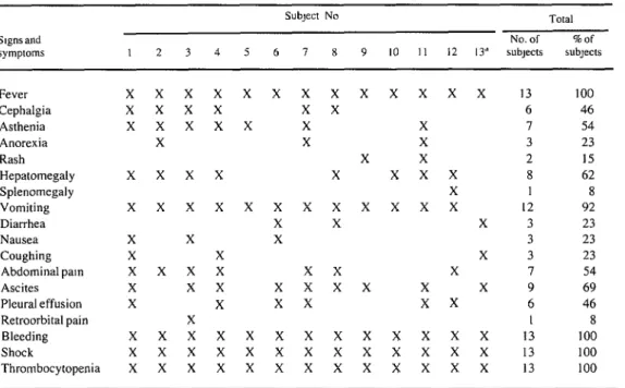
The principal disease signs and symptoms manifested by each subject are shown in Table 2, and the principal hemorrhagic manifestations observed are shown in Table 3. Fever, hemor- rhage, shock, and thrombocytopenia were pre- sent in all cases. Other commonly reported signs and symptoms were vomiting (in 12 cases), as- cites (in nine cases), hepatomegaly (in eight cases), asthenia (in seven cases), and abdominal pain (in seven cases).
Regarding specific hemorrhagic manifesta- tions, those most commonly reported were bleeding from the upper digestive tract (in 12 cases), petechiae (in six cases), bleeding from venipuncture sites (in four cases), and hematuria (in four cases).
Eleven of the 13 subjects experienced shock within six days of the onset of fever, most fre- quently on the second, third, or fourth day. Ten of the subjects died between days three and six, the largest number dying on day six.
Case 1. A white female patient five years of age showed symptoms of catarrh, high fever, asthenia, nausea, coughing, abdominal pain, vomiting, and cephalgia three days before enter- ing the hospital. She was admitted with signs of shock, trembling, convulsions, dark vomit, hepatomegaly, ascites, pleural effusion, and bleeding from the oral, gingival, and nasal mucosae. Her platelet count was 45,000/mm3. She died from cardiorespiratory arrest 24 hours after being admitted.
Case 2. A white male patient five years of age who was asthmatic complained of dengue symptoms three days before entering the hospital He was admitted with bloody vomiting, hepato- megaly, signs of incipient shock, and a platelet count of 59,000/mm3. He died 24 hours after admission with a diagnosis of DHF/DSS.
Case 3. A mulatto female patient 12 years of age had a fever of 38”C, nausea, retroorbital pain, and general malaise four days before enter- ing the hospital. During her hospital stay she experienced fever, cephalgia, asthenia, vomit- ing, abdominal pain, ascites, hepatomegaly,
gingival bleeding, and bleeding from ven- ipuncture sites. Her platelet count was 58,000/ mm3. She lapsed into shock and died on the third day after being admitted.
Case 4. A white female patient five years of age was admitted to the hospital with fever, cephalgia, vomiting, abdominal pain, hepato- megaly, ascites, pleural effusion, upper diges- tive tract bleeding, paleness of the skin and mucosae, hematuria, and signs of shock. Her platelet count was 50,000/mm3. She died from respiratory arrest on the second day after being admitted.
Case 5. A white female patient eight years of age was admitted with fever, severe asthenia, bloody vomiting, enterorrhagia, petechiae on the torso, and erythematous lesions over her whole body. Her platelet count was 12,00O/mm’. She lapsed into shock and died from cardiorespirato- ry arrest on the day of her admission to the hospital.
Case 6. A white female patient four years of age experienced a fever of 39°C and vomiting three days prior to being admitted to the hospital. She was hospitalized with ascites, pleural effu- sion, petechiae, upper digestive tract bleeding, a platelet count of 56,000/mm3, and signs of incipient shock. She remained in that condition and died from cardiorespiratory arrest three days after being admitted.
Case 7. A white male patient eight years of age who was admitted to the hospital experi- enced a fever of 39-4O”C, bloody vomiting, petechiae, bleeding from venipuncture sites, cephalgia, asthenia, ascites, pleural effusion, thrombocytopenia, and shock. He died from car- diorespiratory arrest five days after admission.
Case 8. A white male patient three years of age who was asthmatic experienced a fever of 39”C, cephalgia, vomiting, diarrhea, upper di- gestive tract bleeding, ascites, hepatomegaly, hematuria, and signs of shock. His platelet count was 33,000/mm3. He died from cardiorespira- tory arrest four days after admission.
216 PAHO BULLETIN l vol. 18, no. 3, 1984
* Table 2. Principal signs and symptoms observed in the 13 study subjects.
S~gnsand
symptoms I 2 3 4 5
Subject No Total
No. of %of
6 7 8 9 IO II I2 13” SUbJects SUbJeCts
Fever Cephalgia Asthenia Anorexia Rash Hepatomegaly Splenomegaly Vomiting Diarrhea Nausea Coughing Abdominal pam Ascites Pleural effusion Retroorbital pain Bleeding Shock
Thrombocytopenia
x x x x x x x x x xxxx 13 100
x x x x x x 6 46
x x x x x X X I 54
X X X 3 23
X X 2 I5
x x x x X x x x 8 62
X I 8
x x x x x x x x x x x x 12 92
X X X 3 23
X X X 3 23
X X X 3 23
x x x x x x X 7 54
X x x x x x x X X 9 69
X X x x x x 6 46
X I 8
x x x x x x x x x x x x x 13 100
xxxxxxxxxxxxx 13 100
xxxxxxxxxxxxx I3 100
“The subject from whom dengue-2 wus was isolated
Table 3. Principal hemorrhagic manifestations exhibited by the 13 study subjects.
Hemorrhagic
mamfestations I 2 3 4 5
Subject No. 6 7 8 9
Total No. of %Of 10 II I2 13’ subjects subjecfs
Petechiae Melena
Bleeding from upper digestive tract Gingival bleeding Nasal bleeding Oral bleeding Otic bleeding Bleeding from
venipuncture sites Hematuria Intestinal bleeding Purpura
Pulmonary bleeding
x x x x x x 6 46
X 1 8
x x xxxxxxxxxx 12 92
X X X 3 23
X X X 3 23
X 1 8
X I 8
X X X X 4 31
X X x x 4 31
X I 8
X 1 8
X I 8
ing, melena, ascites, a platelet count of 56,000/ mm3, otic bleeding, and metabolic acidosis. She subsequently lapsed into shock and died from pulmonary hemorrhagic and cardiac arrest.
Case 10. A female patient six years of age was hospitalized because of a fever of 40°C and vomiting, which later became dark. Painful he- patomegaly and distal cyanosis were revealed by physical examination. The patient lapsed into shock and died from cardiac arrest two days after admission. Her platelet count was 87,000/ mm3.
Case 1 I. A mulatto female patient five years of age, with a record of pultaceous tonsillitis treated with antibiotics, was admitted suffering from fever, a rash, vomiting, loss of appetite, and paleness of the skin and mucosae. Her con- dition worsened progressively, and she fell into shock accompanied by bleeding from the upper digestive tract, nostrils, and venipuncture sites. Physical examination revealed painful abdomi- nal distension, pleural effusion, and hepato- megaly. The patient’s leukocyte count was 15 ,250/mm3 and her platelet count was 56,000/ mm3. In addition, microscopic hematuria was observed. The patient died of cardiorespiratory arrest 24 hours after admission.
Case 12. A white female patient six years of age was admitted to the hospital with a history of 39-40°C fever and vomiting, which had con- tinued for four days. An assessment of her gen- eral condition at the time of admission revealed petechiae on her torso and extremities, fever, painful hepatomegaly, splenomegaly, abdomi- nal pain, and pleural effusion. The illness evolved toward shock accompanied by upper di- gestive tract bleeding, hematuria, and metabolic acidosis. The patient lapsed into a deep coma and died from cardiac arrest 24 hours after ad- mission. Her platelet count was 63,OOO/mm’.
Case 13. An unweaned male mulatto infant 45 days old was admitted to the hospital with a two-day history of 38-39°C fever and petechiae on the skin. At the time of admission he had a fever of 38”C, nasal mucus secretions, and dysp- nea. He later developed diarrhea, oral and peri- oral cyanosis, moderate gingival bleeding, upper digestive tract bleeding, and ascites. The patient
lapsed into shock several hours after developing these symptoms and exhibited moderate bleed- ing from the nose and venipuncture sites; he died several hours later. The patient’s leukocyte count was 17,30O/mm’ and his platelet count was 35,000/mm3. Virus isolation efforts using liver tissues from this patient subsequently yielded dengue-2 virus.
Results and Discussion
Anatomicopathologic studies of the foregoing subjects detected visceral or serous bleeding in 13% of the cases-predominantly of the gastric and intestinal mucosae, endocardium, lungs, and pleural cavity. Histopathologic examination revealed mediozonal and focal centrolobular hepatonecrosis in 75% of the cases.
Regarding virus isolation, one of the 3 1 post- mortem specimens processed by ourselves (the liver specimen from case number 13) yielded an isolate. This isolated virus was identified as dengue-2 by the indirect immunofluores- cence technique (I I). Hemagglutination-inhibi- tion testing of the two available serum specimens yielded negative results (<l/20).
Despite the fact that the clinical diagnosis of Grade IV DHF/DSS was confirmed by virus isolation from only one of the 13 study subjects. it is believed that the other patients also had DHF. This conclusion, derived partly from ap- plication of the analysis used by Barnes and Ro- sen in 1974, during a hemorrhagic fever epi- demic on the island of Niue (14). is based on the following observations:
1) The signs and symptoms observed were similar to those reported in Southeast Asia for virologically or serologically confirmed cases. 2) These cases occurred during the 198 1 den- gue-2 epidemic in Cuba, over the course of which a large number of other patients with a similar symptomatology were observed (9. 15).
218 PAHOBULLETIN l vol. 18, no. 3, 1984
Fever, shock, and thrombocytopenia were ob- served in all of the study subjects discussed above; also, all of them manifested one or more kinds of hemorrhagic symptoms, with bleeding from the upper digestive tract being the most common type. In general, the reported clinical pictures were similar to those reported from Fiji in 1977 by Kuberski et al. (16), from Malaysia in 198 1 by George et al. (17), and from Cuba in 1983 by Rojo et al. (18) for subjects with a positive diagnosis of DHF-the principal ob- served signs and symptoms being fever, hemor- rhagic manifestations, and vomiting. In addi- tion, George reported in his work that shock was the cause of death in all the fatal cases studied, this observation being similar to our own; and Eram et al. (19), reporting on rural Indonesian cases in 1979, refers to four patients with DHF/DSS who died of shock on the sixth or seventh day after the onset of illness, circum- stances again resembling those observed in our own study.
It also appears noteworthy that Bhamara- pravati et al. (20), studying autopsy data from 100 DHF/DSS fatalities in Thailand, reported that the most frequently observed pathologic lesions were petechial-type hemorrhages in various or- gans and mucosae, with the stomach being the internal organ most commonly affected. Hepato- necrosis was also common, being found in 65% of the subjects studied. Similarly, in our study we observed that some type of bleeding occurred in 83% of the cases studied, most often in the gastric and intestinal mucosae, and that hepato- necrosis occurred in 75% of the cases.
It is well-known that the frequency of virus isolation from DHF/DSS patients is much lower
than from patients with a classic clinical picture of dengue fever. This fact has been related to the accelerated and high antibody response oc- curring in cases of secondary infection, so that the invading virus particles tend to be bound up in immune complexes (21, 22). In this vein, Nisalak et al. (23) reported in 1970 that the frequency of virus isolation was inversely prop- ortional to the degree of severity of the disease; they obtained only four virus isolates from 103 postmortem sera and only two isolates from 523 postmortem organ specimens obtained from 98 subjects with fatal cases of DHF/DSS.
The low virus isolation rate obtained by our efforts (one isolation from 3 1 postmortem speci- mens for a rate of 3.2%) may well have been due, at least in part, to the foregoing circum- stances. And, conceivably, it could also have been due to use of the unweaned mouse isolation procedure, which although frequently utilized is not the most sensitive isolation method (24). In this vein, it should be noted that Mas (25) ob- tained two dengue-2 isolates from liver and spleen specimens taken from a fatal case of DHF, using a LLCMK, cell culture method for this purpose.
The isolate obtained in our study came from the liver of patient 13, an unweaned infant 45 days old whose serum did not reveal any dengue- 2 antibodies upon hemagglutination-inhibition testing. This serologic finding suggests the re- sponsible infection may have been a primary infection, even though it was not possible to obtain data’conceming the mother’s immunity to dengue virus. Other authors (14) have reported cases of DHF/DSS in patients with primary in- fection.
SUMMARY
The dengue-2 epidemic that struck Cuba in 198 1 and tissue specimens obtained from 13 children who caused some 344.203 cases. For the first time in the died in that epidemic. All 13 had been diagnosed clin- Caribbean, significant numbers of people were af- ically as having Grade IV dengue hemorrhagic fever/ flitted with dengue hemorrhagic feverjdengue shock dengue shock syndrome.
syndrome, and a total of 159 deaths were recorded. These children’s clinical histories showed that all This article reports the results of a postmortem of them manifested fever, hemorrhage, shock, and
vomiting (in 12 cases), ascites (in nine cases), hepa- tomegaly (in eight cases), abdominal pain (in seven cases), and asthenia (in seven cases); 11 of them experienced shock within six days of the onset of fever; and 10 of them died between days three and six.
Postmortem organ specimens from the children, as well as four serum or blood specimens, were sus- pended in an appropriate medium, diluted, and inocu- lated mtracerebrally and subcutaneously into un- weaned mice. This procedure succeeded in isolating
dengue-2 virus from the liver specimen of one sub- ject-a young infant who may have died of a primary dengue infection. This low rate of virus isolation may have been due to the well-known difficulty of isolating the viral agent from DHF/DSS cases, and also to the fact that the isolation method used was not the most sensitive existing method. Despite this circumstance, however, other findings and background factors relat- ing to the 13 fatalities leave little room for doubt that DHF/DSS was indeed the cause of death.
REFERENCES (I) Hammon, W. M. Dengue hemorrhagic fever: Do we know its cause? Am J Trop Med Hyg 22:82-&g,
1973.
(2) Halstead, S. B., J. E. Scanlon, P. Umpaivit, and S. Udomsadki. Dengue and chikungunya virus infection in man in Thailand, 1962- 1964: IV. Epidem- iologic studies in the Bangkok metropolitan area. Am J Trop Med Hyg 18:997- 102 1, 1969.
(3) Russell, P. K. Dengue in the Caribbean, 1977. In: Pan American Health Organization. Dengue in the Caribbean, 1977. PAHO Scientific Publication 375. Washington, D.C., 1979, pp. l-3.
(4) Pittaluga, G. Sobre un brote de dengue en La Habana. Rev Med Trop Parasitol Bacterial C/in Lab (Hnvanu) 1 l:l-3, 1945.
(5) Mas, P. Dengue Fever in Cuba in 1977: Some Laboratory Aspects. In: Pan American Health Organi- zation. Dengue in the Cnribbeun, 1977. PAHO Sci- enttfic Publication 375. Washington, D.C., 1979, pp. 40-43.
(6) Kouri, G., M. G. Guzman, J. Bravo, M. Ca- lunga. N. Cantelar, M. Soler, L. Morier, A. Feman- dez, and R. Femandez. El dengue hemorragico en Cuba. Algunos aspectos epidemiologicos, clinicos y virologicos de la epidemia de fiebre hemorragica del dengue de 198 1. Paper presented at the V Foro de la Academia de Ciencias de Cuba. Havana, 1982.
(7) Lopez Correa, R. H., B. L. Cline, C. Ramirez Ronda, R. Bermudez, G. E. Sather, and G. Kuno. Dengue fever with hemorrhagic manifestations: A re- port of three cases from Puerto Rico. Am J Trop Med Hyg 27:1216-1224, 1978.
(8) Fraser, H. S., W. A. Wilson, E. Rose, E. J. Thomas, and J. G. P. Sissons. Dengue fever in Ja- maica with shock and hypocomplementaemia, hae- morrhagic, visceral and neurological complications. West Indian Med J 27: 106- 116, 1978.
(9) Kouri, G., P. Mas, M. G. Guzman, M. Soler, A. Goyenechea, and L. Morier. Dengue hemorrhagic fever in Cuba, 198 1: Rapid diagnosis of the etiologic agent. Bull Pan Am Health Organ93:414-420, 1982. (IO) Kouri, G. Report presented to the Meeting
of Experts on Yellow Fever and Dengue sponsored by the Pan American Health Organization at Merida, Venezuela, in May 1982.
(II) Yamaguishi, H., and I. Yosmiona. A mod- ified method for the preparation of antigens for the indirect fluorescent antibody technique with Japanese encephalitis virus. Kitusuto Arch Exp Med 50:3-4, 1977.
(22) Clarke, D. H., and J. Casals. Techniques for hemagglutination and hemagglutination-inhibition with arthropod-borne viruses. Am J Trop Med Hyg 7: 561-573, 1958.
(13) World Health Organization. Guide for Diag- nosis, Treatment and Control of Dengue Haemor- rhagic Fever. Technical Advisory Committee on Den- gue Haemotrhagic Fever for the South-East Asian and Western Pacific Regions. Geneva, 1980, p. 8.
(14) Barnes, W. J. S., and L. Rosen. Fatal hemor- rhagic disease and shock associated with primary den- gue infection on a Pacific island. Am J Trop Med Hyg 23:495-506, 1974.
(15) Guzman, M. G., G. Kouri, J. Bravo, M. Soler, and S. Vasquez. Dengue hemorrhagic fever in Cuba, 1981: II. Study of patients clinically diagnosed with dengue hemorrhagic fever and dengue shock syndrome. Trans R Sot Trop Med Hyg (in press).
(16) Kuberski, T., L. Rosen, D. Reed, and J. Mataika. Clinical and laboratory observations on pa- tients with primary and secondary dengue type 1 in- fections with hemorrhagic manifestations in Fiji. Am
J Trop Med Hyg 261775-783, 1977.
(17) George, R., and G. Duraisamy. Bleeding manifestations of dengue haemorrhagic fever in Malaysia. Acta Trap 38:71-78, 1981.
(18) Rojo C., M.; M. Carriles D., C. Coto H.,
L. M. Lahoz B., C. Bosch S., B. Acosta P., M. Calderon S., A. Saavedra M., R. Marrero R., and M. Rodriguez A. Dengue hemorragico: Estudio clinico de 202 pacientes pediatricos. Rev Cubana Pediatriu 54:519-538, 1982.
220 PAHO BULLETIN l vol. 18, no. 3, 1984
demic dengue hemorrhagic fever in rural Indonesia: II. Clinical studies. AmJ Trop Med Hyg 28:71 l-716, 1979.
(20) Bhamarapravati, N., P. Tuchinda, and V. Boonyapaknavik. Pathology of Thailand haemorragic fever: A study of 100 autopsy cases. Am J Trop Med Parnsitol 61:500-510, 1967.
(21) Nimmannitya, S., S. B. Halstead, S. N. Cohen. and M. R. Marglotta. Dengue and chikun- gunya virus infection in man in Thailand, 1962-l 964: 1. Observations on hospitalized patients with hemor- rhagic fever. Am J Trop Med Hvg IS: 954-97 I , 1969.
(22) Halstead, S. B., S. Nimmannitya, and S. N. Cohen, Observations related to pathogenesis of den-
gue hemorragic fever: IV Relation of disease severity to antibody response and virus recovered. Yule J Bwl Med 42:31 l-328, 1970.
(23) Nisalak, A.. S. B. Halstead, P. Singharat, S. Udomsakdi, S W. Nye, and K. Vinijchaikul. Obser- vations related to pathogenesis of dengue hemorrhagic fever: III. Virologlc studies of fatal diseases. Yule J Biol Med 42~293-3 IO, 1970.
(24) Schlesinger, R. W. Dengue viruses. Vrrol Monogr 16:19, 1977.
(25) MBs, P. Instituto Naclonal de Hlglene, Epl- demiologia y Microblologia (Cuba). Personal com- munication. I98 I.
LOW LEVELS OF REYE’S SYNDROME RECORDED IN THE UNITED STATES
The U.S. Centers for Disease Control have received written reports of 222 and I9 I cases of Reye’s syndrome for the respective surveillance periods 1 December I98 l-30 November 1982 and 1 December 1982-30 November 1983; data for the latter (1983) period are preliminary. The sex and age distribution of cases was very similar for the 1982 and 1983 periods; and, as in the past, most patients were hospitalized during the first six months of each period. Although delayed reports were expected to increase the total number of cases in the 1983 period, as of early 1984 the two totals were the lowest recorded since continuous national surveillance of Reye’s syndrome was estab- lished in December 1976.
This relatively low reported incidence of Reye’s syndrome in 1982 and 1983 probably reflects, at least in part, the intensity and/or type of influenza activity. In terms of all available criteria, the intensity of influenza B activity in early 1982 was low. The intensity of influenza activity in 1983 was greater, although not as great as in l980- 198 I , and the predominant isolate was influenza A(H3N2) which has been previously as- sociated with fewer large clusters of Reye’s syndrome than influenza B.
The apparent drop in varicella-associated cases in 1982 (47 cases) and 1983 (25 cases) is currently less well explained by changes in virus activity; 83 such cases were reported in 198 1. From 1977 through 1982 (the years for which data are most recently available), the reported incidence of varicella itself has remained relatively stable.