Bull Pan Am Health Organ 17(Z), 1983.
DENGUE
HEMORRHAGIC
FEVER
IN CUBA,
1981:
RAPID
DIAGNOSIS
OF THE ETIOLOGIC
AGENT1
G. Kouri,*
Pedro M&,3 Maria G. GuzmB;n,* Maritza
Soler,5
A. Goyenechea,G and L. Morier7
The 1981 outbreak of dengue-2 in Cuba, which produced cases of hemorrhagic
fever, shock syndrome, and death,. prompted quick action by Cuban health authorities to diagnose the problem and isolate the responsible disease agent. This article describes that work and its results. It is expected that data relating
to the epidemic, derivedfrom this work and other sources, willprovide valuable
infomation about dengue etiopatholopy.
Introduction
Since 1827 many dengue epidemics have been reported in the Caribbean and other parts of the Americas. Initially, of course, diagnosis was based on the disease’s clinical characteristics (I); but when virologic and serologic methods became available, retro- spective studies could be made (2,3) to confirm earlier clinical observations. Subse-
quently, circulation of the four
dengue
sero-types was demonstrated on a number of Ca- ribbean islands and the American mainland.
Specifically, dengue-3 was found to be cir- culating in the Caribbean in 1963 (4,5), as was dengue-2 in 1968. Dengue-2 predomi- nated in the region in 1969, although dengue-3 was also present (6). In 1977 a dengue-1 epi- demic occurred in Jamaica; this soon spread to other Caribbean islands and the American
‘Also appearing in Spanish in the Boletin de la Oficinu Sanitaria Panamericana 93(5):414-420, 1982.
2Virologist; Director,‘@edro Kouri Institute of Trop- ical Medicine (IPK), Havana, Cuba.
%rologist; ‘Dep&y Direct& of Research; National In- stitute of Hygiene, Epidemiology, and Microbiology (INHEM), Havana, Cuba.
khief, Department of Virology, IPK. %irologist, IPK.
khief, Virology Laboratory, INHEM. %irologist, IPK.
mainland (7). In 1981 dengue-4 was reported circulating on several islands of the Lesser An- tilles-the first time this serotype had been found in the Americas (8). As of early 1981, only isolated cases of hemorrhagic fever and dengue shock syndrome had been reported in the Region (9,10).
With regard to Cuba, during this century clinically diagnosed dengue cases were re- ported in 1944 (II); but no further cases were notified until 1977, when an outbreak of dengue-1 in the Caribbean produced a large- scale epidemic in Cuba; in all, 477,438 cases of classical dengue were reported, and the presence of the virus on the island (12) was confirmed by viral isolation. A serologic sur- vey made in 1975, using dengue-2 antigen, showed 2.6 per cent of the subjects tested to be positive for dengue-2 antibodies. All those yielding positive responses were over 45 years of age (12), a result supporting the view that from 1944 to 1977 there was no dengue activi- ty on the island.
In late May of 1981 the health services of Havana Province8 detected a growing num- ber of patients with symptoms of fever; mus- cular, postorbital, and abdominal pain; rash; intense headache; and asthenia. In many cases
*An area coterminous with Metropolitan Havana.
Kouri et al. l DENGUE DIAGNOSIS IN CUBA, 1981 127
these symptoms were accompanied by hemor- rhagic manifestations with varying degrees of severity, and in some instances by shock and death.
In view of the urgency of the situation and the need to reach immediate diagnostic con- clusions, it was decided to study both single sera from patients experiencing the disease and a group of sera from patients who had had the disease over seven days for the purpose of ascertaining the hemagglutination-inhibition (HI) responses of these sera to dengue, yellow fever, and Chikungunya virus antigens. In addition, serum specimens were immediately collected from patients with acute symptoms for the purpose of virus isolation.
This article reports the results of those ini- tial serologic studies and the circumstances leading to identification of four virus strains isolated within a week of the time the epidemic was recognized. Over the course of a few days, these results made it possible to confirm the presumptive diagnosis initially submitted to the national health authorities. It should also be mentioned that the diagnosis of hemor- rhagic dengue cases was carried out according to the criteria established by the Technical Advisory Committee on Dengue Hemorrhag- ic Fever for the WHO Region of Southeast Asia and the Western Pacific (13).
Materials and Methods Serology
During the first day the epidemic was re- ported, serum specimens were obtained from four pediatric patients with manifestations of hemorrhagic dengue; and, on the following day, 16 sera were collected from people in the area where the epidemic began who reported having symptoms compatible with the disease during the preceding 20 days. (Eleven of these 16 subjects manifested hemorrhagic dengue symptoms.) All the sera were treated with
acetone; and the hemagglutination-inhibition procedure described by Clark and Casals (14), as adapted to micro-methods, was performed using dengue- 1, dengue-2, dengue-3, yellow fever, and Chikungunya virus antigens pre- pared in mouse brains and extracted by the saccharose-acetone technique.
Vim Isolation
Specimens. The serum specimens used for virus isolation were obtained from patients with clinically diagnosed dengue cases. All were collected within 96 hours of the onset of clinical symptoms. The specimens were trans- ported frozen and were maintained in a frozen state until use.
Inoculation in mice. The sera were inoculated into newborn mice by the intracerebral and subcutaneous routes. Pure sera and sera di- luted 1:lO and 1:50 with Medium 199 con- taining 10 per cent calf serum were used for these inoculations. One litter of mice was used for each inoculation at each dilution, an uni- noculated mouse in each litter being left as a control. The inoculated and control mice were then observed for 21 days.
Inoculation in tissue culture. Clone 9 of the LLC-MK2 cell line was obtained from Cuba’s National Institute of Hygiene, Epidemiology, and Microbiology in Havana. This cell line was maintained by serial passages in Medium 199 with 20 per cent calf serum, penicillin (100 I.U. per ml), and streptomycin (100 mi- crograms per ml). The cells were sown in plas- tic flasks, and 0.5 ml of each serum specimen was inoculated onto a confluent cell layer and allowed to remain in contact for 15 minutes at room temperature. Subsequently, the same medium with 1 per cent fetal calf serum was used for maintenance. The incubation tem- perature was 36’C, and the flasks were ex- amined daily for cytopathic effects.
128 PAHO BULLETIN l vol. 17, no. 2, 1983
V&s Identification
Plaque reduction. Virus strains isolated in both mice and tissue cultures were identified by means of the plaque-reduction neutraliza- tion test according to the method of Russell and McCown (1.5). For this purpose we used l:lO, 1:20, 1:40, and 1:80 dilutions of ascitic fluids hyperimmune to dengue virus types 1, 2, 3, and 4 received from the United States Centers for Disease Control (CDC) in Atlan- ta. These various ascitic fluid dilutions were mixed with equal volumes of each virus sus- pension (to yield final concentrations of 30- 100 plaque formation units (PFU) per 0.1 ml) and were placed in a water-bath for 45 min- utes at 37°C. Some of the mixture (0.3 ml) was then inoculated onto LLC-MK2 cell cul- tures and was permitted to remain in contact with the cells for 15 minutes. The cells were then overlaid with agar and incubated for five days, after which they were again overlaid- this time with agar containing a 1:7,500 dilu- tion of neutral red stain. Each culture was then read 24 hours after staining.
Immunofluorescence. This method was used for rapid identification of the virus strains isolated by inoculation into mice. Brain tissue specimens from the inoculated mice were mounted on slides, and the indirect immuno- fluorescence test was performed using 1:20,
1:40, 1:80, and 1: 160 dilutions of ascitic fluids hyperimmune to dengue virus types 1, 2, 3, and 4, and to yellow fever virus (received from
the Military Institute of Epidemiology and Microbiology of Prague) in combination with mouse antiserum conjugated with fluorescein isothiocyanate.
Results
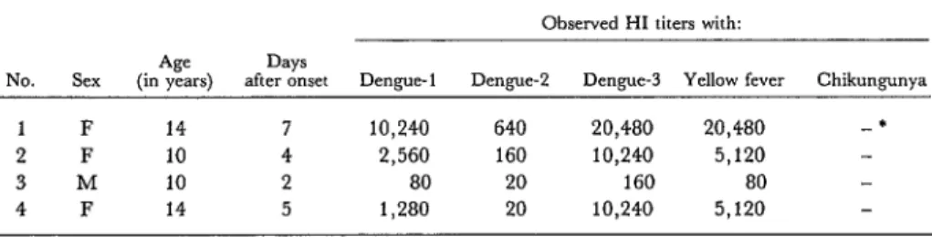
Table 1 shows the HI test results obtained with the single serum specimens from four children manifesting symptoms of hemor- rhagic dengue. As may be seen, the titers were extraordinarily high, with titers to dengue types 1 and 3 and to yellow fever being the highest. No antibodies to Chikungunya virus were detected.
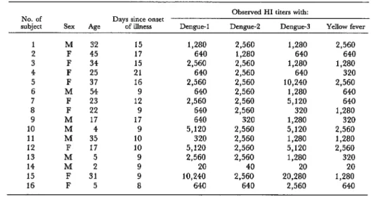
Table 2 shows HI test results obtained with 16 single sera collected approximately 48 hours after the epidemic became known. Most of these single sera, obtained between nine and 21 days after onset of the disease, were found to yield very high titers against the three dengue serotypes tested and against yellow fever virus. They also tended to show a certain predominance of high antibody titers against the dengue-2 serotype. Subject 14, a child two years of age, showed an antigenic response of the primary type (the last previous dengue epidemic in Cuba occurred in 1977). Subject 16, a child five years of age who died with symptoms of shock and hemorrhage, pre- sented a secondary type of antibody response.
Four strains of dengue-2 virus were iso- lated, the first two in newborn mice inoculated
Table 1. HI test results obtained with single sera from four children with manifestations of hemorrhagic dengue. The specimens tested were all collected within a week
of onset of disease symptoms.
Observed HI titers with: Age Days
No. Sex (in years) after onset Dengue-1 D+3lgUd! Dengue-3 Yellow fever Chikungunya
1 F 14 7 10,240 640 20,480 20,480 -*
2 F 10 4 2,560 160 10,240 5,120
3 M 10 2 80 20 160 80
4 F 14 5 1,280 20 10,240 5,120
Kouri et al. l DENGUE DIAGNOSIS IN CUBA, 1981 129
Table 2. HI test results obtained with single sera from convalescents who were examined within approximately 48 hours of the time the epidemic became known. These sera
were proCured during the second and third weeks of illness.
No. of
subject
Observed HI titers with:
Days since onset
Age of illness Dengue-1 Dengue-2 Dengue-3 YelIow fever
1 M 32 15 1,280 2,560 1,280 2,560
2 F 45 17 640 1,280 640 640
3 F 34 15 2,560 2,560 1,280 1,280
4 F 25 21 640 2,560 640 320
5 F 37 16 2,560 2,560 10,240 2,560
6 M 54 9 640 2,560 1,280 640
7 F 23 12 2,560 2,560 5,120 640
8 F 22 9 640 2,560 320 1,280
9 M 17 17 640 320 1,280 320
10 M 4 9 5,120 2,560 5,120 2,560
11 M 35 10 320 2,560 1,280 1,280
12 F 17 10 5,120 2,560 5,120 2,560
13 M 5 9 2,560 2,560 1,280 320
14 M 2 9 20 40 20 20
15 F 31 9 10,240 2,560 20,280 1,280
16 F 5 8 640 640 2,560 640
with serum taken from two patients on the third day of their illness. Strain one was iso- lated from a mouse on the third day following its inoculation, after the mouse showed typical disease symptoms. This mouse had been in- oculated with a 1:50 dilution of the serum. (Subsequently, sickness was observed in other mice that had received this serum pure, di- luted l:lO, and diluted 1:50.) Dengue was therefore diagnosed on the third day after mouse inoculation, and the serotype involved was determined on the fourth day (see Table 4) by means of the indirect immunofluores- cence method. The second dengue-2 strain was isolated 16 days after mouse inoculation from a mouse receiving a 1:50 dilution of se- rum.
and two were women 31 and 25 years of age, respectively, and those yielding strains three and four were boys 14 and 15 years of age, respectively. HI tests with sera from the first two patients failed to detect dengue anti- bodies; sera from the last two patients were not subjected to HI testing.
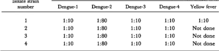
Tables 3 and 4 show data obtained with plaque reduction testing of these four isolates and indirect immunofluorescence testing of the first two. Plaque reduction testing of all four strains yielded very similar results. That is, 50 per cent plaque reduction was produced in each case by a 1:80 dilution of the dengue-2 hyperimmune mouse ascitic fluid, while the yellow fever and other dengue ascitic fluids yielded results that were essentially negative. The third and fourth dengue-2 strains were In a similar fashion, isolates one and two isolated directly in LLC-MK2 cell cultures in- yielded positive immunofluorescence test re- oculated with the sera of patients who had sponses with dengue-2 hyperimmune ascitic been sick for four and two days, respectively. fluid dilutions of 1:160 and 1:80. However, The cytopathic effect, evidenced by a round- much lower dilutions (no greater than 1:20) ing off and detachment of cells, began to ap- were required to produce detectable immuno- pear on the fifth day after inoculation. fluorescence with ascitic fluid sensitized to
130 PAHO BULLETIN a vol. 17, no. 2, 1983
Table. 3 Identification of the four isolated dengue strains by the plaque reduction method.
Isolate strain number
Highest dilutions of hyperimmune ascitic fluid for the indicated virus producing a plaque reduction of at least 50 per cent
Dengue-1 DeIlgW2 Dengue-3 Dengue-4 Yellow fever
1 1:lO I:80 1:lO 1:lO 1:lO
2 1:lO 1:ao 1:lO 1:lO Not done
3 1:lO 1:80 1:lO 1:lO Not done
4 1:lO 1:80 1:lO 1:lO Not done
Table 4. Immunofluorescence test results obtained with the first two dengue-2 strains isolated.
Dengue strain tested
Highest dilutions of as&c fluid for the indicated virus at which immunofluorescence was apparent
Dengue-1 D~~gWZ Dengue-3 Dengue-4 Yellow fever
Isolate 1 1:20 1:160 1:20 Not done
Isolate 2 1:80 1:20 1:20
Dengue-2
(from New Guinea) 1:20 1:640 1:20 Not done
- = Negative result.
Discussion and Conclusions
Overall, these data clearly suggest that the virus causing the epidemic of dengue hemor- rhagic fever which occurred on Cuba in mid- 1981 was dengue-2. As in the 1977 epidemic (121, it was not possible to determine the cir- culating dengue serotype by means of HI test- ing, a difficulty that was confirmed as the epidemic progressed. However, correlation of the clinical, epidemiologic, and serologic find- ings made it possible to determine within hours that we were facing an outbreak of dis- ease cases caused by a new dengue serotype, together with an epidemic of hemorrhagic fever. On the basis of these findings, a cam- paign to control this epidemic and Aedes at- g#ti, the responsible mosquito vector, was launched immediately.
Subsequently, the responsible dengue-2 vi- rus was quickly isolated and identified. As al- ready noted, this was done four days after
mouse inoculation by means of the indirect immunofluorescence test, the results of which were confirmed by plaque reduction.
Kouriet al. l DENGUE DIAGNOSIS IN CUBA, 1981 131
In late May of 1981 health services in Cuba’s Havana Province recorded increasing numbers of disease cases with dengue-like symptoms, together with cases of apparently dengue-related hemor- rhage, shock, and death.
Responding to this threat, health authorities ob- tained sera from patients currently manifesting dengue-like symptoms as well as from others who had had the disease for over seven days. These were subjected to hemagglutination-inhibition (HI) tests for the purpose of disease diagnosis, and to various other procedures designed to isolate and identify the responsible virus.
Sera from four children with hemorrhagic symp- toms yielded high antibody titers against yellow fever, dengue-1, and dengue-3 viruses and rela- tively low antibody titers against dengue-2. How- ever, 15 of 16 convalescent sera showed strongly positive antibody responses to all four viruses.
One of various mice inoculated with patients’ sera in an attempt to isolate the virus became sick three days after inoculation. The following day, im-
SUMMARY
munofluorescence testing of the mouse’s brain tis- sue, using ascitic fluids hyperimmune to dengue virus types 1, 2, 3, and 4 and to yellow fever virus, indicated that dengue-2 was responsible for the epi- demic. Subsequent testing of this and other virus isolates provided confirmation.
Data gathered during the course of this epidemic, where cases of hemorrhagic fever and shock syn- drome occurred during the second of two successive eprdemics caused by different dengue serotypes, are expected to provide valuable information about dengue etiopathology.
Because no other dengue-2 activity had previous- ly been reported in the Americas or in countries with which Cuba maintains close relations, the origins of the epidemic are unclear. However, the outbreak has served to alert health authorities in the Caribbean and other vulnerable portions of the Americas that dengue hemorrhagic fever and shock syndrome have been occurring in an epidemic fashion for the first time in this part of the world.
REFERENCES
(1) Ehrenkranz, N. J., A. K. Ventura, R. R. Cuadrado, W. L. Pond, and J. E. Porter. Pan- demic dengue in Caribbean countries and the southern United States: Past, present, and poten- tial problems. N EnglJ Med 285: 1460-1469, 1971.
(2) Rosen, L. Observations on the epidemiology of dengue in Panama. Am J Hyg 68:45-58, 1958.
(3) Downs, W. G. Immunity patterns produced by arthropod-borne viruses in the Caribbean area. An Inst Hig Med Trap (Lisbon) lG(Supp1 9):88-100,
1959.
(4) Russell, P. K., E. L. Buescher, J. M. Mc- Cown, and J. Ordoiiez. Recovery of dengue vi- ruses from patients during epidemics in Puerto Ri- co and East Pakistan. Am J Trap Med Hyg 15: 573-579, 1966.
(5) Spence, L., A. H. Jonkers, and J. Casals. Dengue type 3 virus isolated from an Antiguan pa- tient during the 1963-1964 Caribbean epidemic. Am J Trot Med Hyg 181584-587, 1969.
(6) Ventura, A. K., and C. M. Hewitt. Recov- ery of dengue 2 and dengue 3 viruses from man in Jamaica. Am J Trap Med Hyg 19:712-715, 1970.
(7) Pan American Health Organization. Dengue in the Caribbean, 1977. PAHO Scientific Publication 375. Washington, D. C., 1979.
(8) United States Centers for Disease Control. Dengue type 4 infections in U. S. travelers to the Caribbean. Morbidity and Mortality Weekly Report 30(21):249, 1981.
(9) Lopez Correa, R. H., B. L. Cline, C. Ra- mirez Ronda, R. BermGdez, G. E. Sather, and C. Kuno. Dengue fever with hemorrhagic manifesta- tions: A report of three cases from Puerto Rico. Am J Trap Med Hyg 27:1216-1224, 1978.
(10) Fraser, H. S., W. A. Wilson, E. Rose, E. J. Thomas, and J. G. P. Sissons. Dengue fever in Jamaica with shock and hypocomplementaemia,
haemorrhagic, visceral, and neurological complica- tions. West Indian MedJ 27~106-116, 1978.
(21) Pittahtga, G. Sobre un brote de “dengue” en La Habana. Rev&a de Medicinu Tropicaly Parasito-
logia, Bacteriologia, C&cay Laboratorio. 11: l-3, 1945. (12) Mas, P. Dengue Fever in Cuba in 1977: Some Laboratory Aspects. In Pan American Health Organization. Dengue in the Caribbean, 1977. PAHO Scientific Publication 375. Washington, D. C., 1979, pp. 40-43.
132 PAHO BULLETIN . vol. 17, no. 2, 1983
East Asian and Western Pacific Region. Geneva, Hyg 7:561-573, 1958.
1980, p. 7. (15) Russell, P. K., and J. M. McCown. Com-
(14) Clarke, D. H., and J. Casals. Techniques parison of dengue 2 and dengue 3 v&us strains by for hemagglutination and hemagglutination-inhi- neutralization tests and identification of a subtype bition with arthropod-borne viruses. Am J Trap Med of dengue 3. Am J Trap Med Hyg 21:97-99, 1972.
SURVEY OF ACUTE RESPIRATORY INFECTIONS IN CHILE Chile’s Institute of Public Health in Santiago has been actively involved in surveillance of acute respiratory infections, including influenza, for sev- eral years. Data for 198 1 indicated that acute respiratory infections in chil- dren were responsible for about 50 per cent of all consultations, the pre- vailing pathogens detected being respiratory syncytial virus and parain- fluenza virus type 3.
A study on viral respiratory infections among children under two years of age was carried out in the period May-December 1982; 199 disease cases were investigated. The highest number of cases was found during the win- ter months (June-August) when an outbreak of respiratory syncytial virus occurred; this outbreak reached a peak during the last week of July.
Positive laboratory results were obtained in 88 of the 199 investigated cases, over half of which occurred in infants under six months old. Respira- tory syncytial virus was the most commonly diagnosed agent. It alone was detected in 56 cases, and in seven more cases it was found in conjunction with other viruses. Parainfluenza virus types 1, 2, or 3 were detected in 10 cases, adenoviruses in nine cases, and herpes simplex and Mycoplasma pneu- moniae in one case each. In 11 cases more than one agent was detected; these multiple infections usually involved respiratory syncytial virus in combina- tion with adenovirus, parainfluenza virus, or enterovirus, or else infection with two or three types of parainfluenza virus.