S
PECIAL REPORT
E
SSENTIAL RADIOLOGY:
THE PAHO-WHO SYSTEM1
Gerald l? Hanson2
He&h services in Latin America, the Caribbean, and deueZoping areas
around the WorZdsufler from a severe shortuge of radiology services. To help
overcome this problem,
PAHOand lts~~o have he&t& to devdop a Basic Ra-
di0ZogicuZ System that offers
gooa
sohtions to the fundamentaZ training,
safety, adcostprobZems invoZved. The foZZowing account describes their ex-
perience to date with that system.
Introduction
Within the Americas the nature of
available diagnostic radiology services varies from country to country, locale
to locale, and often reliable data about these services are lacking. However, as
a general mle the following appears true of these services in Latin America
and the Caribbean (I, 2):
1
In rural and marginal urban areas, most people do not have access
to diagnostic
radiology.
2 About
half of the rural hospitals (defined as those with about 50 beds or less) do not provide diagnostic radiology services.3 Some 80 to 90% of most countries’ installed X-ray equipment is in the capital city or a few other large cities; very few X-ray machines are found in cities of 100,000 inhabitants or less.
4 Of the X-ray equipment that is installed, some 30 to 60% is not in working order. 5 Diagnostic radiology services in most big-city hospitals are saturated, and patient
waiting times for X-ray examinations are long.
6 Many simple X-ray examinations are performed at university or referral hospitals because there is no other alternative.
7 Radiological diagnostic procedures are often conducted without due regard for proper indications, expected diagnostic yields, or adequate performance (includ- ing limitation of the patient’s dose to optimal levels).
8 In most countries, medical students have little or no experience with radiology services before beginning their professional careers. A prime need therefore exists to institute this training, and to include within it appropriate guidance on radia- tion protection.
These general conclusions are sup- ported, among other things, by recent sample surveys conducted in five Latin American countries. Those surveys showed that a very high percentage of all X-ray examinations performed at referral and university hospitals are of a routine nature (3). (Many of these examinations should be conducted at local hospitals, but are not because the necessary staff and equipment are lacking or insufficient.) The surveys also found that only a small fraction of the pa- tients given medical care at local hospitals received X-ray examinations. In some cases the percentage of patients receiving an X-ray examination at large-city hospitals was 15 times greater than the percentage receiving such an examination at local hospitals.
The PAHO/WJXO Basic Radiological System (BRS)
In March 1975 a meeting was held at
PAHO Headquarters in Washington, D . C . , to defme the type of X-ray system that could best serve the radiological needs of developing countries. Specifi- cations developed at this meeting were improved and published by the World Health Organization in 1982 (4-S). The current WHO specifications for the Basic Radiological System’s X-ray unit, plus accessories, may be sum- marized as follows:
1 Output of the generator should be high enough to (a) produce a minimum expo- sure of 0.5 milliroentgens in one second or less at a focus-film distance of 140 cm behind a water phantom of 30 cm thickness, and (b) produce 0.5 milliroentgens in less than 50 milliseconds at 140 cm behind a water phantom of 12 cm thickness.
2 The unit should have a rotating anode X-ray tube with a focal spot of less than one millimeter capable of handling 20 kW for an interval of 0.1 second. 3 The inherent filtration of the tube must be equivalent to at least 2.5 mm of
aluminum.
4 The control panel should indicate the status of the electrical supply, the chosen kilovolt (kV) and milliampere (mA) values, and the object thickness. Only four kV values are possible: 120, 90, 70, and 55 kV. The minimum range of mA values, usable over the entire kV range, is 0.8-200 in 25 steps.
5 The design must ensure that the tube is always connected to the cassette holder in a rigid and stable way, permitting precise centering of the X-ray beam. A fured- focus film distance of 140 cm must be used.
6 A stationary, focused lead/aluminum grid with 40 to 50 lines per centimeter and a ratio of 10: 1 must be included.
7 The tube must be provided with an adequate collimator permitting restriction of the X-ray beam to the size of the films used. ‘Ibe collimator design must prevent any part of the patient from being closer to the X-ray source than 30 cm. The smallest format of the collimator must be no larger than 18 x 24 cm.
8 A movable pointer or other reliable system for centering the beam must be provided.
9 Film sizes should be standardized, and no more than four film sizes should be used. The cassette holder must accept at least the following three formats: 35.5 x 43 cm, 18 x 43 cm, and 24 x 30 cm.
10 The support provided for the patient must be rigid, must have an X-ray permea- ble top, and must be able to support a weight of 110 kg without appreciable distortion.
11 Strict time-temperature control must be used in the film processing. Darkroom equipment must be provided with the X-ray equipment.
12 A standard range of patient protection devices must be provided with the X-ray machine.
13 The cassette holder must incorporate a lead shield with a minimum thickness of 0.5 mm in the back wall.
14 At least one protective apron and one pair of gloves with a minimum thickness equivalent to 0.25 mm of lead must be provided.
15 A protective screen large enough to protect a standing operator must be an inte- gral part of the control panel. The lead equivalent must be at least 0.5 mm, provided that the X-ray beam is never directed at the screen. A leaded glass win- dow no smaller than 30 cm must be incorporated into the screen.
Experience with the Basic Radiological System (BRS).
By agreement
between the Government of Colombia and
PAHO,testing of four BRS-type
X-ray machines manufactured and donated by the General Electric Com-
pany was started in Antioquia in the fall of 1983. In one week, engineers
from the General Electric Company installed four X-ray machines in three
small hospitals serving rural areas and in one large health center serving a
marginal urban area of Medellin (9,
IO).In less than two weeks, two assistant
nurses and one physician from each hospital had been trained; and after five
days of intensive work at Medellin, followed by one or two days of practical
training in the local hospitals, the X-ray operators were obtaining reasonably
satisfactory results with about
100standard X-ray projections.
The Basic Radiological System in-
cludes not only appropriate X-ray equipment but also appropriate training
for personnel and appropriate integration of radiological diagnosis into the
health services sytem. Therefore, the field test included training of operators
who were residents of the local community, as well as training of general
medical staff members to evaluate the most common radiographic proce-
dures required to diagnose local pathologic conditions. Other components of
the system include an appropriate methodology for technical and profes-
sional support and referral, and also organization of an efficient supply and
maintenance network. Supervision (another integral part of the system) was
provided by a professor of radiology and by radiology residents from the Uni-
versity of Antioquia.
In
1985four
WHOBRS-type machines
manufactured by the Siemens Company were installed in Nicaragua and one
One of these Siemens machines was
recently
testedat the Mount Sinai Medical Center in Miami Beach, Florida.
This machine is currently providing services at the General Hospital in
Mex-
ico City. Other field trials of
the BRS have been organized in Africa, Asia,
Europe, and the Middle East; the results of all these trials have been very good (11).
Overall, these generally favorable ex-
periences
have led to the following conclusions:
1 There are very few examinations that cannot be made by the
BRS
operator using the WHO Basic RadidogicaZ System MangaL of Radiographic Techniqzle (12)-a document that was especially prepared for use with the BRS X-ray machine. (The manual excludes contrast studies of the alimentary tract.)2 The quality of the radiographs is excellent, even when judged by the standards of the most developed institutions.
3 An abbreviated training period of approximately two weeks is sufficient to teach the operator how to produce the standard radiographic projections, use the equip- ment, and apply the WHO manual, but is insufficient for proper instruction in darkroom techniques.
4 With the exception of some early problems in Nicaragua, no significant faults have been discovered in any of the WHO BRS-type machines.
5 Continuing on-the-job instruction by experienced radiologists and technicians is an essential part of the system and must be incorporated into any program utilit-
ing the PAHO/ WHO BRS.
Premises Requirements for Installation of the BRS.
Generally, three
rooms will be required for a BRS installation: the radiographic room, a dark-
room, and a combination of&e-storeroom. The location should be chosen
for easy patient access, as sufficient radiation protection is incorporated into
the design of the machine or can be provided easily. Access to water supply
and wastewater drainage facilities are required for the darkroom. The electri-
cal supply needed is 5 amperes at
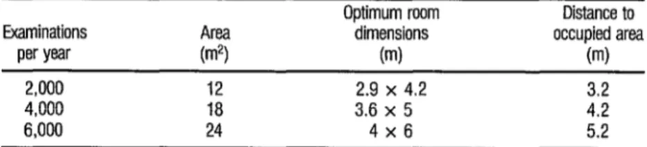
110 or 230 volts and 50 or 60 hertz.Safe operation of a BRS-type unit in-
stalled at almost any location depends upon incorporation of enough lead
into the back of the cassette holder to absorb almost all of the primary radia-
tion after it passes through the patient. Current
WHOspecifications require a
minimum of
0.5mm of lead, enough to provide adequate protection at
a design level of
10milliroentgens per week for the conditions shown
in Table
1.TABLE 1. Design charactetktics of MS X-ray room for satisfactory radiation protection.
Examinations AWl
per year (m2)
2,000 12
4,000 18 6,000 24
Optimum room dimensions
(ml
2.9 x 4.2 3.6 x 5
4x6
Distance to occupied area
(m)
3.2 4.2 5.2
The first essential radiology or BRS
specifications included the following minimum room dimensions:
radiographic room
18 m2,minimum ceiling height 2.5 m
processing room
2 m2, minimum ceiling height 2.25 m
office
8m2, minimum ceiling height
2.2 5m
While the field trials demonstrated
that it was possible to install a machine in less space (e.g., a radiographic
room of about
10m2 with a ceiling height of about 2.2 m, a processing room
of about 4 m2, and no office space), the original room dimension specifica-
tions (or the modifications taking workload into consideration as shown in
Table
1)are highly desirable for purposes of both operation and protection.
If the minimum requirements speci-
fied in the table are met, and if the workloads are typical of those found in
most rural hospitals (e.g., less than
10milliampere-minutes per week) it is
quite probable that no additional shielding, besides that provided by the
room walls, will be required.
Radiation Protection Experience
Radiation protection surveys have
been conducted at the aforementioned test sites in Chile, Colombia, and the
United States in collaboration with personnel of local radiation protection
services pertaining to the ministries of health in Colombia and Chile and the
Mount Sinai Medical Center in the United States (13). The results of these
surveys, which were made on the
WHOBRS-type X-ray machines manufac-
tured by the General Electric, Philips, and Siemens companies, can be sum-
marized as follows:
1 The total permanent filtration of the useful X-ray beam met or surpassed the requirements of the International Commission on Radiological
Protec-
tion
(ICRP) (14).2 The useful X-ray beam was precisely aligned with the X-ray film. Tests performed with a beam alignment test tool demonstrated that the beam-centering was within two degrees of strict coincidence.
3 The collimator limited the useful beam to the area of the film. In all cases the difference between the X-ray field and the film was within the generally accepted limits of 2 % of the source-to-image distance.
4 The movable patient pointer easily enabled the operator to ensure that the area of clinical interest was in the center of the X-ray beam.
6 The amount of radiation scattered to the operator’s position in front of the protec-
tive shield was very small, and it was not possible to detect radiation behind the
protective shield.
7 The reproducibility of the radiation output during repeated exposures at various
selected kilovolt and milliampere values was excellent.
With regard to patient exposure, the
radiation dose that a patient might receive during the most common X-ray
examinations (chest, abdomen, lumbo-sacral spine, and skull) was estimated
utilizing a methodology developed by the U.S. Center for Devices and Ra-
diological Health. The results for the
WHOBRS-type X-ray machines manu-
factured by General Electric, Philips, and Siemens that were used in clinical
field trials in Latin America were then compared with average values ob-
tained in the United States and with ranges of values reported by the
ICRP(15). With only one exception (this being the chest radiographs at one test
site) the patient’s radiation exposure from the
WHOBRS-type machines was
less than the average patient exposure values found in the United States,
where a concerted effort has been made over the past two decades to reduce
the patient’s radiation exposure.
Significantly higher patient exposure
values were obtained on a sample basis from various conventional X-ray de-
partments throughout Latin America that were not using
WHOBRS-type
equipment. In one instance (without control for differences in the film, in-
tensifying screens, and processing) the patient exposures from the four com-
mon examinations mentioned previously were found to be 250% to 700%
higher than patient exposures from a
WHOBRS-type unit installed nearby. In
each of the two X-ray departments involved, the same consultant radiologist
examined the radiographs and judged them to be of satisfactory diagnos-
tic quality.
Conclusions
The Basic Radiological System devel-
oped by
PAHOand
WHO,within the context of extending coverage to under-
served populations, makes use of high-quality components. These (includ-
ing the tube, generator, focused grid, tube stand, and patient examination
table) have been combined in an optimum design configuration that is de-
ceptively simple. As a result of a profound analysis of the X-ray examination
process (an analysis that preceded issuance of the
WHOspecifications), it has
become possible for a health worker with a minimum amount of training to
consistently produce high-quality radiographs.
Due partly to elimination of all elec-
trical components except the X-ray tube and generator, the BRS X-ray ma-
chine is rugged, easy to install, and easy to maintain.
Protection of the patient and X-ray
operator have been incorporated into the design. Results obtained in field
trials have shown that much less radiation exposure is received by the patient
when the BRS machine is used than when similar examinations are per-
formed with conventional X-ray machines.
The
WHOBasic Radiological System
employs the most appropriate available technology for producing a high-
quality radiograph at reasonable cost in small hospitals and in health centers
serving marginal urban areas. In larger referral and university hospitals, the
BRS can perform about
80%to 95O/o of the X-ray examinations required,
thus liberating scarce resources to purchase and install more complex equip-
ment where it is truly needed.
References
1 Gmez Crespo, G., G. Hanson, and P. E. S. Palmer. Planning Data for Essential Radiol- ogy Services in Rural and Marginal-Urban Areas. In: International Symposium on the Planning of Radiolo
on the Planning o B
ical Departments. Book of Papers, Fourth International Symposium RadioLogicaL Departments. (ISPRAD IV, San Juan, Puerto Rico, 29 April-2 May 1984).
2 Hanson, G. Estrategia y mctodos para lograr el acceso universal a unos servicios radiolbgi- cos de alta calidad en AmErica Latina. Paper presented at the VII Spanish Radiology Semi- nar cosponsored by the Mt. Sinai Medical Centef, Miami Beach, and the Inter-American College of Radiology (Miami, Florida, 7-13 Apnl 1985).
3
4
5
G
7
Gdmez Crespq, .G., and G. Hanson. El Sistema OPSlOMS de Radiologia Esencial: C&no dar mejor servlclo con menos gasto. Bo/ Of San& Panam 100(5):548-555, 1986. Chamberlain, R. H. Basic radiology: A worldwide challenge. JAMA 214(9):1687-1992, 1970.
Pan American Health Organization, Department of Health Promotion. A Primaq Care RadioLogical System. (Report of a Meeting Held in Wasbzgton, D. C., on 17-2s March
1975). Washington, D.C., 1975.
Palmer, l? E. S. Radiology and Prima7y Care. PAHO Scientific Publication No. 357. Pan American Health Organization, Washington, D.C., 1978.
World Health Organization. Technical Specifications for the X-ray Apparatus to be Used in a Basic Radiological System (Updated Version of January 1985). WHO document RAD 85.1. Geneva, 1985.
Holm, T. New developments in basic radiographic systems. EurJRadiol’3:291-293, 1983. Palmer, P. E. S., G. Hanson, G. G6me.z Crespo, and 0. Nieto. A Status Report of the Clinical Field Trial of the Basic Radiological System in Colombia: So Far, So Good. Inter- national Symposium on the Planning of Radiological Departments. In: Book of Papers, Fourth International Symposium on the PLanning of RadioLogical’ Departments. (ISPRAD IV, San Juan, Puerto Rico, 29 April-2 May 1984).
a 2
10 Palmer, l? E. S., G. amez Crespo, G. Hanson, and 0. Nieto. El sistema OPSlOMS de radiologia esencial en Colombia. Rev Med Chil114:581-585, 1986.
2 11 Palmer, P. E. S. Basic radiological system. WbrLd Health, Geneva, June 1985.
13 Hanson, G., P. E. S. Palmer, and G. G6mez Crespo. Radiation Protection Considerations for Basic Radiology. In: Book of Papers, Fourth International Symposium on the Pthzing
of Radiologicul Departnzents. (ISPRAD IV, San Juan, Puerto Rico, 29 April-2 May 1984). 14 International Commission on Radiological Protection. Protection Against Ionizing Radti-
tion f;om External Sources Used in Medicine. ICRP Publication 33. Pergamon Press, Oxford, 1982.
15 International Commission on Radiological Protection. Protection of the Patient in Dhg- nostic Radiology. ICRP Publication 34. Pergamon Press, Oxford, 1982.
WHO
Suppats
Intrauterine
Devices
A WHO scientific group convened to look into the modes of action, safety and efficacy of intra- uterine devices (IUDs) recently concluded that they are “probably the most effective and reli- able reversible method of fertility regulation available to women.”
The group emphasized that ft was referdng to the
currently
available copper- and hormone- releasing IUDs, when property used.The experts also noted the particular situation in the United States, where two manufacturers discontinued making and marketing IUDs in re- sponse to increasing legal costs arising from lawsuits in which pelvic infection and subse quent infertifii were claimed to have resulted from IUD use. In their report, the experts stated that the decisions to withdraw the Lippes Loop, Copper-7, and TCu-200 IUDs from the Ameri- can market “were based on commercial and ff- nancial considerations rather than on questions of safety”
In general they considered the IUD to be “an important method of fertility regulation with high continuation rates and significant advantages in convenience of use.” But they also stressed the need to carefully screen women considering us- ing IUDs to ensure that no contraindications such as genital cancers, vaginal bleeding of un- known cause, suspected pregnancy or active pelvic infection were overlooked. The text of the group’s report has been published in WHO Technical Report 753, which is available from the World Heakh Organization, 1211 Geneva 27, Switzerland.
Soum: World Heailh Organization, Press Release WH0/26. 12 Cktober 1967.