(1)Medical and Experimental Mycology Group, Corporación para Investigaciones Biológicas (CIB), Medellín, Colombia. (2)Molecular Diagnosis Laboratory, Hospital Pablo Tobón Uribe, Medellín, Colombia.
(3)Microbiology School, Universidad de Antioquia, Medellín, Colombia.
Correspondence to: Ángel González, Corporación para Investigaciones Biológicas, CIB, Carrera 72A No. 78B-141, Medellín, Colombia. Phone: (574) 441-0855, ext. 219, 221. Fax: (574)
LYSOZYME PLAYS A DUAL ROLE AGAINST THE DIMORPHIC
FUNGUS
Paracoccidioides brasiliensis
Damaris LOPERA(1), Beatriz H. ARISTIZABAL(1,2), Angela RESTREPO(1), Luz Elena CANO(1,3) & Ángel GONZÁLEZ(1,3)
SUMMARY
In order to determine the role of lysozyme, an antimicrobial peptide belonging to the innate immune system, against the dimorphic fungus Paracoccidioides brasiliensis, co-cultures of the MH-S murine alveolar macrophages cell line with P. brasiliensis
conidia were done; assays to evaluate the effect of physiological and inflammatory concentrations of lysozyme directly on the fungus life cycle were also undertaken. We observed that TNF-α-activated macrophages significantly inhibited the conidia to yeast transition (p = 0.0043) and exerted an important fungicidal effect (p = 0.0044), killing 27% more fungal propagules in
comparison with controls. Nonetheless, after adding a selective inhibitor of lysozyme, the fungicidal effect was reverted. When P. brasiliensis propagules were exposed directly to different concentrations of lysozyme, a dual effect was observed. Physiologic
concentrations of the enzyme facilitated the conidia-to-yeast transition process (p < 0.05). On the contrary, inflammatory concentrations impaired the normal temperature-dependant fungal transition (p < 0.0001). When yeast cells were exposed to
lysozyme, irrespective of concentration, the multiple-budding ability was badly impaired (p < 0.0001). In addition, ultra-structural
changes such as subcellular degradation, fusion of lipid vacuoles, lamellar structures and interruption of the fibrilar layer were observed in lysozyme exposed conidia. These results suggest that lysozyme appears to exert a dual role as part of the anti-P. brasiliensis defense mechanisms.
KEYWORDS: Lysozyme; TNF-α; Alveolar macrophages; Paracoccidioides brasiliensis.
INTRODUCTION
In respiratory tract secretions, lysozyme is the most abundant antimicrobial peptide, and its local levels increase tenfold during inflammation31. The bactericidal properties of lysozyme are primarily
ascribed to its enzymic activity, resulting in hydrolysis of β-linkage between the N-acetylglucosamine and N-acetylmuramic of the bacterial peptidoglycan causing cell lysis. However, an increasing body of evidence supports the existence of non-enzymatic and/or non-lytic modes of action still not well characterized11,20. The antimicrobial action
of lysozyme contributes to the innate host defense and acts in concert with the mechanical and phagocytic clearance mechanisms of the host interacting also with other cationic proteins7. Lysozyme is present in
the phagocytic cells’ granules of neutrophils, as well as in macrophages, monocytes and epithelial cells. After its discovery by Alexander Fleming in 1922, the enzymatic action of lysozyme has been extensively characterized; however, its role in the host-parasite interaction has been studied more intensely in bacteria than in fungi.
Although lysozyme is constitutively expressed, providing an ever present defense, pro-inflammatory cytokines, such as tumor necrosis factor alpha (TNF-α), dramatically increase lysozyme levels. TNF-α
is a pleiotropic cytokine capable of increasing the respiratory burst stimulating lysozyme overproduction by human macrophages25,30.
Paracoccidioidomycosis (PCM) is one of the most important endemic mycosis in Latin America. This mycosis affects primarily the lung and is frequently followed by dissemination to mucosa, skin, adrenals and other organs and systems2,27. The natural infection is
acquired by inhalation of Paracoccidioides brasiliensis conidia,
propagules that convert into the invasive yeast form once in the lungs27.
In a murine model of PCM, it was observed that resistant mice (AS/n) infected with P. brasiliensis yeast cells were able to control the
infection when compared with susceptible mice (B10A); additionally, high levels of TNF-α were found in the former indicating an important role of this cytokine in host’s defense22.
It is also known that the mononuclear phagocytic system plays a fundamental role in resistance to P. brasiliensis17. On the same token,
macrophages activated with interferon gamma (IFN-γ) or TNF-α have antifungal properties against P. brasiliensis that can be dependent or
independent of nitric oxide production, respectively12,13. Although
to fungus control is not fully understood. In this study, we attempted to determine if lysozyme participates in the fungicidal/fungistatic mechanismagainst P. brasiliensis conidia or if this enzyme would affect either the conidia-to-yeast transition or the multi-budding process of this pathogenic fungus.
MATERIALS AND METHODS
Reagents: Hen egg white lysozyme, immunochemicals and all reagents employed in this study were obtained from Sigma Chemical Co. (St Louis, USA), unless specified.
Production and collection of P. brasiliensis conidia: P. brasiliensis
strain deposited in the American Type Culture Collection (ATCC No. 60855) and originally isolated from a Colombian patient was used throughout this study28.
The fungus was maintained at 18 ºC in its mycelial form by successive transfers on synthetic McVeigh-Morton (SMV) medium26. Growth was
then transferred to an Erlenmeyer with liquid SMV and incubated for 10-15 days (18 ºC) with constant shaking at 150 rpm (Model G-2 gyratory shaker, New Brunswick Scientific, Co. New Brunswick, N.J.). After this time, the growth was collected, homogenized in a blender (Eberbach container assembly semi-micro press with fit cover) for 15-20 seconds in four intervals of four seconds each, and plated in Petri dishes containing a media that stimulates sporulation, namely, water agar medium and dextrose salts agar3,28. The culture dishes were incubated at 18 ºC for
periods of 3-5 months and inspected periodically for the presence of contaminating bacteria or mold growth.
Culture dishes were washed with 0.85% saline solution plus 0.01% of Tween-20; this suspension was then shaken at 250 rpm for 45 min at 18 ºC in Erlenmeyer flask containing glass beads. The homogenized suspension was sonicated twice for 15 seconds (7Hz) at 4 ºC with one minute intervals (Sonicator model 200, Branson Ultrasonic Co, Danbury,CT)
The conidia were separated from the mycelium fragments through Percoll gradients16 and finally, their viability was determined using
the fluorescein diacetate-ethidium bromide fluorescence method4.
All procedures leading with conidia were carried out with extreme precautions and were manipulated in biological safety cabinets class IIA.
Production and collection of P. brasiliensis yeast cells: Yeast cells cultures were maintained by periodic transfers each four days to Sabouraud dextrose medium, supplemented with 0.2% asparagine (Sigma) and 0.01% thiamine (Sigma) at 36 ºC. The growth was washed with 2 mL of phosphate buffer and adjusted to 25 mL of this buffer. In order to separate the yeast’s buds from the mother cell (≤ 2 buds); the suspension was sonicated twice for 20 seconds (7Hz) at 4 ºC with one minute intervals. After separation, the yeasts were counted in a hemocytometer. The sonication procedure did not alter the yeast’s viability (standardized procedure in our laboratory).
Alveolar Macrophages: The MH-S murine alveolar macrophages cell line, originally collected from BALB/c mice and deposited in the
European Collection of Cell Cultures (EACC, No. 95090612), were cultured at 37 ºC, 5% CO2 in RPMI-1640 medium supplemented with 15% of foetal bovine serum, 2 mM glutamine and 0.05 mM 2-β-mercaptoethanol21.
Monolayer treatments: Confluent MH-S cell monolayers were harvested, counted in a hemocytometer and their viability determined by trypan blue exclusion. Cellular suspension was adjusted at 6.25 x10⁴ viable cells/mL and 0.4 mL of this suspension was deposited in
each eight-well LabTekMR chambers slides (Cat number 177402;
Nalgene Nunc International, Naperville, IL).
Macrophages were treated with 10 ng/mL of TNF-α (Cat number T-7539, Sigma) 24 h before infection12 and/or with a competitive
inhibitor of the enzymatic activity of lysozyme, N, N’, triacetylglucosamine (NAG3), also referred as N, N’, N”-triacetylchitotriose (Cat number T-2144 St, Sigma)1,19. In order to inhibit
the constitutively lysozyme production, the inhibitor was added simultaneously with TNF-α before infection. The final concentration of this trisaccharide was 140 µM, which represents four molecules of the inhibitor per each one of lysozyme. In addition, non-infected and non-treated macrophages were also studied and used as controls.
Infection of macrophages: Conidia were suspended in RPMI containing 30% (vol/vol) fresh mouse serum from BALB/c mice. Conidial suspensions were incubated at 37 ºC for 20 min for opsonization to take place5. Monolayer macrophages were inoculated
with 0.02 mL of the conidial suspension, which gave a conidium-to-macrophage ratio of 1:5. The chambers were incubated at 37 ºC, 5% CO2 for 96 h.
Determination of the conidia -to-yeast transition process: Slides removed from this chambers were fixed with cold absolute methanol, air dried, and stained with silver methenamine (Grocot’s stain) to determine the presence of intracellular fungal propagules12. The
monolayers were observed with light microscopy (40X) and the number of intracellular fungal propagules was counted, distinguishing between conidia (non-transformed) and yeast cells (transformed). More than 200 intracellular propagules were counted and the percent of transformation was calculated as number of intracellular yeast cells/ 200 intracellular fungal cells × 100.
Colony forming units (CFU): Other chamber wells were used to evaluate the CFU’s number. The monolayers were lysed with 200 µL of distilled water for 30 min at 37 ºC and the suspension was harvested and adjusted to a final volume of 1 mL; 350 µL of the latter suspension were plated in agar plates (60 per 15 mm of diameter) with BHI plus 5% of glucose (Cat Number G5400, Sigma), 300 µM EDTA and 4% horse serum (Gibco, Grand Island, NY, USA; Cat Number 16055-122)18.
The inoculated agar dishes were incubated at 36 ºC for 15 days, and colonies per plate were counted after eight to 10 days of incubation.
Fungal cultures in presence of lysozyme: A stock solution of lysozyme was dissolved in sterile distilled water at a concentration of 16 mg mL-1 and sterilized by passage through a 0.22 µm Millipore
filter MFS 25 (Millipore Co., Bedford, MA, USA).
mL were transferred to each of six tubes containing 1.0 mL of filtered BHI supplemented with 1% glucose (Cat Number G5400, Sigma), 0.01% thiamine and 0.02% L-cystine (Cat number C-8755, Sigma). Each tube received a different lysozyme volume, reaching final concentrations of 0, 50, 100 µg/mL (physiological concentrations) and 200, 400 and 800 µg/mL (inflammatory concentrations)24. Aliquots of
200 µL were plated by triplicate in 96 wells microplates (Nunclon Surface, Nunc Brand) and incubated at 36 °C ± 0.5 ºC for 96 hours.
Dimorphic transition and budding changes determination: To determine if lysozyme induced morphological changes in P. brasiliensis
propagules, we evaluated the capacity of the conidia to convert into yeast, and also yeast budding.
After 24, 48, 72, and 96h of incubation, the kinetics of conidium-to-yeast transition was determined. Morphological evaluation was made in an inverted microscope (Lietz Diavert 903, Germany). The number of single or multi-budding yeast cells was counted in each well to evaluate the transition process.
In order to evaluate lysozyme effects on the budding process, yeast cells obtained after the sonication process were exposed to the same concentrations of lysozyme and plated to a final concentration of 1 x 10³ yeasts per well. After 24, 48, and 72 hours post-incubation the
number of multi-budding yeast cells were counted.
Ultrastructural changes determined by transmission electron microscopy: Conidia treated with lysozyme at concentrations of 50 µg/mL and 800 µg/mL were harvested by centrifugation after 96h of incubation, washed once in phosphate buffer and fixed in 3% glutaraldehyde in 0.1M phosphate buffer (pH 7.2) overnight. Samples were processed according to standard protocols9. In brief, the fixed
samples were treated with 2% OsO4 at room temperature for one hour, dehydrated slowly with ethanol and embedded in Epon resin. Thin-sections were observed in a Zeiss EM electron microscope.
Statistical analyses: Results are expressed as the mean ± standard deviation for at least three experiments by duplicate (n = 6). Comparisons between groups were analyzed by the Student t test using
the GraphPad Prism program, version 4.0 (California, USA). A value of p < 0.05 was considered statistically significant.
RESULTS
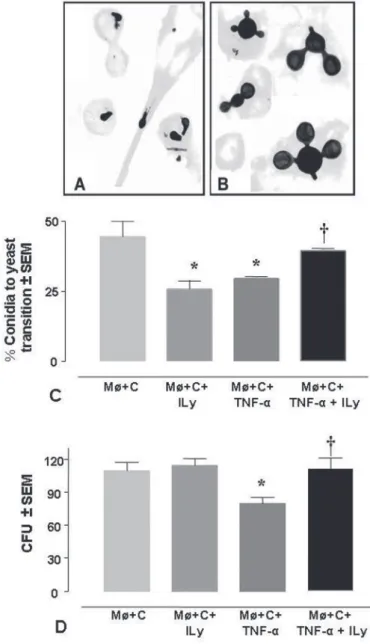
Intracellular transition of P. brasiliensis conidia after lysozyme inhibition: As shown in Fig. 1A and 1B, 96h after infection of the macrophages with P. brasiliensis conidia, fungal propagules appeared
internalized by phagocytes. In non-activated and non-lysozyme treated macrophages (control group), we observed that approximately 50% of these propagules had undergone intracellular transition. The addition of TNF-α resulted in significant (p = 0.0043) inhibition of the
conidium-to-yeast transition process in comparison with the control group (Fig. 1C). Nonetheless, this effect was reverted in the presence of the lysozyme inhibitor (p = 0.0022), a reagent that also inhibited the
transition process in the non-activated macrophages exposed to the same inhibitor, in a way similar to that TNF-α.
Lysozyme participation in the fungicidal/fungistatic effect
exerted by TNF-α-activated macrophages infected with P.
brasiliensis conidia: The ability of the alveolar macrophages to destroy fungal propagules was evaluated by determining CFU’s number. As shown in Fig. 1D, only macrophages activated with TNF-α exerted a
Fig. 1 - P. brasiliensis intracellular transition and CFU of conidia after lysozyme inhibition in normal or TNF-α activated Møs. A) Morphology of intracellular P. brasiliensis conidia, and B) yeast cells observed after 96 h of co-culture with alveolar macrophages (Magnifications 1000X). C) Intracellular transition of P. brasiliensis conidia, after lysozyme inhibition in activated or non-activated macrophages with TNF-α for 96 h at 37 ºC (n = 6). Bars show the mean ± SEM. *p < 0.01 between the control group (non-activated Møs infected with
fungicidal effect on P. brasiliensis conidia (p = 0.0044). This group of
phagocytes destroyed 27% more propagules than the non-activated macrophages, an effect that was reverted in the presence of the lysozyme inhibitor (p = 0.0054).
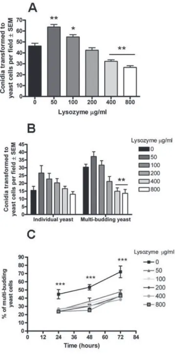
Effect of lysozyme on the fungal transition: Fig. 2A and 2B show the effect of different concentrations of lysozyme in the transition of
P. brasiliensis conidia, and the capacity of this new yeast cells to bud. The susceptibility of conidia was dependent on lysozyme concentration. Physiological concentrations of the enzyme (50 and 100 µg/mL) (Fig. 2A) facilitated the conidia-to-yeast transition process, p < 0.001 and p < 0.05, respectively. On the contrary, inflammatory concentrations
of lysozyme (400 and 800 µg/mL) impaired fungal transition (p < 0.001), as well as its budding ability (p < 0.001) (Fig. 2A and 2B).
Effect of lysozyme on the ability of yeast to bud: In order to confirm the effect of lysozyme on the multi-budding capacity described above, the effect of lysozyme directly on the tissue yeast form was evaluated. As observed in Fig. 2C, yeasts incubated for 72 hours in the presence of different lysozyme concentrations presented a smaller number of blastoconidia in all groups exposed to this enzyme, in contrast to control yeast (no lysozyme) which exhibited multiple budding. This inhibition became apparent from the first 24h when only 25% of the yeasts exposed to the different concentrations of lysozyme were able to begin their budding, in contrast with 45% observed in the control yeast. Similar findings were observed after 48 and 72h post-incubation being more evident in the latter. After 72h post-incubation multiple-budding increased to 72% in the control group in contrast to yeasts exposed to lysozyme which rose only less than 50%. Concentrations of lysozyme were not statistically significant different from each other.
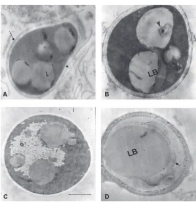
Ultrastructural changes: Transmission electron micrograph of untreated P. brasiliensis conidia is presented in Fig. 3A. Conidia show their characteristic oval or pear shape, homogenous intracytoplasmic lipid bodies and well delimited cell wall. These propagules had a clear thin layer near the plasma membrane that delimited the periplasmic space. The cellular wall was surrounded by a fibrillar material that, in some cases, appeared discontinuous in the wall; none of untreated P.
brasiliensis conidia studied shown an apparently change on their
ultrastructure.
Nonetheless, propagules exposed to physiologic and inflammatory concentration of lysozyme (50 µg/mL and 800 µg/mL, respectively) showed similar changes. The most evident ultrastructural changes were observed in the cytoplasm where fusion of lipid bodies and formation of lamellar structures as a product of subcellular degradation were recorded (Fig. 3B and 3D). In some cells it was evident cytoplasm reduction (Fig. 3D). Although the interruption of fibrillar layer was also observed in control cells, it was more evident in conidia exposed to lysozyme (Fig. 3C).
DISCUSSION
These results reveal for the first time that lysozyme probably participates in the restriction and control of the pathogenic fungus P. brasiliensis cooperating with TNF-α-activated macrophages in their
fungicidal/fungistatic mechanisms exerted against the infective propagules.
TNF-α-activated macrophages impaired both the transition process and the viability of conidia, effects that could be reverted in the presence of the lysozyme inhibitor, suggesting that this enzyme contributes to
the defense mechanisms exerted by the macrophages activated with TNF-α.
We observed that when the lysozyme inhibitor was added to non-activated macrophages the transition process was impaired. Nonetheless, the viability of the fungus remained unaffected as shown by the CFU. Since we did not find a direct negative effect of the lysozyme inhibitor on P. brasiliensis growth (data not shown), new
approaches are needed to identify the effect observed on the intracellular transition process.
TNF-α is involved in multiple steps in the antifungal and inflammatory responses against P. brasiliensis. This cytokine activates
macrophages and is required for the sustained recruitment of inflammatory cells into granulomatous lesions22,32. In addition, previous
studies done by our group have demonstrated that TNF-α-activated macrophages exert an antifungal effect independent of nitric oxide production12. According with the above results, the present study
suggests that lysozyme probably contributes to the mechanisms exerted by TNF-α-activated macrophages.
In addition, in this study we observed that inflammatory concentrations of lysozyme were capable of inhibiting both conidia-to-yeast transition and budding processes.
However, a contrary effect was noticed when physiological concentration of lysozyme resulted in the increase of the transitional process. This effect should be explored further in order to understand the differential behavior according to lysozyme concentrations.
On the other hand, in vitro assays have shown that the budding
process was inhibited by lysozyme even at concentrations lower than 50 µg/mL, suggesting that minimal amounts of this enzyme are sufficient to control the development of the fungus in its tissue form. The absence of a dose-dependent effect on budding suggests that inflammatory (higher) concentrations of this antimicrobial peptide would not be more effective against the fungus.
CANTOR et al.6, using an animal model of emphysema showed
that when hamsters were exposed to aerosolized lysozyme, this enzyme enhanced elastolysis, suggesting that deposition of lysozyme inthe lung’s extracellular matrix may enhance the progression ofpulmonary emphysema. Studies performed in our laboratory have shown that in mice infected with P. brasiliensis conidia marked lysozyme expression is induced in the lungs during first four days post-infection (unpublished data). In addition, recently we observed marked elastolysis during this early inflammatory infiltration14.
Additionally, in the TEM studies the most evident ultrastructural changes observed in lysozyme-treated conidia occurred in the cytoplasm where fusion of lipid bodies and lamellar structures were apparent. The mechanism responsible for these changes is not well understood and it would be difficult to attribute such effect to the sole enzymatic activity. It is known that the target of lysozyme is the polysaccharide exposed on the cell wall; however, two additional hypotheses suggested that a variety of cationic peptides (including lysozyme) might render bacteria non-viable by activating their autolytic wall enzymes leading to bacteriolysis11; other possibility to be considered is that lysozyme
has antibacterial activities independent of its enzymatic action, probably due to its positive charge15.
It is important to emphasize that results obtained from in vitro
studies are not necessarily predictive of their activity in vivo, because
this enzyme may be increased by certain cofactors such as lactoferrin, hydrogen peroxide, defensins and cathelisicidins7,10,33. Thus, it is
possible that the effect observed in vitro by lysozyme could be
potentiated in vivo by these other reactants; however, more studies to
confirm the participation of these cofactors are needed.
Studies on the antifungal action of lysozyme have focused on
Candida albicans. NISHIYAMA et al.24 showed that lysozyme inhibited
the separation of apparently mature C. albicans yeast cells from each
other, and recorded also accumulation of wall-like material in the periplasmic space indicating that cell-wall components may be possible targets for this enzyme. Additionally, they found a synergistic action between lysozyme and lanoconazole, an antifungal drug.
In other study, WU et al.34 showed that lysozyme had a dual action
on C. albicans, killing the organism at higher concentrations and
modulating the production of aspartyl proteinases, a putative virulence factor of C. albicans. SAMARANAYAKE et al.29, reported that although
generally the Candida species such as C. glabrata, C. albicans, C. tropicalis and C. krusei, were susceptible to lysozyme, differences in
their susceptibility were noted among the different isolates within the same species. Fungi such as Aspergillus fumigatus, Rhizopus orizae8
and Histoplasma capsulatum23 also showed certain susceptibility to
lysozyme, compound that mediated a fungistatic mechanism.
In this study, we observed that lysozyme induced ultrastructural and morphological changes in P. brasiliensis propagules; nonetheless,
more studies are necessary to determine the mechanisms involved in the changes brought about by this enzyme on the fungal structures.
Finally, the knowledge gained concerning the molecules implied in the microbicidal activity generated by activated macrophages would allow identification of key molecules that may contribute to improve fungal control. Therefore, we consider that lysozyme could be a target for further developments aimed at defining its importance in the host defense against P. brasiliensis infection.
RESUMO
A lisozima desempenha um papel duplo contra o fungo dimórfico Paracoccidioides brasiliensis
Com a finalidade de determinar o papel da lisozima, um peptídeo antimicrobiano que pertence ao sistema imune inato, contra o fungo dimórfico Paracoccidioides brasiliensis, foram feitas co-culturas de
uma linha de macrófagos alveolares murinos (MH-S) com as conídias do fungo na presença ou não do TNF-α e/ou um inibidor da lisozima; também foram feitos ensaios que avaliaram o efeito das concentrações fisiológicas e inflamatórias de lisozima diretamente sobre o ciclo de vida do fungo. Observamos que os macrófagos ativados com a citoquina tiveram um efeito significativo na inibição da transição conídia/levedura (p = 0,0043) e exerceram um efeito fungicida importante (p = 0,0044), matando mais de 27% das propágulas do fungo em comparação com os macrófagos não ativados. No entanto, após ser o inibidor seletivo da lisozima adicionado, o efeito fungicida foi revertido. Quando os propágulos do fungo foram expostos diretamente a diferentes concentrações da lisozima, um duplo efeito foi observado. Assim, as concentrações fisiológicas da enzima facilitaram o processo de transição conídia-levedura (p < 0,05). Contrariamente, as concentrações inflamatórias prejudicaram a transição fúngica (p < 0,0001). Quando as leveduras foram expostas a qualquer concentração de lisozima, sua capacidade de multi-brotação foi gravemente prejudicada (p < 0,0001). Além disso, mudanças ultra-estruturais, como a sub degradação, a fusão dos vacúolos dos lípidos, estruturas lamelares e interrupção da camada fibrilar foram observadas em conídios expostos à lisozima. Estes resultados sugerem que a lisozima poderia exercer um duplo papel no mecanismo antifúngico contra P. brasiliensis.
ACKNOWLEDGMENTS
This work was supported by Fundación para la Promoción de la Investigación y la Tecnología, Banco de la República, Bogotá, Colombia (grant 1652); Comité para el Desarrollo de la Investigación, CODI, Universidad de Antioquia, Medellín, Colombia and the Young
Researchers Program, Instituto Colombiano para el Desarrollo de la Ciencia y la Tecnología, Francisco José de Caldas, COLCIENCIAS, Bogotá, Colombia.
We also thank to the Instituto Nacional de Salud (Bogotá, Colombia) for the electron microscopy processes and analyses.
REFERENCES
1. BANERJEE, S.K.; HOLLER, E.; HESS, G.P.; RUPLEY, J.A. - Reaction of N-acetylglucosamine oligosaccharides with lysozyme. Temperature, pH, and solvent deuterium isotope effects; equilbrium, steady state, and pre-steady state measurements. J. biol. Chem., 250: 4355-4367, 1975.
2. BORGES-WALMSLEY, M.I.; CHEN, D.; SHU, X. & WALMSLEY, A.R. - The pathobiology of Paracoccidioides brasiliensis. Trends Microbiol., 10: 80-87, 2002.
3. BUSTAMANTE-SIMON, B.; McEWEN, J.G.; TABARES, A.M.; ARANGO, M. & RESTREPO-MORENO, A. - Characteristics of the conidia produced by the mycelial form of Paracoccidioides brasiliensis. Sabouraudia, 23: 407-414, 1985.
4. CALICH, V.L.G.; PURCHIO, A. & PAULA, C.R. - A new fluorescent viability test for fungi cells. Mycopathologia (Den Haag), 66: 175-177, 1979.
5. CALICH, V.L.G.; KIPNIS, T.L.; MARIANO, M.; FAVA-NETO, C. & DIAS, W.D. - The activation of the complement system by Paracoccidioides brasiliensis “in vitro”: its opsonic effect and possible significance for an in vivo model of infection. Clin. Immunol. Immunopath., 12: 20-30, 1979.
6. CANTOR, J.O.; SHTEYNGART, B.; CERRETA, J.M. & TURINO, G.M. - The effect of lysozyme on elastase-mediated injury. Exp. Biol. Med., 227: 108-113, 2002. 7. COLE, A.M.; LIAO, H.I.; STUCHLIK, O. et al. - Cationic polypeptides are required for
antibacterial activity of human airway fluid. J. Immunol., 169: 6985-6991, 2002.
8. DIAMOND, R.D.; KRZESICKI, R.; EPSTEIN, B. & JAO, W. - Damage to hyphal forms of fungi by human leukocytes in vitro. A possible host defense mechanism in aspergillosis and mucormycosis. Amer. J. Path., 91: 313-328, 1978.
9. DIAS, M.F.; MESQUITA, J.; RODRIGUES, N.; FILGUEIRA, A.L. & DE SOUZA, W. -Viability of yeast form cells of Paracoccidioides brasiliensis after sonication. Med. Mycol., 42: 43-49, 2004.
10. GANZ, T. - Antimicrobial polypeptides in host defense of the respiratory tract. J. clin. Invest., 109: 693-697, 2002.
11. GINSBURG, I. - Bactericidal cationic peptides can also function as bacteriolysis-inducing agents mimicking beta-lactam antibiotics? It is enigmatic why this concept is consistently disregarded. Med. Hypotheses, 62: 367-374, 2004.
12. GONZÁLEZ, A.; ARISTIZÁBAL, B.H.; GÓMEZ, E.; RESTREPO, A. & CANO, L.E. -Short report: inhibition by tumor necrosis factor-alpha-activated macrophages of the transition of Paracoccidioides brasiliensis conidia to yeast cells through a mechanism independent of nitric oxide. Amer. J. trop. Med. Hyg., 71: 828-830, 2004.
13. GONZALEZ, A.; DE GREGORI, W.; VÉLEZ, D.; RESTREPO, A. & CANO, L.E. -Nitric oxide participation in the fungicidal mechanism of gamma interferon-activated murine macrophages against Paracoccidioides brasiliensis conidia. Infect. Immun., 68: 2546-2552, 2000.
14. GONZALEZ, A.; LENZI H.L.; MOTTA, E.M. et al. - Expression and arrangement of extracellular matrix proteins in the lungs of mice infected with Paracoccidioides brasiliensis conidia. Int. J. exp. Path., 89: 106-116, 2008.
16. JIMENEZ, M. DEL P.; RESTREPO, A.; GARCIA, L.F. & CANO, L.E. - Separation of
Paracoccidioides brasiliensis conidia through percoll gradients. Med. Mycol., 42:
349-353, 2004.
17. KASHINO, S.S.; FAZIOLI, R.A.; MOSCARDI-BACCHI, M. et al. - Effect of macrophage blockade on the resistance of inbred mice to Paracoccidioides brasiliensis infection.
Mycopathologia (Den Haag), 130: 131-140, 1995.
18. KURITA, N.; OARADA, M.; MIYAJI, M. & ITO, E. - Effect of cytokines on antifungal activity of human polymorphonuclear leucocytes against yeast cells of
Paracoccidioides brasiliensis. Med. Mycol., 38: 177-182, 2000.
19. LEHRER, S.S. & FASMAN, G.D. - Fluorescence of lysozyme and lysozyme substrate complexes. Separation of tryptophan contributions by fluorescence difference methods. J. biol. Chem., 242: 4644-4651, 1967.
20. MASSCHALCK, B. & MICHIELS, C.W. - Antimicrobial properties of lysozyme in relation to foodborne vegetative bacteria. Crit. Rev. Microbiol., 29: 191-214, 2003. 21. MBAWUIKE, I.N. & HERSCOWITZ, H.B. - MH-S, a murine alveolar macrophage cell line: morphological, cytochemical, and functional characteristics. J. Leuk. Biol., 46: 119-127, 1989.
22. NASCIMENTO, F.R.; CALICH, V.L.; RODRIGUEZ, D. & RUSSO, M. - Dual role for nitric oxide in paracoccidioidomycosis: essential for resistance, but overproduction associated with susceptibility. J. Immunol., 168: 4593-4600, 2002.
23. NEWMAN, S.L.; GOOTEE, L.; GABAY, J.E. & SELSTED, M.E. - Identification of constituents of human neutrophil azurophil granules that mediate fungistasis against
Histoplasma capsulatum. Infect. Immun., 68: 5668-5672, 2000.
24. NISHIYAMA, Y.; NAKAOKA, C.; HIRATANI, T. et al. - Synergy of lysozyme and
lanoconazole on the morphology of Candida albicans. J. electron Microsc. (Tokyo), 50: 41-49, 2001.
25. PULSEN, S.M.; ENGSTAD, R.E. & ROBERTSEN, B. - Enhanced lysozyme production in Atlantic salmon (Salmo salar L.) macrophages treated with yeast beta-glucan and bacterial lypopolysaccharide. Fish Shellfish Immunol., 11: 23-27, 2001.
26. RESTREPO, A. & JIMÉNEZ, B.E. - Growth of Paracoccidioides brasiliensis yeast phase in a chemically defined culture medium. J. clin. Microbiol., 12: 279-281, 1980. 27. RESTREPO, A. - Paracoccidioidomycosis. In: DISMUKES, W.E.; PAPPAS, P.G. &
SOBEL, J.D., ed. Clinical Mycology. New York, Oxford University Press, 2003. p. 328-345.
28. RESTREPO, A.; SALAZAR, M.E.; CANO, L.E. & PATIÑO, M.M. - A technique to collect and dislodge conidia produced by Paracoccidioides brasiliensis mycelial form. J. med. vet. Mycol., 24: 247-250, 1986.
29. SAMARANAYAKE, Y.H.; MACFARLANE, T.W.; SAMARANAYAKE, L.P. & AITCHISON, T.C. - The in vitro lysozyme susceptibility of Candida speciescultured in sucrose supplemented media. Microbios, 74: 23-28, 1993.
30. SEDGWICK, J.D.; RIMINTON, D.S.; CYSTER, J.G. & KÖRNER, H. - Tumor necrosis factor: a master-regulator of leukocyte movement. Immunol. Today, 21: 110-113, 2000.
31. SKERRETT, S.J. - Lysozyme in pulmonary host defense: new tricks for an old dog. Amer. J. resp. crit. Care Med., 169: 435-436, 2004.
32. SOUTO, J.T.; FIGUEIREDO, F.; FURLANETTO, A. et al. - Interferon-gamma and tumor necrosis factor-alpha determine resistance to Paracoccidioides brasiliensis infection
in mice. Amer. J. Path., 156: 1811-1820, 2000.
33. TRAVIS, S.M.; SINGH, P.K. & WELSH, M.J. - Antimicrobial peptides and proteins in the innate defense of the airway surface. Curr. Opin. Immunol., 13: 89-95, 2001. 34. WU, T.; SAMARANAYAKE, L.P.; LEUNG, W.K. & SULLIVAN, P.A. - Inhibition of
growth and secreted aspartyl proteinase production in Candida albicans by lysozyme. J. med. Microbiol., 48: 721-730, 1999.