Ana Filipa Nogueira Tavares
Dissertation presented to obtain the Ph.D degree in Biochemistry
Instituto de Tecnologia Química e Biológica | Universidade Nova de Lisboa
Oeiras,
April, 2013
agents against human pathogens
I
II
III IV
O2•-
O2
H2O
CO-RM CO
SOD
H2O2 Fe2+ +
OH•
S S SH SH
DNA
Thiol-containing proteins Fe-S
proteins
Protein Protein
Nitrofurans
Nitrofurans activated
NtrA
GSNO
Ana Filipa Nogueira Tavares
Dissertation presented to obtain the Ph.D degree in Biochemistry
Instituto de Tecnologia Química e Biológica | Universidade Nova de Lisboa
Oeiras, April 2013
of antimicrobial agents against
human pathogens
ii
From left to the right: Maria de Fátima Lopes, Mónica Oleastro, Lígia Saraiva, Ivo Boneca, Mário Ramirez, Ana Filipa Tavares and Carlos Romão
2nd April 2013
Second Edition, April 2013
Molecular Genetics of Microbial Resistance Laboratory
Instituto de Tecnologia Química e Biológica
Universidade Nova de Lisboa
iii I would like to formally express my sincere gratitude to the people that
contributed and supported my work:
To Dr. Lígia Saraiva for receiving me in her laboratory and accepting to be my
supervisor. Thank you for your constant support, encouragement and endless capacity to always see the bright side, even in hard and more challenging
moments. For the trust and confidence in me, for your friendship and 24 h availability whenever I needed to ask a question or communicate the result of
an experiment! Thank you for your great advices and for the careful revision of
this thesis. A big thank you for everything!!!
To Prof. Miguel Teixeira, for the collaboration on the work presented in
Chapter 4, especially for performing the EPR experiments, and for the very
interesting and helpful discussions.
To Prof. Carlos Romão, for the collaboration on the work presented in Chapter
4, for the enthusiastic discussions and all the lessons about CO-Releasing Molecules chemistry.
To Dr. Mónica Oleastro, from Instituto Dr. Ricardo Jorge, for the collaboration
on the work presented in Chapter 5, for receiving me in her laboratory and for
the helpful advices. I would like also to thank João Benoliel for his help during
iv
thesis commission, for their suggestions and helpful advices.
To Dr. Colin McVey, from Structural Genomics lab at ITQB for reviewing the
abstract of this thesis, accordingly with ITQB intern rules.
To all past and present colleagues of MGMR lab´s for making it such an
enjoyable and pleasant place to work. A very special thank you to Lígia Nobre,
for all the help during the course of this work, for always have time to listen my new ideas or results, for the valuable discussions and for being a good
friend. To Marta Justino, for the great discussions, all the help in statistical
analyses and for always willing to give me new ideas and suggestions. To
Margarida Parente, for the help provided in the last part of the experimental
work. To Susana Lobo, for her support, friendship and also for the help with
the cover of this thesis! To Mafalda Figueiredo, for her companionship and for
being a great friend. To Sara Sousa,for her friendship and good moments. To
Joana Baptista,forbeing a special friend, for all the funny moments inside and
outside the lab, for being my “shoulder friend” when I need to laugh or cry!
To former members of the group, especially to Cláudia Almeida, for her help
in my early days at the lab, and to Vera Gonçalves, for the good talks and
laughs.
Thank you all for so many moments of happiness and so many experiences shared in the last years.
v
Releasing Molecules. In special to João Seixas, for the collaboration on the
work presented in Chapter 4.
To all my colleagues in other labs at the institute, especially to those from the
2008 class of the ITQB PhD course and from the InTeraQB. Thank you for
providing me good moments, for the experiences changed and friendly words.
To my colleagues from University that became very especial friends:
Margarida Augusto, Joana Medeiros and Simão Luz, for all the fun and
friendship. To Inês Lima, for her comprehension and support, a true and
special friendship full of precious and unforgotten moments throughout these 10 years!
Aos meus amigos de sempre e a todos os outros, muito obrigada por todo o apoio e amizade.
À minha família pelo vosso interminável e incondicional apoio e carinho e a
quem devo aquilo que sou.
Aos meus pais, obrigada por acreditarem em mim e me darem força para ser
sempre melhor e para tentar ir sempre mais longe.
À minha irmã Xana pela amizade, pelo apoio e por estares sempre presente
em todos os momentos da minha vida!
E aos meus avós, por me acompanharem e ajudarem durante toda a vida.
vi
inesgotável. Obrigada por estares sempre ao meu lado em todos os momentos e por me fazeres feliz!
Fundação para Ciência e Tecnologia for the financial support crucial for the
accomplishment of this work and by awarding a PhD grant
(SFRH/BD/38457/2007).
vii
This thesis comprises the research work performed at the Molecular Genetics of Microbial Resistance Laboratory from the Instituto de Tecnologia Química e Biológica, Universidade Nova de Lisboa under the supervision of Dr. Lígia M. Saraiva.
The thesis is divided in three main parts: Part I consists of a general introduction organized in two chapters, one focusing on bacteria and their resistance to antibiotics and to the innate immune system, and a second one concerning an overview on the general aspects of carbon monoxide and its relation with biological systems and bacteria. Part II comprises the experimental results obtained during this work, which is divided in three chapters based on two original publications and one manuscript in preparation. Part III presents a general discussion of all the work performed in this thesis.
viii
ix
Thesis
Publications
The work presented in this thesis is based on following original publications,
listed by chronological order:
Tavares AFN, Nobre LS, Melo AM & Saraiva LM (2009) A Novel Nitroreductase of Staphylococcus aureus with S‐Nitrosoglutathione Reductase Activity. Journal of Bacteriology 191: 3403‐3406.
Tavares AFN, Teixeira M, Romão CC, Seixas JD, Nobre LS & Saraiva LM (2011) Reactive oxygen species mediate bactericidal killing elicited by Carbon Monoxide‐Releasing Molecules. Journal of Biological Chemistry 286: 26708‐ 26717.
Tavares AFN, Nobre LS & Saraiva LM (2012) A role for reactive oxygen species in the antibacterial properties of Carbon Monoxide‐Releasing Molecules. FEMS Microbiology Letters, 336:1‐10.
Manuscript in preparation, based on results presented in Chapter 5:
Tavares AFN, Parente M, Justino MC, Oleastro M, Nobre LS & Saraiva LM (2013) Fighting Helicobacter pylori with Carbon Monoxide‐Releasing Molecules.
x
Nobre LS, Todorovic S, Tavares AFN, Oldfield E, Hildebrandt P, Teixeira M & Saraiva LM (2010) Binding of azole antibiotics to Staphylococcus aureus flavohemoglobin increases intracellular oxidative stress. Journal of Bacteriology 192: 1527‐1533.
xi
Nowadays, the growing increase of antibiotic resistance represents a global public health concern. Hence, it is crucial to understand the mode of action of antimicrobial agents and to develop new strategies to control bacterial infections. The work presented in this thesis contributes with new insights into the mechanisms that underpin the action of antimicrobial agents against pathogenic bacteria through: (i) the study of a putative nitroreductase of Staphylococcus aureus and its involvement in nitrofurans activation; (ii) elucidation of the mechanisms that sustain the antibacterial activity of the recently discovered Carbon Monoxide‐Releasing Molecules (CO‐RMs); and (iii) evaluation of the bactericidal effect of CO‐RMs on Helicobacter pylori.
The bactericidal effect of nitrofuran antibiotics is dependent on the reduction of their nitro group by bacterial nitroreductases. Sequence analysis of the S. aureus genome allowed identification of a novel putative nitroreductase, SA0UHSC_00833, herein named NtrA. To analyse the NtrA contribution to nitrofurans activation, a mutant strain lacking this gene was constructed and its resistance to nitrofurans was evaluated. Furthermore, the biochemical characterization of NtrA was performed. To achieve this, the ntrA
gene was cloned, and the recombinant protein expressed, purified and analysed. The results revealed that: (i) the S. aureus strain lacking the ntrA
xii
metabolism of S‐Nitrosoglutathione (GSNO), a biological nitric oxide (NO) donor. Several lines of evidence supported this conclusion, namely: (i) the high induction of the ntrA gene transcription observed in cells of S. aureus exposed to GSNO; (ii) the higher susceptibility to GSNO killing of the S. aureus strain deleted in ntrA gene; and (iii) the ability of the recombinant NtrA to detoxify GSNO with a significant activity of 1.5 µmol.min‐1.mg protein‐1.
Altogether, S. aureus NtrA showed to be a bifunctional enzyme that besides promoting nitrofurans activation also protects bacteria from the GSNO deleterious effects. Importantly, a phylogenetic analysis based on the alignment of several nitroreductase amino acid sequences revealed that NtrA is member of a novel family of bacterial nitroreductases.
The second part of the work was devoted to the study of CO‐RMs, which were previously reported to efficiently eliminate pathogens such as
xiii
damage, and a strain deleted in recA, a gene product involved in DNA repair, exhibited higher susceptibility to these compounds; (iv) upon CORM‐2 treatment, the intracellular levels of free iron increased by four times, with the iron‐sulphur centres being the most probable source of iron; and (v) CORM‐2 promoted oxidation of free thiols groups. Moreover, CO‐RMs were shown to generate hydroxyl radicals per se in aqueous solution, which was abolished upon scavenging of the CO molecule. In conclusion, this work revealed that CO‐RMs mediate bacterial cell death through ROS formation.
In the last part of this thesis, the bactericidal action of CO‐RMs toward
H. pylori was demonstrated. Indeed, CO‐RMs were shown to cause a significant decrease in H. pylori viability. Moreover, this effect was not only observed for a laboratory strain but also for several clinical isolates. Interestingly, one of the mechanisms of CO‐RMs bactericidal action on H. pylori was found to occur through inhibition of urease, an enzyme that is essential for survival and pathogenesis of this bacterium.
Additionally, CORM‐2 was proven to enhance the H. pylori
susceptibility to the antibiotics commonly used to eradicate the pathogen, namely, metronidazole, clarithromycin and amoxicillin. Actually, the minimal inhibitory and bactericidal concentration of these antibiotics to H. pylori
strains significantly decreased in the presence of sub‐lethal doses of CORM‐2. Importantly, the viability of CORM‐2‐treated H. pylori in mammalian cells, such as macrophages, was found to be lower.
xiv
xv
Actualmente, o aumento de resistência por parte das bactérias aos antibióticos representa um problema de saúde pública global. Por isso, é fundamental entender o modo de acção dos agentes antimicrobianos e desenvolver novas estratégias para controlar as infecções bacterianas. O trabalho apresentado nesta tese contribuiu com novos dados sobre os mecanismos que sustentam a acção de agentes antimicrobianos contra bactérias patogénicas através de: (i) o estudo de uma nitroredutase hipotética de Staphylococcus aureus e do seu envolvimento na activação
dos antibióticos nitrofuranos; (ii) a elucidação dos mecanismos que estão na base da actividade antibacteriana das recém‐descobertas moléculas libertadoras de monóxido de carbono (CO‐RMs); e (iii) a avaliação do efeito bactericida dos CO‐RMs em Helicobacter pylori.
O efeito bactericida dos nitrofuranos é dependente da redução do seu grupo nitro por nitroredutases bacterianas. Ao analisar o genoma de S. aureus, identificou‐se uma hipotética nitroredutase, SA0UHSC_00833, aqui
xvi
respectivamente. Estes valores estão dentro da gama de actividades normalmente observados para as nitroredutases canónicas.
Foi ainda demonstrado que a nitroredutase NtrA de S. aureus está envolvida no metabolismo da S‐nitrosoglutationa (GSNO), um dador biológico de óxido nítrico (NO). Várias evidências apoiam esta conclusão, nomeadamente: (i) a indução da transcrição do gene ntrA observada em células de S. aureus expostas ao GSNO; (ii) a maior susceptibilidade da
estirpe mutada no gene ntrA ao tratamento com GSNO; e (iii) a capacidade da NtrA recombinante para destoxificar GSNO com uma actividade de 1.5 μmol.min‐1.mg proteina‐1.
Em conclusão, a nitroredutase NtrA de S. aureus mostrou ser uma enzima bifuncional que para além de promover a activação dos nitrofuranos também protege as bactérias contra os efeitos nocivos do GSNO. É importante notar que a análise filogenética feita com base no alinhamento de várias sequências de amino ácidos de nitroredutases revelou que a NtrA é membro de uma nova família de nitroredutases bacterianas.
xvii
compostos. O presente trabalho demonstrou que as células de E. coli
expostas aos CO‐RMs contêm níveis mais elevados de espécies reactivas de oxigénio (ERO) intracelulares. Além disso, mostrou‐se que: (i) a suplementação do meio de cultura com os antioxidantes glutationa e cisteína aboliu o efeito bactericida dos CO‐RMs; (ii) as estirpes mutadas nos genes que codificam sistemas de destoxificação de ERO, designadamente a catalase e a superóxido dismutase, têm maior susceptibilidade ao CORM‐2; (iii) o CORM‐2 induz danos no DNA, e uma estirpe mutada no gene recA, cujo produto está envolvido na reparação do DNA, exibiu maior susceptibilidade a estes compostos; (iv) após tratamento com CORM‐2 os níveis intracelulares de ferro livre aumentaram quatro vezes, sendo os centros de ferro‐enxofre a fonte mais provável de ferro; e (v) o CORM‐2
promoveu a oxidação de grupos tióis livres. Além disso, em solução aquosa os
CO‐RMs provaram ser capazes de gerar per se o radical hidroxilo, o que não se
observou quando o grupo CO não está presente na molécula. Este trabalho
permitiu então concluir que os CO‐RMs medeiam a morte celular bacteriana
através da formação de ERO.
Na última parte desta tese, a acção bactericida do CO‐RMs em H. pylori
foi demonstrada. De facto, os CO‐RMs causam uma redução significativa na
viabilidade de H. pylori. Este efeito não foi somente observado para uma
estirpe de laboratório mas também para os vários isolados clínicos que foram
estudados. Foi interessante observar que um dos mecanismos da acção
bactericida dos CO‐RMs em H. pylori ocorre através da inibição da urease, uma
enzima que é essencial para a sobrevivência e patogenicidade desta bactéria.
Adicionalmente, o CORM‐2 aumentou a susceptibilidade de H. pylori
aos antibióticos habitualmente utilizados para a sua erradicação, como o
xviii
diminui significativamente na presença de doses sub‐letais de CORM‐2. É
importante notar que em células de mamíferos, tais como macrófagos, foi
menor a viabilidade de H. pylori quando previamente tratada com CORM‐2.
Em suma, o trabalho apresentado nesta tese contribuiu para uma
melhor compreensão da base molecular de acção dos nitrofuranos e dos CO‐
RMs. Em particular, o papel da nitroredutase bifuncional NtrA de S. aureus na
acção dos nitrofuranos e na destoxificação de GSNO foi desvendado. Em
relação ao CO‐RMs, a formação de ERO revelou ser um factor importante nas
suas propriedades antibacterianas. Finalmente, foi mostrado, pela primeira vez,
que os CO‐RMs são capazes de eliminar H. pylori, o que pode vir a representar
uma nova estratégia para controlar estas importantes infecções bacterianas em
humanos.
xix
Abbreviations
Δ Deletion
ALF062 Pentacarbonyl bromide
ALF021 Bromo(pentacarbonyl)manganese
ALF492 Tricarbonyldichloro(thiogalactopyranoside) ruthenium(II)
AMX Amoxicillin
BCA Bicinchoninic acid method
BHI Brain heart infusion
BMPO 5‐tert‐butoxycarbonyl 5‐mwthyl‐1‐pyrroline N‐oxide ßCD ß‐cyclodextrin
CFU Colony forming units CH Clarithromycin
CO Carbon monoxide
CO2 Carbon dioxide
CO3 •‐
Carbonate
CoA Coenzyme
CODH Carbon Monoxide dehydrogenase COHb Carboxy‐haemoglobin
CO‐RM Carbon Monoxide‐Releasing Molecules
CORM‐1 Dimanganese decacarbonyl
CORM‐2 Tricarbonyldichloro ruthenium (II) dimer CORM‐3 Tricarbonyldichloro(glycinato) ruthenium (II)
CORM‐A1 Sodium boranocarbonate
DCFH‐DA 2´,7´‐dichlorofluorescein diacetate DMEM Dulbecco´s Modified Eagle medium DMSO Dimethyl sulfoxide
DNA Deoxyribonucleic acid
DNIC Dinitrosyl‐iron‐dithiol complexes
DTNB 5,5´Dithiobis‐(2‐nitrobenzoic acid)
e‐ Electron
xx
FAD Flavin adenine dinucleotide
FALDH Glutathione‐dependent formaldehyde dehydrogenase
FCS Fetal calf serum
FDP Flavodiiron protein
Fe Iron
Fe‐S Iron‐sulphur cluster
FI Fluorescence intensity
FMN Flavin mononucleotide g EPR g‐factor
GDH Glutamate dehydrogenase
GOGAT Glutamate synthase
GSH Glutathione
GSNO S‐Nitrosoglutathione GSNOR GSNO reductase
H+ Proton
Hb Haemoglobin
HBA Horse blood‐agar
Hcp Hybrid‐cluster protein
Hmp Flavohaemoglobin
HO Haem oxygenase
H2O2 Hydrogen peroxide
HOCl Hypochlorous acid
HUS Haemolytic uremic syndrome I Identity
IC50 Half‐maximal inhibitory concentration
iCO‐RM Inactive form of CO‐RM (depletes of CO groups) iCORM‐2 Tetrakis(dimethylsulfoxide dichlororuthenium(II) iNOS Inducible nitric oxide synthase
IL‐1 Interleukin 10
IL‐6 Interleukin 6 INF‐γ Interferon‐γ
xxi
KCa Large conductance calcium‐activated potassium
kcat Catalytic constant
KM Michaelis‐Menten constant
Da Dalton
LB Luria‐Bertani
LPS Lipopolysaccharides
M Molar
MALT Mucosa‐associated lymphoid‐tissue
MAPK Mitogen‐activated protein kinase MBC Minimal bactericidal concentration MIC Minimal inhibitory concentration MCC Metal carbonyl complexes
Mo Molybdenum
MOI Multiplicity of infection MPO Myeloperoxidase
MRSA Methicillin‐resisitant S. aureus
MS Minimal salts
MTZ Metronidazole NAC N‐acetyl cysteine
NADH ß‐nicotinamide adenine dinucleotide, reduced form
NADP ß‐nicotinamide adenine dinucleotide phosphate, oxidized form NADPH ß‐nicotinamide adenine dinucleotide phosphate, reduced form
ND Not determined
NH3 Ammonia
NH4
+
Ammonium ion
nNOS Neural nitric oxide synthase
NO Nitric oxide
NO2 ‐
Nitrite
•
NO2 Nitrogen dioxide
N2O3 Nitrogen trioxide
NorV or FlRd Flavorubredoxin
xxii
ns non significant
NTR Nitroreductase
O2 Dyoxygen
O2
•‐
Superoxide anion
•
OH Hydroxyl radical
OHOO‐ Peroxynitrite
OD Optical density
PAMP Pathogen‐associated molecular pattern
PBS Phosphate buffer
PCR Polymerase chain reaction Phox NADPH oxidase
PPI Proton pump inhibitor
ppm Parts per million
RNA Ribonucleic acid
RNS Reactive nitrogen species ROS Reactive oxygen species
RT‐PCR Reverse transcriptase‐polimerase chain reaction
Ru Ruthenium
S Similarity
SDS‐PAGE Sodium dodecyl sulfate polyacrylamide gel electrophoresis
SE Standard error
sGC Soluble guanylate cyclase
SMX Sulfamethoxazole
SNAP S‐nitroso N‐acetyl DL‐penicillamine
Sod Superoxide dismutase
SSTI Skin and soft‐tissue infection
t1/2 Half‐life
TMP Trimethoprim
TNB 5‐thio‐2‐nitrobenzoic acid
TNF‐α Tumor necrosis factor‐α TSA TSB‐agar
xxiii
UV Ultraviolet
Vmax Maximum velocity
VISA Vancomycin intermediate‐resistant S. aureus
VRSA Vancomycin resistant S. aureus
WHO World Health Organization
wt Wild type
Latin abbreviations
i.e. id est, that is to say
e.g. exempli gratia, for example
et al. et alia, and other people
c. circa, around
Strains
B. anthracis Bacillus anthracis
B. subtilis Bacillus subtilis
C. diphtheria Corynebacterium diphtheria
C. jejuni Campylobacter jejuni
C. tetani Clostridium tetani
E. coli Escherichia coli
H. pylori Helicobacter pylori
M. tuberculosis Mycobacterium tuberculosis
N. meningitidis Neisseria meningitidis
P. aeruginosa Pseudomonas aeruginosa
R. rubrum Rhodospirillum rubrum
S. aureus Staphylococcus aureus
S. typhimurium Salmonella typhimurium
xxiv
Aminoacids
A Ala Alanine C Cys Cysteine D Asp Aspartic acid E Glu Glutamic acid F Phe Phenylalanine G Gly Glycine
H His Histidine I Ile Isoleucine K Lys Lysine L Leu Leucine
M Met Methionine N Asn Asparagine P Pro Proline Q Gln Glutamine R Arg Arginine S Ser Serine
T Thr Threonine V Val Valine W Trp Tryptophan Y Tyr Tyrosine
xxv
Table
of
Contents
Acknowledgments iii
Thesis Outline vii
Thesis Publications ix
Abstract xi
Resumo xv
Abbreviations xix
Table of Contents xxv
Introduction
Chapter 1| Clinical relevance of bacteria and resistance to the innate immune
system
1.1 Pathogenic bacteria 5
1.1.1 Escherichia coli 5
1.1.2 Staphylococcus aureus 8
1.1.3 Helicobacter pylori 11
1.2 Nitrofurans as antimicrobial agents 16
1.2.1 Nitroreductases 17
1.3 Innate immune system response against bacteria 21
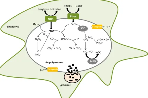
1.3.1 Oxidative stress 22
1.3.2 Nitrosative stress 26 1.3.3 Bacterial defense mechanisms 28
1.4 References 32
Chapter 2| Carbon Monoxide: the new face of an old molecule
2.1 Chemical, physical and biological properties of carbon monoxide 51
2.2 Endogenous production and physiological role of carbon monoxide 54
xxvi
2.4.1 Carbon monoxide metabolism 63 2.4.2 Carbon monoxide sensing 64 2.4.3 Production of carbon monoxide 65
2.5 Carbon monoxide and Carbon Monoxide‐Releasing Molecules as bactericides 67
2.5.1 Transcriptional response of bacteria to Carbon Monoxide‐
Releasing Molecules 70
2.6 References 74
Results
Chapter 3| A novel nitroreductase of Staphylococcus aureus with S‐
nitrosoglutathione reductase activity
3.1 Introduction 88
3.2 Results and discussion 89
3.3 Acknowledgments 97
3.4 References 98
3.5 Supplementary data 99
Chapter 4| Reactive oxygen species mediate bactericidal killing elicited by
Carbon Monoxide‐Releasing Molecules
4.1 Introduction 104
4.2 Material and methods 106
4.3 Results 112
4.4 Discussion 125
4.5 Acknowledgments 128
4.6 References 128
4.7 Supplementary data 130
xxvii
Molecules
5.1 Introduction 134
5.2 Material and methods 135
5.3 Results and discussion 140
5.4 Acknowledgments 151
5.5 References 151
5.6 Supplementary data 153
Discussion
Chapter 6| General discussion
6.1 The role of a novel S. aureus nitroreductese 161
6.2 Dissecting the bactericidal action mechanism of Carbon Monoxide‐
Releasing Molecules 167
6.3 Carbon Monoxide‐Releasing Molecules as antimicrobial agents
against H. pylori 176
Chapter 1|
Clinical relevance of bacteria and
resistance to the innate immune system
_____________________________________________________________
1.1 Pathogenic bacteria 5
1.1.1Escherichia coli 5
1.1.2 Staphylococcus aureus 8
1.1.3Helicobacter pylori 11
1.2 Nitrofurans as antimicrobial agents 16
1.2.1 Nitroreductases 17
1.3Innate immune system response against bacteria 21
1.3.1 Oxidative stress 22
1.3.2 Nitrosative stress 26
1.3.3 Bacterial defense mechanisms 28
4
5
C
h
a
p
ter
1
1.1 Pathogenic bacteria
The human body is exposed to millions of microorganisms, being
estimated that it contains 10 times more bacterial than human cells
(Shanahan, 2002; Tlaskalova-Hogenova et al., 2004). The majority of bacteria
are not harmful and some of them have developed beneficial and essential
relationships with the host, constituting the normal microflora (Hooper &
Gordon, 2001). However, some bacteria are able to cause damage or illness to
the host, being considered as pathogens. The ability to cause disease results
from the outcome of host-pathogen interactions, which will depend on the
damage that microorganism will inflict to the host, and the defense
mechanisms of both host and bacteria (Casadevall & Pirofski, 2000; Medzhitov
et al., 2012). When the host is not able to counteract a situation of bacterial
infection, the use of antibiotics is required. However, the development and
spread of antibiotic-resistant bacteria represents one of the most significant
global health challenges for this century (Bush et al., 2011).
1.1.1 Escherichia coli
The Gram-negative bacterium E. coli was first identified from feces of
neonate and breast-fed infant by Theodor Escherich in 1885 (Escherich, 1989).
This rod-shaped bacteriumis by far the most well studied model organism and
one of the first to have its genome completely sequenced, with approximately
4.6 million base pairs of DNA (Blattner et al., 1997). Most E. coli strains are
commonly regarded as non-pathogenic and along with other facultative
anaerobe species have been identified in the normal microflora of the
anaerobic environment of the colon (Eckburg et al., 2005; Tenaillon et al.,
2010). In fact, E. coli cells typical colonize the gastrointestinal tract of human
6
mutual benefits for decades. E. coli strains may benefit from the dietary fibers
degraded by anaerobes and on other hand, limit oxygen content of the
intestines and produce vitamin K (Ramotar et al., 1984; Kaper et al., 2004;
Jones et al., 2007; Tenaillon et al., 2010). Nevertheless, some E. coli strains
have virulence factors, that can cause a broad spectrum of diseases that fall
into two categories: intestinal pathologies, such as diarrhea, and
extraintestinal pathologies including urinary tract infections (UTIs), haemolytic
uremic syndrome (HUS), sepsis and meningitis (Finlay & Falkow, 1997; Russo &
Johnson, 2003; Kaper et al., 2004; Belanger et al., 2011). It is estimated that
pathogenic strains of E. coli are responsible for more than 2 million human
deaths worldwide per year, particularly due to diarrhea among young children
and septicemia, a bloodstream infection derived from urinary tract infections
(Kosek et al., 2003; Russo & Johnson, 2003). The most notorious pathogenic
strain is the enterohaemorrhagic E. coli 0157:H7, which is a potentially fatal
food-borne pathogen that causes haemorrhagic diarrhea and HUS, that may
lead to acute renal failure (Karmali et al., 1983; Riley et al., 1983). According to
the World Health Organization (WHO), up to 10% of patients with
enterohaemorrhagic E. coli infections may develop HUS, with a fatality rate
ranging from 3 to 5%. In 2011, an outbreak of haemorrhagic gastroenteritis
and HUS caused by E. coli 0104:H4 occurred in Germany (Askar et al., 2011).
Data collected by WHO show that from May to end of June 2011, 896 patients
(including 33 deaths) developed HUS and 3241 patients presented
enterohaemorrhagic gastroenteritis worldwide (including 17 deaths) (Askar et
al., 2011; Wu et al., 2011).
Urinary tract infections are among the most common humans bacterial
infectious diseases, being in its majority (70-95%) caused by uropathogenic E.
7
C
h
a
p
ter
1
the combination of trimethoprim and sulfamethoxazole antibiotics
(TMP/SMX), which inhibits sequential steps in bacterial tetrahydrofolic acid
synthesis, has been used as first-line drug of choice to treat UTIs (Warren et
al., 1999; Nicolle, 2003; Vouloumanou et al., 2011). However, there is a
concern about the emergence of E. coli resistance to TMP/SMX. In fact,
increasing values of antibiotic resistance over 20% have been reported in
several European countries and in United States of America (Huovinen et al.,
1995; Huovinen, 2001; Mazzei et al., 2006). Hence, fluoroquinolone, an
antibiotic that interferes with DNA replication, has been used as an alternative
antimicrobial agent (Hooper, 1999; Guay, 2008). However, in some countries
resistance to fluoroquinolones exceeds now 10-25%, and fosfomycin, an
inhibitor of cell wall synthesis, and nitrofurantoin (see section 1.2) are used as
alternative treatments (Guay, 2001, , 2008; Michalopoulos et al., 2011). Some
β-lactams drugs such as aminopenicillins and cephalosporins are also active against E. coli by blocking cell wall synthesis (Nickel, 2005). However, these
antibacterial agents are not usually recommended because, in general, they
are less effective than non-β-lactams such as TMP/SMX or fluoriquinolones. Furthermore, E. coli exhibits high levels ofresistance to β-lactams antibiotics (Nicolle, 2002; Nickel, 2005; Guay, 2008; Gagliotti et al., 2011). Accordingly
with European Antimicrobial Resistance Surveillance System
(www.ecdc.europa.eu/en/activities/surveillance/EARS-Net), the number of
bloodstream infections caused by E. coli increased remarkably by 71% in the
last years (2002 to 2009), associated with an alarming increase of
8
1.1.2 Staphylococcus aureus
S. aureus are Gram-positive spherical bacteria usually arranged in
pairs, short chains or in clusters, which were first isolated from pus from an
abscess by Alexander Ogston in the late 1800s (Ogston, 1882; Madigan et al.,
2000). This facultative anaerobic pathogen is an opportunistic commensal
bacterium, being mainly found in
upper respiratory tract, especially in
nose and pharynx, and in skin surface
of healthy individuals (Figure 1.1)
(Wertheim et al., 2005). The
asymptomatic colonization provides a
reservoir from which infection may
occur when host defenses are
breached (Kluytmans et al., 1997;
Wertheim et al., 2005). Upon
breaching the epithelium, this
versatile pathogen is capable of
causing from skin and soft-tissue infections (SSTIs), such as pimples, boils, acne
and cellulites, most treatable with antibiotics, to life-threatening diseases
including pneumonia, meningitis, arthritis, osteomyelitis, septicemia and toxic
shock syndrome (Lowy, 1998; Stevens et al., 2005; Klevens et al., 2007;
Gordon & Lowy, 2008). S. aureus infections have been recognized as a major
worldwide health concern due to the emergence of antibiotic resistant strains.
Actually, soon after the introduction in 1941 of the ß-lactam penicillin,
antibiotic resistant strains rapidly spread, initially in hospitals and
subsequently into the community (Rammelkamp & Maxon, 1942; Lowy, 2003).
By the late of 1960s, more than 80% of both community and hospital-acquired Figure 1.1|S. aureuscarriage r ates per body site in adults from general
population. Adapted from (Wertheim
et al., 2005)
Nose 27%
Pharynx 10-20% Neck 10%
Skin chest 15%
Ankle 10%
Perineum 22% Vagina 5% Hand
27% Forearm
20% Axilla 8%
Skin abdomen
9
C
h
a
p
ter
1
staphylococcal isolates were resistant to penicillin (Chambers, 2001; Lowy,
2003). Interestingly, the community-acquired isolates were often resistant
only to penicillin, whereas hospital strains were typically resistant to several
antibiotics (Chambers, 2001). In 1961, the penicillin derivative - methicillin was
introduced in clinical practice to treat staphylococcal infections; again less
than one year later, methicillin-resistant S. aureus strains (MRSA) were
reported (Barber, 1961). Although until 1990s, MRSA remained a problem
restricted to hospitals and other health care-associated settings, in the
following years MRSA arose in patients without prior healthcare contact and
became a widespread cause of community infections (Lyon et al., 1984; Udo et
al., 1993; Herold et al., 1998). Outbreaks of community acquired MRSA
infections have been reported worldwide, and although usually described as a
cause of superficial mild SSTIs (around 90% cases), some strains appear to be
especially virulent, causing severe infections such sepsis, necrotizing fasciitis,
necrotizing pneumonia and endocarditis (Chambers, 2001; Francis et al., 2005;
Gonzalez et al., 2005; Seybold et al., 2006; Chambers & Deleo, 2009; Kallen et
al., 2009). Fortunately, the prevalence of community acquired MRSA is still
low in Portugal (Tavares et al., 2010). Although methicillin is no longer in
clinical use, the term MRSA remains to describe S. aureus strains resistant to
β-lactam antibiotics (Foster, 2004; Grundmann et al., 2010). Unlike
healthcare-associated MRSA, which typically are resistant to multiple antibiotics as
macrolides, tetracyclines, gentamicin and lincosamides, the community
isolates are often resistant only to ß-lactam antibiotics (Chambers, 2001; Feng
et al., 2008; LaPlante et al., 2008; Dryden et al., 2010). In fact, antimicrobial
compounds, such as TMP/SMX, clindamycin, tetracyclines, fluoroquinolones
and linezoid have been used to successfully treat community acquired
10
Figure 1.2| Target sites of the main antibiotics used againstS. aureusinfections. PABA (p-aminobenzoic acid), DHF (dihydrofolate) and THF (tetrahydrofolate).
ß-lactams Glicopeptides (vancomycin)
Aminoglycosides(kanamycin and gentamicin) Tetracyclines DNA mRNA Daptomocin Trimethoprim Sulfonamides Rifampicin Quinolones PABA DHF THF DNA gyrase DNA-directed RNA polymerase Acid folic metabolism Macrolides Lincosamides (clindamycin) Oxazolidinione (linezoid) Fusidic acid Protein syntesis Cell wall synthesis Cytoplasmatic membrane structure Ribossomes 50 30 50 30 50 30
severe nosocomial S. aureus infections have been treated with linezolid,
daptomycin, and combinations of tetracycline plus rifampicin or fusidic acid
and clindamycin plus rifampicin (Dryden et al., 2010). The glycopeptide
vancomycin has been shown to be the most effective therapeutic agent and is
used as the “last resort” against infections caused by MRSA multidrug-resistant. Nevertheless in 1997, the first vancomycin intermediate-resistant S.
aureus (VISA) was identified in Japan, and subsequently also reported in other
countries, reveling that emergence of vancomycin resistance was globally
spread (Hiramatsu et al., 1997; Smith et al., 1999). More recently, reports of
complete vancomycin resistant S. aureus (VRSA) arose, which is even more
alarming (Appelbaum, 2006). Combinations of vancomycin with
aminoglycoside, fusicid acid or rifampicin have also been used to treat more
serious infections caused by S. aureus (Dryden et al., 2010). Figure 1.2
summarizes the target sites of the main antibiotics used against
11
C
h
a
p
ter
1
1.1.3Helicobacter pylori
H. pylori was first isolated from the gastric epithelium of a patient with
chronic active gastritis by Warren and Marshall in the early 1980s (Warren &
Marshall, 1983). In 2005, these two researchers won the Nobel Prize in
Physiology or Medicine for the discovery and role of H. pylori in gastric
diseases. H. pylori is a Gram-negative bacterium and usually presents a
spiral-shaped morphology; however, after prolonged cultivation in vitro and in
response to stresses such as starvation and antibiotic treatment, bacteria may
be converted to a coccoid shape, which is described to be viable but
nonculturable (Berry et al., 1995; Kusters et al., 1997; Enroth et al., 1999;
Kusters et al., 2006).
This microaerophilic flagellated pathogen colonizes the stomach of
more than half world´s population causing gastritis and being a predisposing
condition to the development of severe upper gastrointestinal diseases
including duodenal or gastric ulcers (1 to 10% of infected patients), gastric
cancer (in 0.1 to 3%) and gastric mucosa-associated lymphoid-tissue (MALT)
lymphoma (in <0.1%) (Dunn et al., 1997a; Everhart, 2000; Farinha & Gascoyne,
2005; Houghton & Wang, 2005; McColl, 2010; Sachs & Scott, 2012). Hence, H.
pylori is classified by the WHO as a class I carcinogen since 1994 (Conference,
1994).
H. pylori is unique in its capacity to efficiently colonize the acidic gastric
environment and despite the host immune response, bacteria can persist for
the lifetime of the host unless eradicated with antimicrobials. The diverse
outcome of H. pylori infection is attributed to bacteria virulence factors, host
genomic predisposition and environmental factors (Graham & Yamaoka, 2000;
Clyne et al., 2007; Wu et al., 2008). In particular, several studies have reported
12
enter in the periplasm space. A mmonia is also able to diffuse through the outer membrane to neutralize the medium acidity. Cytoplasmatic ammonia can also neutralize protons entering the cytoplasm. Flagella and spiral-shaped morphology of the bac terium allow the penetration through the mucus layer to reach gastric epithelial cells, to which it sticks via specialized adhesins. OM-outer membrane and IM- inner membrane. Adapted from (Algood & Cover, 2006; Zanotti & Cendron, 2010; Scottet al., 2007)
Figure 1.3| Colonization
factors ofH. pylori.During infection, the bacterium enters the gastric lumen; via urease bacteria survives the acidic environment by producing ammonia (NH3), which is
then converted to ammonium (NH4+) by
accepting protons (H+) that
Host epithelium pH 5-7
pH 1-3
Mucus
Urea
Lumen
Urea Urease CO2 + 2NH3
NH4+
NH3 OM IM
H+
NH4+
NH3+ H+
flagella, are unable to colonize animal models. These findings reveal the
extreme importance of both virulence factors in enabling the adherence of
pathogen to the gastric epithelium (Eaton et al., 1991; Eaton et al., 1992;
Eaton & Krakowka, 1994; Tsuda et al., 1994; Andrutis et al., 1995). Indeed,
urease allows bacterial survival in the acidic pH of the gastric lumen (described
below), whereas flagella and the spiral-shaped morphology let bacteria bore
into the gastric mucus to reach stomach's epithelial cell layer, where bacterial
adhesins mediate close interaction with the host cells (Figure 1.3) (Marshall et
13
C
h
a
p
ter
1
The role of urease in mediating H. pylori acid resistance is attributable
to its ability to hydrolyze urea, present in gastric juice, to carbon dioxide (CO2)
and ammonia (NH3) (Marshall et al., 1990). The ammonia generated by urease
activity consumes protons (H+) neutralizing the gastric juice, enabling H. pylori
to survive and multiply in the stomach (Figure 1.3) (Eaton et al., 1991; Scott et
al., 2007; Zanotti & Cendron, 2010). Importantly, ammonia also has a cytotoxic
effect on gastric epithelial cells (Smoot et al., 1990).
H. pylori urease is a 1.1 MDa spherical assembly of 12 catalytic units,
each composed of UreA-UreB subunits (Ha et al., 2001). Besides these two
structural subunits, four accessory proteins (UreEFGH) are involved in urease
assembly by mediating incorporation of nickel into the apoenzyme (Cussac et
al., 1992; Burne & Chen, 2000; Zanotti & Cendron, 2010). This
nickel-containing enzyme represents 6% of total H. pylori cell proteins and can be
found in the cytoplasm as well as on bacterial surface upon autolysis of
neighboring bacteria (Hu & Mobley, 1990; Phadnis et al., 1996; Dunn et al.,
1997b; Ha et al., 2001; Stingl & De Reuse, 2005). Although, it is well
established that cytoplasmatic urease protects H. pylori against gastric acidity,
the role of surface urease during H.pylori gastric colonization remains under
debate. Some authors argue that extracellular urease does not contribute to
acid resistance of H. pylori since the purified enzyme is irreversible inactivated
at a pH<4. On the other hand, others reported that bacteria without
extracellular urease activity are susceptible to an external pH of 3, and that
urease supramolecular assembly plays a critical role in the activity of the
enzyme as it protects the active sites, remaining the activity unaffected down
to pH of 3 (Bauerfeind et al., 1997; Krishnamurthy et al., 1998; Ha et al., 2001;
14
Due to the key role of urease in host stomach´s colonization, urease
inhibitors constitute potential drugs for treatment of gastric infections
(Follmer, 2010; Kosikowska & Berlicki, 2011). In fact, new strategies to
eliminate H. pylori infections are urgently needed since a dramatic fall in
eradication rates has been reported worldwidely in the last years, mainly due
to the increased emergence of antibiotic resistance (Selgrad & Malfertheiner,
2011).
H. pylori eradication is usually done by the so-called triple therapy,
which combines two antibiotics (clarithromycin, amoxicillin and
metronidazole) with a proton pump inhibitor (PPI, e.g. omeprazole) to reduce
gastric acid production (Coelho et al., 2000; Gisbert et al., 2005; Chey & Wong,
2007; Malfertheiner et al., 2007; Wolle & Malfertheiner, 2007). However,
triple therapy has become progressively less effective with treatment success
rates being, in most countries currently lower that 80%, mainly due to
antibiotic resistance (Megraud, 2004; Wolle & Malfertheiner, 2007; Graham &
Fischbach, 2010; Yakoob et al., 2010; Rimbara et al., 2011). Therefore, triple
clarithromycin-based therapy should be avoided unless infection is known to
be susceptible to clarithromycin or in areas with proven low macrolide
resistance rates (i.e. ≤20%) (Chuah et al., 2011; Rimbara et al., 2011).
Importantly, resistance to clarithromycin cannot be overcome by increasing the dose or duration of therapy (Rimbara et al., 2011). Clarithromycin
resistance is mainly associated with mutations in the 23S ribosomal RNA inside
50S ribosomal subunits, which results in conformational changes that leads to
a decrease binding affinity of the drug to H. pylori ribossomes, allowing normal
bacterial protein synthesis (Versalovic et al., 1996; Occhialini et al., 1997;
Taylor et al., 1997). In case of low resistance to clarithromycin and in
PPI-15
C
h
a
p
ter
1
clarithromycin-metronidazole is recommended (Malfertheiner et al., 2007).
Nevertheless, metronidazole resistance is also problematic and although it can
be partially overcome by increasing the dose of antibiotic and the duration of
therapy, it may cause intolerable side effects and pill burden, which
compromises treatment (Rimbara et al., 2011). Metronidazole action is similar
to that described to nitrofurans (see section 1.2 and 1.2.1), since this
nitroimidazole antibiotic also needs to be converted to cytotoxic forms
through reduction of the nitro group to exert antimicrobial effects (Lindmark
& Muller, 1976). Several studies have shown that oxygen
insensitive-nitroreductase rdxA, NAD(P)H flavin oxidoreductase frxA and ferredoxin-like
protein fdxB contribute to the activation of metronidazole in H. pylori. In
agreement, metronidazole resistance is usually associated with mutations in
these genes (Goodwin et al., 1998; Kwon et al., 2000; Jenks & Edwards, 2002;
Mendz & Megraud, 2002). On the contrary, amoxicillin resistance is not a
major concern since the resistance rate remains so far low (Yakoob et al.,
2010).
Due to the increase inefficiency of the triple therapy, the use of four-drug regiments has been recommended. Such treatment include quadruple or sequential therapy comprising PPI plus three antibiotics (clarithromycin, metronidazole and amoxicillin), or a bismuth-containing quadruple therapy
(PPI plus bismuth salts, metronidazole and tetracycline). So far, these
treatments have provided better eradication rates (Fischbach & Evans, 2007;
Chuah et al., 2011; Rimbara et al., 2011; Selgrad & Malfertheiner, 2011;
16
1.2 Nitrofurans as antimicrobial agents
Nitrofurans are a class of synthetic antimicrobials that include
nitrofurantoin and nitrofurazone. These compounds remain clinically effective
against a wide range of bacteria, including those responsible for UTIs as E. coli
(see section 1.1.1) (Chamberlain, 1976; Johnson et al., 1993; Guay, 2001).
Although, S. aureus is a relatively uncommon cause of UTIs in the general
population (<1%), it is quite usual among patients submitted to urinary tract
catheterization and to genitourinary procedures (Barrett et al., 1999;
Goldstein, 2000; Muder et al., 2006; Guay, 2008). Importantly, the majority of
staphylococcal isolates from patients with catheter-associated UTIs are MRSA
(Muder et al., 2006).
Nitrofurantoin is currently used as an oral antibiotic for the treatment
of genitourinary infections and although,
nitrofurazone is primarily utilized as a topical
antibiotic in burns and skin grafts, a
nitrofurazone-impregnated catheter can be
utilized to prevent catheter-associated urinary
tract infections (Johnson et al., 1993; Guay,
2001). Although the bactericidal action of
nitrofurans has yet to be fully elucidated,
reduction of nitro group by nitroreductases
(NTR, see section 1.2.1) is essential for its activation (McCalla et al., 1970;
Bryant & DeLuca, 1991; McOsker & Fitzpatrick, 1994). Therefore, mutations in
genes encoding nitroreductases result in increased bacterial resistance to
nitrofurans (McCalla et al., 1978; Bryant et al., 1981; Whiteway et al., 1998).
Following activation, the actual antimicrobial action of these prodrugs remains
largely unknown, but it is believed to be due to the inhibition of bacterial Figure 1.4| Chemical structures
of nitrofurantoin (A) and
nitrofurazone (B). Adapted from (Mottier et al., 2005).
A
17
C
h
a
p
ter
1
enzymes involved, at least, in DNA and RNA synthesis and carbohydrate
metabolism (McCalla et al., 1970; Streeter & Hoener, 1988; Shah & Wade,
1989; McOsker & Fitzpatrick, 1994). The multiple target sites of nitrofuran
antibiotics may explain the reduced ability of bacteria to develop resistance
toward these drugs (Guay, 2001). In particular, several reports have
demonstrated that MRSA isolates from catheter-associated UTIs are
susceptibility to both nitrofurantoin and nitrofurazone (Shah & Wade, 1989;
Flournoy & Robinson, 1990; Johnson et al., 1993; Johnson et al., 1999). The
observations that S. aureus are still susceptibility to nitrofurans even after
several decades of use are of clinical importance (Chamberlain, 1976; McOsker
& Fitzpatrick, 1994; Guay, 2001).
1.2.1 Nitroreductases
Nitroreductases are the enzymes responsible for nitrofurans activation
(McCalla et al., 1978; McCoy et al., 1981; Roldan et al., 2008). These enzymes
that reduce the nitro group are divided into two groups based on oxygen
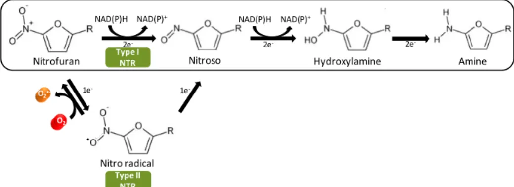
sensitivity: type I-oxygen insensitive and type II-oxygen sensitive enzymes
(Peterson et al., 1979).
Type I-oxygen insensitive nitroreductases are the best-studied
enzymes, and although usually found in bacteria, they rarely occur in
eukaryotes (Knox et al., 1993; Ueda et al., 2003; Roldan et al., 2008). This type
of nitroreductases are FMN binding proteins and NAD(P)H-dependent
enzymes. Nitroreductases catalyze the sequential reduction of nitro group
through the addition of two electrons from NAD(P)H to yield a nitroso
intermediate, which is reduced to amine via a hydroxilamine derivative. In
itself, hydroxylamine has toxic, carcinogenic and mutagenic properties (Figure
18
Type II-oxygen sensitive nitroreductases are FAD- or FMN-containing
enzymes that mediate one-electron reduction of the nitro group forming a
nitro anion radical, which in the presence of oxygen produces superoxide
anion in a futile cycle, regenerating the parental nitro-compound. Thus, these
enzymes only mediate reduction of nitrofurans under anaerobic conditions
(Figure 1.5) (Mason & Holtzman, 1975a, 1975b; Peterson et al., 1979). The
former type of nitroreductases is found in E. coli and several Clostridium
strains (McCalla et al., 1975; Peterson et al., 1979; Angermaier & Simon,
1983). In general, nitrofuran reduction in eukaryotic cells is mediated by this
type of nitoreductases (Wolpert et al., 1973; Adams & Rickert, 1995).
Figure 1.5| General scheme of nitrofurans reduction by nitroreductases
(NTR). The oxygen-insensitive nitroreductases (type I) catalyze the reduction of the nitro group of nitrofurans by addition of a pair of electrons forming the nitroso and hydroxylamino intermediates and finally the amine formation. The oxygen-sensitive nitroreductases (type II) catalyze the single-electron reduction of the nitro group to produce a nitro anion radical, which can be reoxidized aerobically to the original structure with the concomitant production of superoxide anion (O2•-) in a futile cycle.
Adapted from (Koderet al., 2002; Hallet al., 2011; Roldanet al., 2008).
Type I NTR
Nitrofuran
NAD(P)+
Nitroso Hydroxylamine
•
Amine
Type II NTR
Nitro radical NAD(P)H
1e- 1e
-2e- 2e- 2e
-O2
O2•
-NAD(P)+
19
C
h
a
p
ter
1
Although bacteria may contain both types of nitroreductases, as is the
case of E. coli, it seems to be those from type I that underpin nitrofurans
action. In fact, bacterial resistance to nitrofurans is usually attributed to
mutations in genes encoding oxygen insensitive nitroreductases (McCalla et
al., 1970; McCalla et al., 1975; McCalla et al., 1978; Sastry & Jayaraman, 1984;
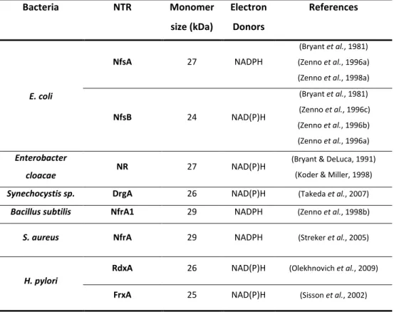
Whiteway et al., 1998). Table 1.1 summarizes the most relevant bacterial
oxygen-insensitive (type I) nitroreductases reported to promote reduction of
nitrofuran antibiotics.
Table 1.1| Bacterial oxygen-insensitive nitroreductases (NTR) mediating
nitrofuran reduction.
Bacteria NTR Monomer
size (kDa)
Electron
Donors
References
E. coli
NfsA 27 NADPH
(Bryant et al., 1981) (Zenno et al., 1996a) (Zenno et al., 1998a)
NfsB 24 NAD(P)H
(Bryant et al., 1981) (Zenno et al., 1996c) (Zenno et al., 1996b) (Zenno et al., 1996a)
Enterobacter
cloacae
NR 27 NAD(P)H (Bryant & DeLuca, 1991)
(Koder & Miller, 1998)
Synechocystis sp. DrgA 26 NAD(P)H (Takeda et al., 2007)
Bacillus subtilis NfrA1 29 NADPH (Zenno et al., 1998b)
S. aureus NfrA 29 NADPH (Streker et al., 2005)
H. pylori
RdxA 26 NAD(P)H (Olekhnovich et al., 2009)
FrxA 25 NAD(P)H (Sisson et al., 2002)
20
Bacterial oxygen insensitive nitroreductases are divided into two main
groups according to their similarity with E. coli nitroreductases, NfsA and NfsB,
which share very low identity (Zenno et al., 1996a; Zenno et al., 1996c, 1996b;
Roldan et al., 2008). The NfsA group, composed by B. subtilis NfrA1 and S.
aureus NfrA is usually NADPH-dependent whereas NfsB group (Enterobacter
NR, Synechocystis DrgA, and H. pylori RdxA and FrxA) may use both NADH and
NADPH as electron donors (Table 1.1) (Bryant et al., 1981; Zenno et al., 1996a;
Zenno et al., 1996c, 1996b; Whiteway et al., 1998; Zenno et al., 1998a; Roldan
et al., 2008). In spite of low degree of amino acid sequence similarity, the two
groups of bacterial insensitive nitroreductases share similar structure and
biochemical properties as all occur as homodimers (24-30 kDa subunits) with a
characteristic α+ß-fold, contain FMN as cofactor and catalyze, besides nitrofurans, the reduction of a broad range of substrates (Zenno et al., 1996a;
Zenno et al., 1996c; Parkinson et al., 2000; Kobori et al., 2001; Haynes et al.,
2002; Race et al., 2005; Roldan et al., 2008).
More interestingly, several bacterial nitroreductases have been
described to protect against oxidative stress, which together with nitrosative
stress are imposed to bacteria by mammalian immune system (see section
1.3). In particular, it was shown that E. coli nfsA is induced by paraquat as a
member of the soxRS regulon, which is involved in the control of oxidative
stress response in E. coli (Liochev et al., 1999). Moreover, B. subtilis nfrA1 and
S. aureus nfrA are also induced under oxidative stress conditions (Mostertz et
al., 2004; Streker et al., 2005). Interestingly, B. subtilis NfrA1 is able to rapidly
scavenge hydrogen peroxide (H2O2) (Cortial et al., 2010). Furthermore, S.
aureus NfrA nitroreductase exhibits disulfide reductase activity, which allows a
thiol-disulfide balance that is quite important since oxidation of thiols resulting