Pro ductio n and characte rizatio n o f
m o no clo nal antibo die s to Brazilian
iso late s o f bo vine viral diarrhe a virus
1Laboratório de Virologia, Departamento de Medicina Veterinária Preventiva,
Microbiologia e Parasitologia, Universidade Federal de Santa Maria, Santa Maria, RS, Brasil
2Department of Veterinary and Biomedical Sciences,
University of Nebraska at Lincoln, Lincoln, NE, USA
3Faculdade de Agronomia e Medicina Veterinária,
Universidade de Passo Fundo, Passo Fundo, RS, Brasil L.C. Kreutz1,3, R. Donis2,
L.H.V. Gil2, M. Lima1,
A.N. Hoffman1,
D.C. Garcez1,
E.F. Flores1
and R. Weiblen1
Abstract
Three Brazilian isolates of bovine viral diarrhea virus (BVDV), anti-genically distinct from the standard North American isolates, were selected to immunize BALB/c mice in order to obtain hybridoma cells secreting anti-BVDV monoclonal antibodies (mAbs). Two hybri-doma clones secreting mAbs, reacting specifically with BVDV-in-fected cells (mAbs 3.1C4 and 6.F11), were selected after five fusions and screening of 1001 hypoxanthine-aminopterin-thymidine-resistant clones. These mAbs reacted in an indirect fluorescent antibody (IFA) assay with all 39 South and North American BVDV field isolates and reference strains available in our laboratory, yet failed to recognize other pestiviruses, namely the hog cholera virus. The mAbs reacted at dilutions up to 1:25,600 (ascitic fluid) and 1:100 (hybridoma culture supernatant) in IFA and immunoperoxidase (IPX) staining of BVDV-infected cells but only mAb 3.1C4 neutralized virus infectivity. Fur-thermore, both mAbs failed to recognize BVDV proteins by IPX in formalin-fixed paraffin-embedded tissues and following SDS-PAGE and immunoblot analysis of virus-infected cells, suggesting they are probably directed to conformational-type epitopes. The protein speci-ficity of these mAbs was then determined by IFA staining of CV-1 cells transiently expressing each of the BVDV proteins: mAb 3.1C4 reacted with the structural protein E2/gp53 and mAb 6.F11 reacted with the structural protein E1/gp25. Both mAbs were shown to be of the IgG2a isotype. To our knowledge, these are the first mAbs produced against South American BVDV isolates and will certainly be useful for research and diagnostic purposes.
Co rre spo nde nce
L.C. Kreutz
Faculdade de Agronomia e Medicina Veterinária
Universidade de Passo Fundo Campus I, Bairro São José 99001-970 Passo Fundo, RS Brasil
Fax: + 55-54-316-8152 E-mail: lckreutz@ upf.tche.br
Research supported by MCT, CNPq, CAPES and FINEP (PRO NEX em Virologia Veterinária, No. 215/96) and FAPERGS (No. 96/1471.6). E. Flores and R. Weiblen are recipients of CNPq fellowships (Nos. 352386/96 and 520011/95, respectively).
Received January 19, 2000 Accepted September 19, 2000
Ke y wo rds
·Bovine viral diarrhea virus
·Monoclonal antibodies
·Antigenic diversity
·Diagnostic reagents
Intro ductio n
Bovine viral diarrhea virus (BVDV) is a small enveloped virus ubiquitous among cattle populations. The viral genome con-sists of a single-stranded, positive-sense RNA molecule of approximately 12.5 kb in length
non-structural viral proteins (1,2). A small and basic protein is thought to form the viral nucleocapsid, and three glycoproteins, namely E0/gp48, E1/gp25 and E2/gp53 are inserted into the viral envelope (2). The non-structural protein NS23/p125 is considered the most important protein involved in virus replication (3). Because of unique features of the viral genome, the NS23/p125 protein may be occasionally cleaved originating two additional products, NS2/p54 and NS3/p80 (2,3). The production of NS3/p80 by some BVDV isolates is correlated with the pro-duction of cytopathology in cultured cells; such isolates are classified as cytopathic bio-type. In contrast, most BVDV isolates ex-press only the NS23/p125 polypeptide and do not produce cytopathology, being classi-fied as non-cytopathic (3,4). In addition, according to the nucleotide sequence of the 5' untranslated region of the viral genome, BVDV isolates might be classified as geno-type 1 and 2. Both cytopathic and non-cyto-pathic biotypes are found within each geno-type (5,6).
BVDV is the type species of the genus
Pestivirus, within the family Flaviviridae, along with the hog cholera virus (HCV) and border disease virus of sheep (7). Clinical manifestations of BVDV infection include fever, mild acute or chronic diarrhea, respi-ratory disease, and hemorrhagic disease (8). Infection of seronegative pregnant cows with a non-cytopathic BVDV biotype may lead to embryonic or fetal deaths, abortion or fetal mummification, congenital malformations, stillbirths or the birth of weak or apparently healthy calves, many of which may be per-sistently infected with the virus (8). Persis-tently infected animals are the main source of BVDV infection to other animals in that they continuously shed viruses. In addition, mucosal disease, one of the most severe clinical manifestations of BVDV infection, occurs in persistently infected animals usu-ally within the first two years after birth (8).
The classification of BVDV isolates into distinct biotypes and genotypes has been unequivocal, yet the grouping of field iso-lates according to their antigenic profile has been quite difficult. Several cross-neutral-ization studies have demonstrated a marked antigenic diversity among geographically distinct isolates (9-12). The antigenic vari-ability among BVDV has been better de-fined by monoclonal antibody (mAb)-based studies (13,14). However, most existing mAbs have been produced against North American or European isolates of BVDV; consequently, it has been rather difficult to characterize South American BVDV isolates. In addition, the unavailability of mAb against Brazilian isolates has hampered the charac-terization and phenotypic studies of BVDV in Brazil. Thus, local isolates of BVDV that were antigenically distinct from the standard BVDV strains (12), as determined by char-acterization with a panel of internationally available mAbs, were used to immunize BALB/c mice with the main objective of producing hybridoma cells secreting anti-BVDV mAbs.
Mate rial and Me tho ds
Ce lls and viruse s
BVDV isolates (UFSM-1, 63 and SV-153.1) that were shown to be antigenically distinct from the standard BVDV laboratory strains (12) were chosen for mouse immuni-zation.
Antige n pro ductio n and mo use immunizatio n
The BVDV isolates were biologically cloned three times by limiting dilution and used to infect MDBK cells to produce anti-gen for mouse immunization. Cells were infected at a multiplicity of infection of 0.1 to 1.0 cell culture infectious dose (CCID50)/
cell and the cell supernatant was collected at 72 h postinfection. Following centrifugation at 25,000 g for 10 min to remove cell debris, the virus suspension was mixed with a solu-tion of polyethylene glycol (PEG, MW 8000; Sigma) and NaCl to achieve a final concen-tration of 7% and 0.4 M, respectively, and stirred at 4o
C for 4 h. The viral particles were pelleted from the solution by centrifugation at 65.000 g for 30 min at 4o
C, and resus-pended in MEM at 1/100 of the initial vol-ume. The concentrated virus was titrated on MDBK cells and aliquots were stored at -70o
C. Concentrated virus was mixed with an equivalent volume of adjuvant and in-jected intraperitoneally (ip) into BALB/c mice (200 µl in Freunds complete adjuvant at day 0 and 200 µl in Freunds incomplete adjuvant at 14, 28, 42, 56 and 70 days after the first injection). Two hundred microliters of concentrated virus without adjuvant was injected ip 3 days prior to removal of the mouse spleen and cell fusion.
Ce ll fusio n, hybrido ma se le ctio n and
scre e ning
Three days prior to fusion, the mice were boosted with an ip injection of adjuvant-free concentrated virus. The spleen was removed and minced and the obtained lymphocytes were mixed with Sp2 cells at a proportion of 10:1. Cell fusion was induced with 50%
PEG (MW 1500; Sigma) for 1 min at 37o
C, followed by slow addition of RPMI medium. The cells were then plated onto 96-well plates containing hypoxanthine-aminopterin-thymi-dine medium (Sigma) supplemented with 15% FBS and 20% conditioned media. Ex-panding hybridomas were detected 7 to 10 days after fusion and the supernatant was tested for the presence of mAbs by immu-noperoxidase (IPX) or indirect fluorescent antibody (IFA) staining. Hybridoma cells secreting BVDV-specific mAbs were then cloned and their reactivity to several BVDV isolates was determined by IFA.
IPX and IFA staining
The presence of mAbs in the supernatant of expanding hybridomas was tested on MDBK cells infected with BVDV isolate UFSM-1. IPX was performed on 96-well plates containing BVDV-infected cell mono-layers fixed with 35% acetone. One hundred microliters of undiluted hybridoma superna-tant was incubated with infected and non-infected cells for 1 h at 37oC. After washing,
horseradish peroxidase-conjugated anti-mouse immunoglobulin (Sigma) was added and incubated as above. After removal of the conjugate, color development was observed following the addition of the substrate solu-tion (3-amino-9-ethylcarbazole; Sigma). For IFA staining, MDBK cells infected with BVDV isolate UFSM-1 and non-infected MDBK cells were individualized by trypsin-ization and dropped on 12-spot Teflon slides. Following adhesion, cells were fixed with acetone, dried and incubated with undiluted medium collected from expanding hybrido-mas for 1 h at 37oC. Fluorescein
isothiocya-nate (FITC)-labeled anti-mouse immunoglo-bulin (Sigma) was added and incubated as above.
Pro ductio n o f ascitic fluid
mAbs, 10 BALB/c mice were primed with Freunds complete adjuvant (Sigma) and 7 days later were injected ip with approxi-mately 106
hybridoma cells. Ten days later the ascitic fluid was collected from the mice, cleared by low speed centrifugation, titrated, aliquoted and stored at -70o
C.
Characte rizatio n o f the mAbs
The BVDV-specific mAbs secreted by hybridoma clones 3.1C4 and 6.F11 were characterized regarding immunoglobulin class, capacity to neutralize virus infectivity, spectrum of reactivity with different BVDV isolates, and ability to react with BVDV antigens in routinely fixed histological sec-tions and in Western immunoblot. The im-munoglobulin class and subclass of mAbs 3.1C4 and 6.F11 were determined using a commercially available kit, according to the manufacturers recommendations (Mouse Type Isotyping kit, Bio-Rad, Hercules, CA, USA). To investigate the capacity of the mAbs to neutralize virus infectivity, serial 2-fold dilutions of the respective ascitic fluid were mixed with 100 CCID50 of BVDV
UFSM-1 isolate and incubated for 1 h at 37oC. A suspension of MDBK cells was then
added to the wells and the plates were incu-bated at 37ºC in the presence of 5% CO2.
Virus neutralization or growth was moni-tored by IFA staining of cells, as described above. The spectrum of reactivity of mAbs 3.1C4 and 6.F11 was assessed by staining cell monolayers infected with BVDV field isolates and reference strains and by staining 2 HCV isolates with IFA (Table 1). The ability of the mAbs to react with BVDV antigens after routine formalin fixation and paraffin embedding was investigated by IPX staining according to previously described protocols, using mAb 15c5 as positive con-trol (15). The ability of these mAbs to recog-nize viral proteins resolved by SDS-PAGE and immobilized on nitrocellulose mem-branes was investigated by Western
immu-noblot analysis of BVDV-infected cell ly-sates, performed according to standard pro-tocols (16).
Pro te in spe cificity o f mAbs 3.1C4 and 6.F11
To determine the mAb protein specific-ity, CV-1 cells transfected with plasmid con-structs that express BVDV proteins (see be-low) were fixed with acetone:methanol (1:1) and incubated with ascitic fluid diluted 1:100 or 1:500 for 1 h at room temperature. The ascitic fluid was then washed and the cells were flooded with FITC-labeled goat anti-mouse antibody (Sigma). Staining of infected cells was observed by fluorescence micros-copy.
Transie nt e xpre ssio n o f BVD V pro te ins
Confluent CV-1 cells (African Green monkey kidney, ATCC-CCL 70) were in-fected with a recombinant vaccinia virus expressing phage T7 RNA polymerase (vTF7-3) (17) at an input multiplicity of 5 for 45 min at 37o
C. DNA-lipid complexes were prepared at room temperature using lipofec-tamine (Gibco-BRL) and DNA at a weight ratio of 12:1, as recommended by the manu-facturer. Immediately following infection, CV-1 cells were washed three times with Dulbeccos minimal essential medium (DMEM, Gibco) without serum and trans-fected with 0.50 ng of each of five plasmid constructs that comprise the entire polypro-tein of BVDV strain NADL (18). The DNA-lipofectamine complexes were incubated with cells at 37o
C in the presence of 5% CO2
for 4 h. After lipofection, the DNA-lipid complexes were removed and the cells were supplemented with DMEM and 10% FBS for an additional 13 h, when the cells were fixed for IFA analysis.
Re sults
hy-Table 1 - Reactivity of mAbs 3.1C4 and 6.F11 w ith BVDV isolates by IFA.
cp: Cytopathic; ncp: non-cytopathic; cp/ncp: mixture of cytopathic and non-cytopathic viruses; nd: not determined; n/a: not applicable. aLaboratório de Virologia, Universida-de FeUniversida-deral Universida-de Santa M aria; bLaboratório de Virologia, Universidade Federal do Rio Grande do Sul; cCentro de Pesquisas Veterinárias Desidério Finamor; dInstituto Biológico de São Paulo; eInstituto Nacional de Tecnologia Agropecuaria (INTA), Castelar, Argentina; fNational Animal Disease Center, Ames, IA, USA; gCornell Univer-sity, NY; hclassical sw ine fever virus; (+) positive reaction; (-) negative reaction.
Isolate Origin Biotype Genotype mAb 3.1C4 mAb 6.F11
UFSM .1a Farroupilha, RS ncp 1 + +
UFSM .2 Alegrete, RS ncp 1 + +
UFSM .3 Pelotas, RS ncp 1 + +
UFSM .4 São Sepé, RS ncp nd + +
UFSM .5 São Sepé, RS ncp nd + +
SV 123.4 Santa M aria, RS ncp 2 + +
SV 126.1 Santa M aria, RS ncp 1 + +
SV 126.8 Farroupilha, RS ncp 1 + +
SV 126.14 Santa M aria, RS ncp 1 + +
SV 152 Farroupilha, RS ncp 1 + +
SV 153.1 Lavras do Sul, RS ncp 1 + +
SV 153.15 Lavras do Sul, RS ncp 1 + +
SV 153.19 São F. de Assis, RS ncp 1 + +
SV 63 Santa M aria, RS ncp 2 + +
SV 260 Lages, SC ncp 2 + +
SV-228/98 Carazinho, RS ncp nd + +
LV85/96b Viamão, RS ncp 2 + +
EVI-006c Eldorado do Sul, RS ncp 1 + +
IBSP-1d Jaboticabal, SP ncp 1 + +
IBSP-2 Jaboticabal, SP cp/ncp 1 + +
IBSP-4 Ribeirão Preto, SP cp/ncp 1 + +
IBSP-5 Ribeirão Preto, SP cp/ncp 1 + +
INTA 1e Argentina cp/ncp 1 + +
INTA 2 Argentina ncp 1 + +
INTA 3 Argentina cp/ncp 1 + +
INTA 4 Argentina cp/ncp 1 + +
INTA 5 Argentina ncp 1 + +
INTA 6 Argentina ncp 1 + +
INTA 7 Argentina ncp 1 + +
1 R Argentina cp/ncp nd + +
34 P Argentina cp/ncp nd + +
34 B Argentina cp/ncp nd + +
NADLf USA cpj 1 + +
SINGER USA cp 1 + +
OREGON USA cp 1 + +
BVDV 890 USA cp/ncp 2 + +
VS-253 USA cp 2 + +
NY-93g USA ncp 2 + +
VS-191 USA ncp 2 + +
CSFVh USA ncp n/a -
-CSFV Brazil ncp n/a -
-bridoma cells secreting anti-BVDV mAbs were performed using virus antigens from cells infected with the isolate UFSM-1 (12). Following five fusions of lymphocytes and myeloma cells, several expanding hybridoma cells were found to be reactive to BVDV-infected cells. However, only two hybridoma clones (3.1C4 and 6.F11) were stabilized and found to secrete specific mAbs against BVDV proteins; several other hybridoma clones secreted mAbs that were also reactive to cellular components. These mAbs have not been further characterized.
Both mAbs were reactive by IFA to all 32 South American isolates and several US iso-lates of BVDV, including viruses from both genotypes (BVDV type 1 and 2), but not to two HCV isolates (Table 1). The BVDV-specific mAb-secreting hybridoma cells were then cloned and were used to produce mAbs either in cell culture or by injection of the hybridoma cells into BALB/c mice to obtain ascitic fluid.
Hybridoma supernatant and ascitic fluid derived from clones 3.1C4 and 6.F11, that were found to react specifically with BVDV-infected cells by IFA and IPX, yielded a positive signal by IFA even when diluted up to 100 (supernatant) and 25,600 times (as-citic fluid). Both mAbs were shown to be-long to the IgG2a isotype. Only mAb 3.1C4 had neutralizing activity (1:80) against BVDV parental virus (UFSM-1, data not shown). This mAb also showed neutralizing activity against the BVDV Singer strain and against a BVDV type 2 cytopathic virus, although at lower titers. The protein speci-ficity of the mAbs could not be determined by Western immunoblot analysis of lysates obtained from BVDV-infected cells. The mAbs did not react with viral proteins sepa-rated by SDS-PAGE and transferred to nitro-cellulose membranes (data not shown), sug-gesting they are probably directed to confor-mational-type epitopes. Likewise, the mAbs failed to react with viral proteins by IPX in tissues submitted to routine formalin
Table 2 - Determination of the BVDV protein specificity of mAbs 3.1C4 and 6.F11.
Protein specificity w as determined by indirect fluorescent antibody assay of CV-1 cells expressing each protein, using the hybridoma supernatant as primary antibody. (-) Negative reaction; (+) positive reaction.
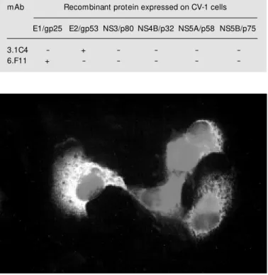
mAb Recombinant protein expressed on CV-1 cells
E1/gp25 E2/gp53 NS3/p80 NS4B/p32 NS5A/p58 NS5B/p75
3.1C4 - + - - -
-6.F11 + - - - -
-reacts with E2/gp53 and mAb 6.F11 -reacts with E1/gp25 (Table 2 and Figure 1).
D iscussio n
The clinical syndromes associated with BVDV infection result in severe economic losses to the cattle industry around the world. In endemic areas, the prevalence of BVDV antibodies among adult cattle may reach up to 70 to 80% (19). Because of their RNA genome and ubiquitous nature, a marked antigenic and genetic heterogeneity is ob-served among field BVDV isolates,
repre-senting a problem for diagnosis and vaccine development (10). Thus, to identify poten-tial vaccine candidate strains, it is necessary to perform phenotyping studies in order to identify the isolates that are representative of the viruses circulating among a given cattle population. These local isolates are more likely to elicit an immune response that is protective to most isolates prevalent in the area than viral strains derived from other geographic locations. Phenotyping studies of BVDV were made possible only after the production of mAbs to laboratory reference strains. Furthermore, mAbs are considered a powerful tool for research and diagnostic purposes.
Recently, several reports have demon-strated that BVDV is highly prevalent in Brazilian cattle (12,16,20), and as such, it is probably associated with significant eco-nomic losses to the national livestock indus-try. These reasons prompted us to initiate epidemiological studies on BVDV infection followed by antigenic and molecular charac-terization of BVDV circulating in Brazilian cattle. Although the production of mAbs to several BVDV isolates has already been re-ported, most of these mAbs have been pro-duced against North American and Euro-pean strains (9,13,14,21). In addition, most of these mAbs were produced against labo-ratory reference strains rather than against field virus isolates. In a recent study, we have demonstrated that the BVDV viruses isolated in Brazil display marked antigenic differences when compared to North Ameri-can reference strains (12,16). We under-stand that the production of mAbs to Brazil-ian BVDV field isolates, antigenically dis-tinct from the standard strains, will help in better defining the antigenic properties of these isolates and in designing more effec-tive diagnostic tools and vaccines.
The low number of hybridoma clones secreting BVDV-specific mAbs obtained in the present study may be attributed to prob-lems inherent to the technique per se, and to
the peculiar biological properties of the pestiviruses as well. These include the in-ability of some viral isolates to replicate to high titers in cell culture, the poor antigenic-ity of BVDV to BALB/c mice and the diffi-culties in achieving a pure and concentrated suspension of viral particles for mouse im-munization. The two mAbs obtained so far failed to recognize viral protein following SDS-PAGE and immunoblot analysis of in-fected cells, suggesting that they recognize conformational-type epitopes. Consequently, the identification of the protein specificity of these mAbs was made possible only by using a transient protein expression system in which BVDV proteins were expressed individually on CV-1 cells. Both mAbs reacted with viral structural proteins: mAb 6.F11 reacted with glycoprotein E1/gp25 and mAb 3.1C4 re-acted with glycoprotein E2/gp53.
Most mAbs against BVDV and other pestiviruses produced to date fall into two major groups: the first group comprises the pan-pestivirus mAbs, which recognize most pestivirus isolates and are mostly directed at the non-structural polypeptide NS23/p125 (9,13,14,21). NS23/p125 is a multifunctional protein ultimately involved in viral RNA replication, and is highly conserved among pestiviruses (3). The other group comprises the type-specific mAbs, which are able to distinguish between strains or virus clusters possessing slight antigenic differences. Most of these mAbs are directed to the envelope glycoproteins E0/gp48 and E2/gp53 (9,13, 14,22). Glycoprotein E2/gp53 is the major envelope glycoprotein and is believed to play a major role in the initial interactions of virions with cell membrane proteins during virus attachment and penetration (3). This viral glycoprotein contains at least three highly variable regions and is a major target for neutralizing antibodies (3,21,22).
Cellular receptor-binding proteins usu-ally contain highly conserved domains that mediate specific interactions with cellular components (23,24). These conserved
re-gions, usually inaccessible to antibody bind-ing, are often surrounded by variable regions that allow viruses to escape from the im-mune response (23,24). Thus, mAbs directed at these variable regions usually fail to rec-ognize a considerable number of field iso-lates (9,13,14,21,22). Interestingly, mAb 3.1C4 was able to recognize all BVDV iso-lates tested so far. This suggests that it binds to a highly conserved epitope within E2/ gp53, which is possibly involved in an im-portant biological function and therefore is under strong variability constraint. Further-more, the epitope recognized by mAb 3.1C4 seems to be required for initiation of infec-tion since viral infectivity was substantially reduced following virus-neutralization as-says. To date, only a few broadly reactive mAbs against E2/gp53 have been described (14). Nevertheless, testing mAb 3.1C4 with a higher number of BVDV isolates will be necessary in order to unequivocally ascer-tain whether the epitope recognized is in-deed thoroughly conserved.
objec-tive of identifying persistently infected ani-mals in a herd.
Ackno wle dgm e nts
We thank Dr. Claudio Canal (UFRGS, Porto Alegre, RS) for determining the immu-noglobulin class, Dr. Paulo M. Roehe
(CPVDF, Eldorado do Sul, RS), Dr. Valéria Moojen (Faculdade de Veterinária, UFRGS, Porto Alegre, RS), Dr. Maristela Pituco (Instituto Biológico de São Paulo), Dr. Anselmo Odeon (INTA Balcarce, Argen-tina), and Dr. Elba Laura Weber (INTA Castelar, Argentina) for providing some of the BVDV isolates used in this study.
Re fe re nce s
1. Collett M S, Larson R & Belzer SK (1988). Proteins encoded by bovine viral diarrhea virus: t he genom ic organizat ion of a pestivirus. Virology, 165: 200-208. 2. M eyers G & Thiel HJ (1996). M olecular
characterization of pestiviruses. Advances in Virus Research, 47: 53-118.
3. Donis RO (1995). M olecular biology of bovine viral diarrhea virus and its interac-tions w ith the host. Veterinary Clinics of North America, 11: 393-424.
4. Donis RO & Dubovi EJ (1987). Differences in virus-induced polypeptides in cells in-fected by cytopathic and noncytopathic biotypes of bovine virus diarrhea-mucosal disease virus. Virology, 156: 168-173. 5. Pellerin C, Hurk JVD & Lecomte J (1994).
Identification of a new group of bovine viral diarrhea virus strains associated w ith severe outbreaks and high mortalities. Vi-rology, 203: 260-268.
6. Ridpath JF, Bolin SR & Dubovi EJ (1994). Segregation of bovine viral diarrhea virus into genotypes. Virology, 205: 66-74. 7. Wengler G, Bradley DW, Collet M S, Heinz
FX, Schlesinger RW & Strauss JH (1995). Flaviviridae. Virus Taxonomy: Sixth Report of the International Committee on Tax-onomy of Viruses. Archives of Virology (Suppl 10): 415-427.
8. Baker JC (1995). The clinical manifesta-tions of bovine viral diarrhea infection. Veterinary Clinics of North America,11: 425-446.
9. Edw ards S & Paton D (1995). Antigenic differences among pestiviruses. Veteri-nary Clinics of North America, 11: 563-578.
10. Dubovi EJ (1992). Genetic diversity and BVD virus. Comparative Immunology, M i-crobiology and Infectious Diseases, 15: 155-162.
11. How ard CJ, Brow nlie J & Clarke M C (1987). Comparison by the neutralization assay of pairs of non-cytopathogenic and cytopathogenic strains of bovine virus di-arrhoea virus isolated from cases of mu-cosal disease. Veterinary M icrobiology, 13: 361-369.
12. Botton AS, da Silva AM , Brum M CS, Weiblen R & Flores EF (1998). Antigenic characterization of Brazilian bovine viral diarrhea virus isolates by monoclonal anti-bodies and cross-neutralization. Brazilian Journal of M edical and Biological Re-search, 31: 1429-1438.
13. Paton DJ, Low ings JP & Barrett ADT (1992). Epitope mapping of the gp53 en-velope protein of bovine viral diarrhea vi-rus. Virology, 190: 763-772.
14. Corapi WV, Donis RO & Dubovi EJ (1990). Characterization of a panel of monoclonal antibodies and their use in the study of the antigenic diversity of bovine viral diar-rhea virus. American Journal of Veterinary Research,51: 1388-1394.
15. Odeon AC, Kelling CL, M arshall DJ, Estela ES, Dubovi EJ & Donis R (1999). Experi-mental infection of calves w ith bovine vi-ral diarrhea virus genotype II (NY-93). Jour-nal of Veterinary Diagnostic Investigation, 11: 221-228.
16. Botton SA, Gil LHVG, Silva AM , Flores EF, W eiblen R, Pit uco EM , Roehe PM , M oojen V & Wendelstein AC (1998). Ca-racterização preliminar de amostras do vírus da diarréia viral bovina (BVDV) isoladas no Brasil. Brazilian Journal of Vet-erinary Research, 18: 84-92.
17. Fuerst TR, Niles EG, Studier FW & M oss B (1986). Eukaryotic transient-expression system based on recombinant vaccinia vi-rus that synthesizes bacteriophage T7 RNA polymerase. Proceedings of the
Na-tional Academy of Sciences, USA, 83: 8122-8126.
18. Vassilev V, Collet M S & Donis RO (1997). Authentic and chimeric full-length genom-ic cDNA clones of bovine viral diarrhea virus that yield infectious transcripts. Jour-nal of Virology, 71: 471-478.
19. Baker JC (1987). Bovine viral diarrhea vi-rus: a review . Journal of the American Vet erinary M edical Associat ion, 190: 1449-1458.
20. Oliveira G, Oliveira EAS, Silva LTH, Vieira LA, Hoffmann VL, Fernandes GV, Silva TC, Caldas APF & Roehe PM (1996). Presença de pestivirus e anticorpos con-tra pestivirus em soros e cultivos celula-res. Arquivos Brasileiros de M edicina Veterinária e Zootecnia, 48: 513-521. 21. Bolin SR, M oennig V & Gourley NE (1988).
M onoclonal antibodies w ith neutralizing activity segregate isolates of bovine viral diarrhea virus into groups. Archives of Vi-rology, 99: 117-123.
22. Deregt D, van Rijn PA, Wiens TY & van den Hurk J (1998). M onoclonal antibodies to the E2 protein of a new genotype (type 2) of bovine viral diarrhea virus define three antigenic domains involved in neu-tralization. Virus Research, 57: 171-181. 23. Wharton SA, Weis W, Skehel JJ & Wiley
DC (1989). Structure, function and antige-nicity of the hemagglutinin of influenza virus. In: Krug RM (Editor), The Influenza. Plenum Press, New York, 153-173. 24. M cKeating JA, M oore JP, Ferguson M ,