738
Srp Arh Celok Lek. 2012 Nov-Dec;140(11-12):738-745 DOI: 10.2298/SARH1212738D
ОРИГИНАЛНИ РАД / ORIGINAL ARTICLE UDC: 611.718
INTRODUCTION
The key concepts related to bone functional adaptation to mechanical stress were develo-ped even in the 19th century [1, 2]. Since then a number of anatomical and anthropological stu-dies have focused on the investigation of bone morphology as the reflection of bone mechani-cal loading history [3]. Due to a rising problem of senile hip fracture in modern populations, research attention was particularly paid to hip bone mineral density, external geometry, and internal architecture as possible determinants of fracture susceptibility [4, 5, 6]. In contrast, the developing femur was less investigated [7-11]. In particular, there are insufficient studi-es invstudi-estigating thstudi-ese morphological featurstudi-es comparing it with the children’s femur.
The central concept of skeletal biology is the idea that bone form reflects mechanical loading history [1, 2, 3]. Namely, the proximal femur adapts its structure to loads to which it is expo-sed in the sense that bone trabeculae tend to ori-entate along the directions of principal stresses [2, 12]. In this way maximum stiffness and bone
strength are achieved [13, 14]. In the “traditional concept” of femoral mechanics, both compressi-ve and tensile stresses occur in the proximal fe-mur [2]. Load which is represented by body we-ight applied to the femoral head tends to bend the femoral neck which causes tension in the superior and compression in the inferior aspect of the neck. However, in contrast to traditional interpretations, recent studies on stress distribu-tion in the proximal femur have revealed that, when capsular, muscle and ligament forces are also considered, stresses occurring in the proxi-mal femur are predominantly compressive [9, 15, 16]. In that sense, both “principal compressi-ve” and “principal tensile” groups of trabeculae actually correspond to compression stresses, transmitting forces from the femoral head into the shaft. It is now considered that loads origina-ting from muscle contractions are greater than the effect of gravity, due to the disadvantageous positioning of muscle attachments on bony lever [17]. Recent literature suggests the significan-ce of shear stresses (which are not included in Wolff ’s concept), as shear may be a dominant failure mode for the proximal femur [18] with
Morphological Characteristics of the Developing
Proximal Femur: A Biomechanical Perspective
Marija Djurić1, Petar Milovanović1, Danijela Djonić1, Arsa Minić2, Michael Hahn31Laboratory for Anthropology, Institute of Anatomy, School of Medicine, University of Belgrade, Belgrade,
Serbia;
2European University, American School of Medicine, Belgrade, Serbia;
3Department of Osteology and Biomechanics, University Medical Center Hamburg-Eppendorf, Hamburg,
Germany
SUMMARY
Introduction In contrast to a plethora of studies on the proximal femur in adults, its external and internal morphology in growing children has not been sufficiently analyzed.
Objective We analyzed changes in external and internal morphology of the proximal femur during growth and development to interpret the links between them and concepts of the human femoral biomechanics.
Methods We assessed external geometry, internal trabecular and cortical arrangement, and bone mineral density (BMD) of the proximal femur in 29 children (age at death from 1 month to 14 years) from archaeological context by using microscopic and radiographic methods.
Results The results showed that both the femoral neck width and length increased with age, with the femoral neck becoming more elongated, while the collo-diaphyseal angle decreased. A strong relationship between age and adjusted areal BMD was found, showing continuous increase during childhood. Parallel trabecular pattern at birth changed to mature three distinct trabecular groups (longitudinal – principal compressive, transversal – tensile and randomly scattered) starting from the age of 8 months. In older children the superior and inferior aspects of the femoral neck differently changed with growth, with medial neck having thicker cortex and trabeculae.
Conclusion In the light of bone adaptation principle, the observed changes in external and internal morphology are governed by mechanical forces acting on the developing femur. Our findings on the development of trabecular pattern and cortical distribution are compatible with recent views on the femoral biomechanics which point out the predominance of compressive stresses in the femoral neck, adaptation to shear stresses, multiaxial loading perspective, prevalence of muscle effects over body weight, and existence of adaptational eccentricity.
Keywords: proximal femur; growth; bone adaptation; mechanical loading
Correspondence to:
Marija DJURIĆ Institute of Anatomy School of Medicine
4/2 Dr Subotića, 11000 Belgrade Serbia
shear stress being the main stimulus for trabecular bone developmental adaptation [19]. Nowadays, it is believed that observation of single loading condition (even if it is represented by resultant force) is too simplified and cannot completely explain the femoral structure, so multidirectio-nal loading history has to be considered [13]. Specifically, extreme loading directions corresponding to extreme posi-tions in range of joint excursions are reported to determine trabecular directions [9].
OBJECTIVE
In this study we used specimens of proximal femora of non-adults derived from archaeological context to investi-gate bone trabecular pattern, external geometry and bone mineral density with respect to the growth of individu-als. Our aim was to find the links between the changes in external and internal morphology of the proximal femur during growth and development and classical vs. more recent understandings of human femoral biomechanics.
METHODS
The material used in this study consisted of 29 right proxi-mal femora derived from archaeological context. The skele-tal remains belong to non-adults, age-at-death from 1 mon-th to 14 years, from mon-the late medieval graveyard of Stara To-rina (Serbia). The criterion for inclusion of the specimens in the study was complete preservation of the right femur (with no signs of breakage of the cortical bone, cortical erosion or other macroscopic bone damage). Sex-specific analysis of skeletons was not performed because of uncertainty of sex determination in non-adults. Age determination was based on maximum femoral diaphyseal length [20, 21, 22].
External geometry
Each specimen of the proximal femur was halved in the coronal plane, and three linear measurements were obta-ined directly from the frontal sections: femoral neck axis length (FAL), femoral neck width (FW), and collo-diap-hyseal (neck-shaft) angle (Q). FAL represents the length of the femoral neck axis from the base of the lateral part of the greater trochanter to the femoral head. FW is the length of the narrowest cross-section of the femoral neck. Q is an angle between the long axis of the femoral neck and the shaft of the femur. The neck index (NI), representing a ratio between the femoral neck width and femoral neck axis length, was introduced here by the authors in order to describe the general shape of the femoral neck.
Bone mineral density
In vitro DXA scans (dual X-ray photon absorptiometry) were performed using a HOLOGIC 1000 W
densitome-ter (Hologic QDR 1000/W; Hologic, Waltham, MA). The femoral specimens were submerged into a water bath in the standard position. Using the standard hip analysis software, areal bone mineral density (aBMD, g/cm2) was determined for the femoral neck region, intertrochanteric region, Ward’s triangle and total hip region.
Trabecular pattern
High-resolution digital X-ray imaging was performed to investigate the internal organization of proximal femora. Antero-posterior radiographs of all specimens were taken by Visaris digital X-ray system (Model Digraf C).
Each frontal section of proximal femora was photograp-hed and qualitative analysis of specimens was undertaken to investigate the trabecular pattern (orientation of trajec-tories, intersections).
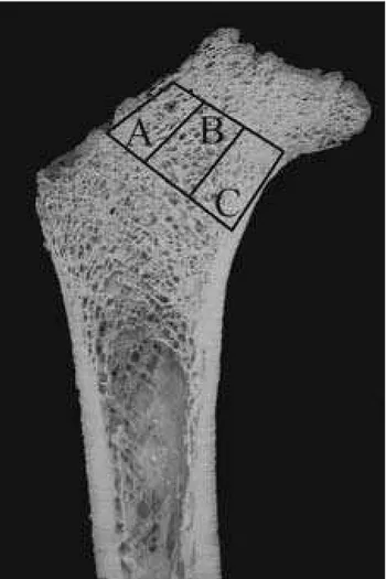
In order to perform microscopic analysis of the thic-kness and distribution of trabecule in different regions of the femoral neck (medial and lateral neck and Ward trian-gle), undecalcified bone samples embedded in methylmet-hacrylate were cut into 100 μm thick slides in frontal plane using a Leica diamond saw (Leica SP 1600). On the cross sections, three regions of interest were defined. Zone A represented the lateral neck region, zone B comprised the central portion of the neck (including Ward’s triangle), while zone C was composed of the medial part of the neck.
740
The regions were situated between two parallel lines drawn perpendicularly to the axis of the femoral neck (Figure 1). Those regions were analyzed on unstained 100 μm thick ground sections of undecalcified bone using a polarized-light microscope.
Statistical analysis
The one-sample Kolmogorov-Smirnov test was used to check for the normality of the distribution in the observed external geometric and densitometric parameters. Since DXA measurements (aBMD) are size dependent, i.e., me-asurementsare from a two-dimensional image projection of a three-dimensionalstructure, in a growing child this causes inaccuracies when interpreting measurements [23]. Therefore, to control the effects of the third dimension, aBMD was adjusted to the femoral neck diameter. Linear regression analysis was used to assess the age-dependence of adjusted bone mineral density and external geometric parameters, while in case of the neck index geometric cu-rve better fitted the data. The Pearson’s correlation was used to determine the level of association among external geometric parameters. SPSS statistical package (version 15) and MedCalc (version 9) were used for the analysis, and the results were considered significant at 0.05 level.
RESULTS
External geometry
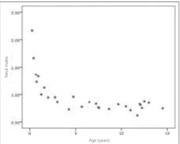
During childhood the femoral neck width and femoral neck axis length showed increase. Nevertheless, the obser-ved age-dependent decrease of the neck index indicates that the femoral neck generally elongates with age (Table 1). However, the decrease of the neck index is not gra-dual; NI decreases dramatically from birth to the end of the first year and then decreases only slightly achieving a plateau at about the age of 3 years (Figure 2). The collo-diaphyseal angle showed a negative age trend, with values ranging between 138 and 112 degrees (Table 2). It did not correlate significantly with the femoral neck axis length, femoral neck width or the neck index (Pearson correlation; p=0.177, p=0.263, p=0.242, respectively).
Bone mineral density
Adjusted areal bone mineral density demonstrated conti-nuous increase during childhood in all regions of interest (Figure 3, Tables 1 and 2).
Table 2. Descriptive statistics for external geometry and densitometric parameters of growing femora
Parameter Mean SD Min. Max.
Femoral neck width (cm) 2.3 0.7 1.3 3.5
Femoral neck axis length (cm) 2.7 1.2 0.6 4.4
Neck index (dimensionless) 0.960 0.332 0.619 2.167
Neck-shaft angle (degrees) 129.1 6.4 112 138
Adjusted BMD neck (g/cm2) 0.315 0.044 0.225 0.389
Adjusted BMD intertrochanteric
(g/cm2) 0.371 0.054 0.285 0.514
Adjusted BMD ward (g/cm2) 0.317 0.048 0.222 0.448
Adjusted BMD total (g/cm2) 0.344 0.048 0.263 0.436
Table 1. Linear regression analysis for age dependence in densitometric and geometric parameters
Parameter R R2 p
Neck-shaft angle -0.305 0.093 0.114
Neck index § -0.910 0.828 <0.001
Neck axis length 0.961 0.923 <0.001
Neck width 0.959 0.920 <0.001
aBMD neck 0.474 0.224 0.011
aBMD intertrochanteric 0.559 0.312 0.002
aBMD Ward 0.515 0.265 0.005
aBMD total 0.543 0.295 0.003
§ non-linearregression
Regression equation: Log (neck index) = 0.0966 - 0.2145 × Log (age)
Djurić М. еt al. Morphological Characteristics of the Developing Proximal Femur: A Biomechanical Perspective
Figure 2. Changes in the neck index as a function of age Age (years)
Neck index
Figure 3. Increase of femoral adjusted areal bone mineral density (BMD) with age of individuals
Trabecular pattern
Qualitative analysis of frontal sections of proximal femora showed that trabecular pattern changed from the parallel trabeculae after birth to three distinct trabecular groups in the second year (Table 3, Figures 4–7), while the upper end of the medullary canal came closer to the trochanter region.
Radiography demonstrated more clearly visible trabe-cular pattern in younger individuals than frontal sections; starting with the age of 8 months in all individuals princi-pal compressive and tensile trabecular groups of trabecu-lae were well defined (Figure 6, Table 3).
Histological observation revealed that starting from the age of 8 months all specimens demonstrated three groups of bone trabeculae; longitudinal (principal compressive in the medial neck), transversal (principal tensile in the late-ral neck) and randomly scattered (Ward’s triangle) (Table 3, Figures 4–9). The thickest were longitudinal and the thinnest were randomly scattered trabeculae (Figure 8). Bone marrow spaces were the smallest between the lon-gitudinal trabeculae and the largest in the Ward’s triangle (Figure 8).
DISCUSSION
In our sample, observation of specimens of different age revealed outstanding changes in bone size and shape, as well as in internal organization, and BMD.
Figure 4. Low power microscopic view of the proximal femur aged one month shows longitudinal bone trabeculae and thin medial cortex (arrow) (100 µm undecalcified section, unstained)
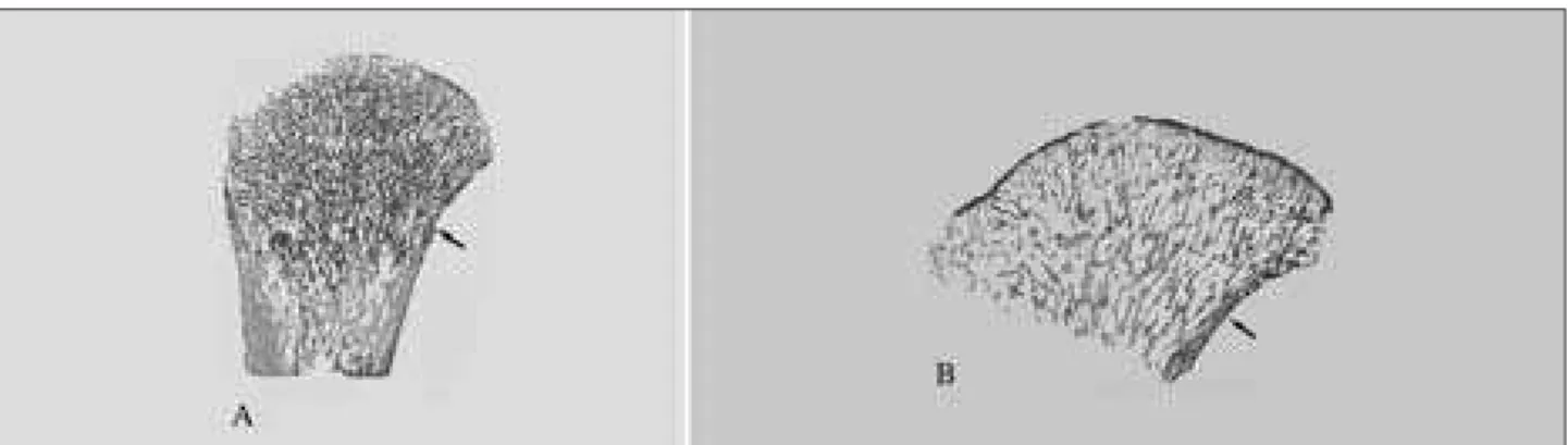
Figure 5. Low power microscopic view (100 µm undecalcified section, unstained) of proximal femora aged 3 months (A) and 5 months (B) showing primarily longitudinal bone trabeculae. Observe transversal growth and thicker medial cortex (arrow) in comparison to the previous Figure.
Figure 6. The proximal femur of an 8-month old individual: A. Frontal section; B. Radiography; C. Low power microscopic view showing longitudinal (right), transversal (left up) and random (left below) bone trabeculae. Medial cortex is marked by arrow.
742
Growth-related changes in external geometry
Our results demonstrated that the neck-shaft angle decre-ased with growth, which is in close agreement with other studies [24]. The decrease in the collo-diaphyseal angle du-ring growth could represent the result of changes in body proportions followed by adaptation of the hip joint to vertical posture and gait changed conditions [25, 26]. Namely, it is proposed that modelling of the femoral neck is governed by the balance between vertical compressive forces originating from gravity and the contractions of the iliopsoas muscle, and the tension caused by abductor muscles attached to the greater trochanter [27], with certain morphological differen-ces between different hominid taxa [28]. It is considered that
Table 3. Trabecular pattern of growing femora
Case No Age Macroscopic observation Radiographic appearance Histological appearance
1 1 month Straight trabeculae running parallel to the bone long axis Straight parallel trabeculae Longitudinal bone trabeculae (Figure 4)
2 3 months Randomly oriented trabecular network without
distinct pattern No distinct pattern
Longitudinally oriented trabeculae thicker in central and medial areas (Figure 5A)
3 5 months Randomly oriented trabecular network without
distinct pattern No distinct pattern
Longitudinally oriented trabeculae thicker in central and medial areas (Figure 5B)
4 8 months
Trabeculae at the upper end of medullar canal from the medial and the lateral side run obliquely toward midline – “ish bone” pattern; principal tensile group of trabeculae parallel to the superior surface of the neck is slightly visible (Figure 6A)
Principal compressive and tensile groups are well deined
(Figures 6B and 7B)
3 groups of trabeculae: longitudinal (principal compressive), transversal (principal tensile) and randomly scattered (Ward’s triangle) (Figures 6C, 7C and 8); progressive growth of trabeculae and medial cortex 5 9 months Observable principal tensile group
6 11 months Observable principal tensile group
7 1 year 3 months
Observable principal compressive group of trabeculae
8 1 year 7 months
All groups of trabeculae are observable on cross sections
9 2 years All groups of trabeculae are observable on cross sections
10 2 years 8 months
Groups of trajectories and Ward’s triangle are visible on cross sections (Figure 7A) 11-29
Older than 2 years and 8 months
Figure 8. Medium power microscopic view of bone trabeculae and marrow spaces of proximal femur aged twelve years. A. Principal compressive bone trabeculae, medial neck; B. Principal tensile bone trabeculae, lateral neck (left) and Ward’s triangle bone trabeculae (center and right).
Figure 9. Main groups of trabeculae in adult femur: 1 – Principal compressive; 2 – Principal tensile; 3 – Greater trochanteric; 4 – Ward’s triangle; 5 – Secondary tensile; 6 – Secondary compressive
fitting of the femoral head to the acetabulum is influenced by the neck-shaft angle. This angle represents a beneficial structural adaptation which keeps the lower extremity away from the pelvis and allows more rotation of the hip joint [29]. Inclination of the hip resultant force towards the vertical pla-ne and increase in the magnitude of trochanteric resultant force with maintaining its direction during the growth peri-od cause a decrease in the neck-shaft angle [27].
Although the values of the neck-shaft angle have been reported to correlate positively with the length of the fe-moral neck in adult population [29], our results indicate that it is not the case during the growth period, i.e., in children there was age-related increase in the neck length while the neck-shaft angle tended to decline.
While in our specimens both the femoral neck width and femoral neck axis length increased with age, the observed age-related increase of the neck index indicates that the growth in length is more rapid causing femoral neck to beco-me elongated as age increases. This adaptation allows a con-siderable range of movements of the hip joint [30] which is necessary for daily activities and adaptation to biological and social functions of childhood and approaching adolescence.
Trabecular pattern – classical vs. recent view on mechanics of the proximal femur
In fetal and early postnatal period, the whole metaphysis is filled by primary trabecular bone [7] and it is considered that increased mechanical loading in the first year leads to conversion of primary to secondary cancellous bone with two distinct groups of intersecting vault-like trabe-culae (compressive and tensile groups) [8, 9, 10]. Town-sley [8] has pointed out that the growth-related changes in bone morphology, as an adaptation to habitual loading conditions, are closely related to the beginning of walking which introduces body weight load on the femur; this is consistent with traditional interpretation of stresses wit-hin the proximal femur, since weight-bearing stresses are concentrated in the primary compressive system of trabe-culae during gait [7, 9, 31]. In our sample, at the age of one month, the proximal femur displayed almost straight longitudinal trabeculae, which is in agreement with data from other studies [7, 8, 10]. With further growth, we no-ticed more distinctive pattern of trabecular arrangement. However, “principal compressive” and “tensile” groups of trabeculae in our sample appeared at the age of 8 months (visible on radiography Figure 6B and histology Figure 6C), which does not fit in the classical concept since it is befo-re the age at which a child begins to walk. Thebefo-refobefo-re, our findings speak more in favor of some recent models of the stresses in the femoral neck. Namely, in contrast to traditi-onal interpretation, the stresses in the proximal femur are considered to be predominantly compressive [15, 16]. The hip muscles, joint capsule and ligaments pull the femoral head into the acetabulum contributing to the compression applied to the femoral head [15]. Therefore, the early appe-arance of the trabecular groups in our sample could reflect increased activity of the muscles inserting into the greater
and lesser trochanter, even before a child starts to walk. This is supported by observations that the largest physiolo-gic loads placed on children’s bones originate from muscle contractions, being even routinely greater than the effect of gravity [17]. Further development and reinforcement of such distinctive trabecular organization in later age could further correspond to stress changes caused by upright sit- ting at about 6 months, crawling at about 9 months, as well as transitory standing and beginning of walking.
Our findings are also compatible with a further changed picture about the types of stresses in the proximal femur with recent emphasis on the importance of shear stress. The bone is the weakest in shear when compared to tension and compression, and the femur is habitually loaded “off-axis” which augments shear stresses between the layers of the femoral neck [32, 33]. Therefore, in order to maintain its stability, the bone has to adapt itself to accommodate shear coupling. In our study, contrary to the findings of Osborne et al. [7], we consistently found secondary medial and late-ral groups of trabeculae in the specimens. This “fish bone” pattern which was observable as early as at the age of 8 months resembled the Hert’s model of trabecular organiza-tion in case of multiaxial loading, and those two groups of trabeculae could be considered to originate from extreme angle load cases which cause large bending moments in the femur [13]. Contrary to Wolff ’s descriptions, and in line with some other authors [9, 12, 32, 33], trabecular inter-sections in our sample were notably non-orthogonal. Such non-ortogonality in trabecular arrangement is considered to be the most favorable organization in case of multiaxial joint loading [9, 32, 33], particularly as it could represent an important adaptive response since it has been shown to reduce shear coupling effects [32]. In that way, the bone stability would be encouraged by reducing shear stresses. In our other specimens, all groups of trabeculae were present and their pattern changed slightly with further growth and gait maturation, which is compatible with microCT data by Ryan and Krovitz [10].
Regional differences in bone amount: mechanical perspective
744
those parts [4, 6, 34]. Namely, in the normal human femo-ral neck, the major part of the load is concentrated in the medial aspect [31], while the lateral neck is under-loaded.
CONCLUSION
The observed growth-related changes in proximal fe-moral external and internal morphology are compatible with bone functional adaptation principle. The changes in shape of the proximal femur could reflect the changing complex loading pattern during growth. Quite homoge-nous bone in the youngest individuals changed differently between the medial and lateral aspect of the femoral neck
during the growth process. The differential trabecular arrangement and cortical distribution are compatible with recent changes in understanding of proximal femur bio-mechanics; predominance of compressive stresses in the femoral neck, significance of adaptation to shear stresses, multiaxial loading conditions perspective, the prevalence of the effects of muscle actions over the effect of body weight, and the existence of bone adaptational eccentricity
ACKNOWLEDGEMENTS
The authors acknowledge the support from the Ministry of Science of the Republic of Serbia (project number: 45005).
Djurić М. еt al. Morphological Characteristics of the Developing Proximal Femur: A Biomechanical Perspective
REFERENCES
1. Roux W. Der Kampf der Teile im Organismus. Leipzig: Wilhelm Engelmann; 1881.
2. Wolff J. Das Gesetz der Transformation der Knochen. Berlin: Verlag von August Hirschwald; 1892.
3. Ruff C, Holt B, Trinkaus E. Who’s afraid of the big bad Wolff?: “Wolff’s law” and bone functional adaptation. Am J Phys Anthropol. 2006; 129(4):484-98.
4. Djuric M, Djonic D, Milovanovic P, Nikolic S, Marshall R, Marinkovic J, et al. Region-specific sex-dependent pattern of age-related changes of proximal femoral cancellous bone and its implications on differential bone fragility. Calcif Tissue Int. 2010; 86(3): 192-201.
5. Milovanovic P, Djonic D, Marshall RP, Hahn M, Nikolic S, Zivkovic V, et al. Micro-structural basis for particular vulnerability of the superolateral neck trabecular bone in the postmenopausal women with hip fractures. Bone. 2012; 50(1):63-8.
6. Djonic D, Milovanovic P, Nikolic S, Ivovic M, Marinkovic J, Beck T, et al. Inter-sex differences in structural properties of aging femora: implications on differential bone fragility: a cadaver study. J Bone Miner Metab. 2011; 29(4):449-57.
7. Osborne D, Effmann E, Broda K, Harrelson J. The development of the upper end of the femur, with special reference to its internal architecture. Radiology. 1980; 137(1I):71-6.
8. Townsley W. The influence of mechanical factors on the development and structure of bone. Am J Phys Anthropol. 1948; 6(1):25-46. 9. Hert J. A new attempt at the interpretation of the functional architecture of the cancellous bone. J Biomech. 1994; 27(2): 239-42.
10. Ryan TM, Krovitz GE. Trabecular bone ontogeny in the human proximal femur. J Hum Evol. 2006; 51(6):591-602.
11. Salle BL, Rauch F, Travers R, Bouvier R, Glorieux FH. Human fetal bone development: histomorphometric evaluation of the proximal femoral metaphysis. Bone. 2002; 30(6):823-8.
12. von Meyer GH. Die Architekur der Spongiosa. Arch Anat Physiol Wissenschaf Med. 1867; 34:615-28.
13. Miller Z, Fuchs MB, Arcan M. Trabecular bone adaptation with an orthotropic material model. J Biomech. 2002; 35(2):247-56. 14. Pedersen P. On optimal orientation of orthotropic materials. Struct
Optim. 1989; 1(2):101-6.
15. Rudman K, Aspden R, Meakin J. Compression or tension? The stress distribution in the proximal femur. Biomed Eng Online. 2006; 5(1):12. 16. Kalmey JK, Lovejoy CO. Collagen fiber orientation in the femoral
necks of apes and humans: do their histological structures reflect differences in locomotor loading? Bone. 2002; 31(2):327-32. 17. Frost HM. Muscle, bone, and the Utah paradigm: a 1999 overview.
Med Sci Sports Exerc. 2000; 32(5):911-7.
18. Ford CM, Keaveny TM. The dependence of shear failure properties of trabecular bone on apparent density and trabecular orientation. J Biomech. 1996; 29(10):1309-17.
19. Keaveny T, Morgan E, Niebur G, Yeh O. Biomechanics of trabecular bone. Annu Rev Biomed Eng. 2001; 3:307-33.
20. Maresh MM. Linear growth of the long bones of extremities from infancy through adolescence. Am J Dis Child. 1955; 89:725-42. 21. Maresh MM. Measurements from roentgenograms, heart size, long
bone lengths, bone, muscles and fat widths, skeletal maturation. In: McCammon RW, editor. Human Growth and Development. Springfield: Charles C. Thomas; 1970. p.155-200.
22. Scheuer JL, Black SM. Developmental Juvenile Osteology. London: Academic Press; 2000.
23. Rauch F, Schoenau E. Changes in bone density during childhood and adolescence: an approach based on bone’s biological organization. J Bone Miner Res. 2001; 16(4):597-604.
24. Hefti F. Deviations in the axes of the lower extremities. Orthopade. 2000; 29(9):814-20.
25. Rafferty KL. Structural design of the femoral neck in primates. J Hum Evol. 1998; 34(4):361-83.
26. Bulandra AM, Gielecki JS, Leciejewska I, Karaszewski P, Sieron D. Digital-image analysis of the femoral shaft/neck angle in human foetuses. Folia Morphol (Warsz). 2003; 62(4):415-7.
27. Heimkes B, Posel P, Plitz W, Zimmer M. Age-related force distribution at the proximal end of the femur in normally growing children. Z Orthop Ihre Grenzgeb. 1997; 135(1):17-23.
28. Holliday TW, Hutchinson VT, Morrow MMB, Livesay GA. Geometric morphometric analyses of hominid proximal femora: Taxonomic and phylogenetic considerations. HOMO J Comp Hum Biol. 2010; 61(1):3-15. 29. Isaac B, Vettivel S, Prasad R, Jeyaseelan L, Chandi G. Prediction of the
femoral neck-shaft angle from the length of the femoral neck. Clin Anat. 1997; 10(5):318-23.
30. Fox JC, Keaveny TM. Trabecular eccentricity and bone adaptation. J Theor Biol. 2001; 212(2):211-21.
31. Lotz J, Cheal E, Hayes W. Stress distributions within the proximal femur during gait and falls: implications for osteoporotic fracture. Osteoporos Int. 1995; 5:252-61.
32. Pidaparti R, Turner C. Cancellous bone architecture: advantages of nonorthogonal trabecular alignment under multidirectional joint loading. J Biomech. 1997; 30:979-83.
33. Skedros JG, Baucom SL. Mathematical analysis of trabecular ‘trajectories’ in apparent trajectorial structures: the unfortunate historical emphasis on the human proximal femur. J Theor Biol. 2007; 244(1):15-45.
КРАТАК САДРЖАЈ
Увод За раз ли ку од оби ља сту ди ја ко је су ана ли зи ра ле гор-њи окра јак бут не ко сти код од ра слих осо ба, ње го ва спо-ља шња и уну тра шња мор фо ло ги ја код де це ни су до вољ но ис тра жи ва не.
Циљ ра да Ис пи ти ва ли смо про ме не спо ља шње и уну тра шње мор фо ло ги је гор њег окрај ка бут не ко сти ко је на ста ју то ком ра ста и раз во ја ра ди утвр ђи ва ња њи хо ве по ве за но сти с би-о ме ха нич ким чи ни би-о ци ма кби-о ји де лу ју на бут ну кби-ост чби-о ве ка. Ме то де ра да На гор њим окрај ци ма бут не ко сти 29 осо ба (уз ра ста од ме сец да на до 14 го ди на) из ар хе о ло шког кон-тек ста ис пи ти ва ни су спо ља шња ге о ме три ја, уну тра шња гра ђа тра бе ку лар не и кор ти кал не ко сти, као и ми не рал на гу сти на ко сти, при ме ном ма кро скоп ских, ми кро скоп ских и ра ди о ло шких ме то да.
Ре зул та ти С уз ра стом де те та до ла зи до по ра ста ши ри не и ду жи не вра та бут не ко сти, с тим да он по ста је у це ли ни из ду-же ни ји, као и сма ње ња ко ло ди ја фи зар ног угла. По сто ји ја ка
по зи тив на по ве за ност уз ра ста де те та и стан дар ди зо ва не ми не рал не гу сти не ко сти. Па ра лел ни рас по ред тра бе ку ла ко ји по сто ји на ро ђе њу ме ња се та ко да се од осмог ме се ца мо гу пре по зна ти три по себ не гру пе тра бе ку ла (ком пре сив-на гру па, тен зи о сив-на гру па и сив-на су мич не гру пе). Гор њи и до-њи сег мент вра та бут не ко сти код де це ста ри јег уз ра ста се раз ли чи то ме ња ју, та ко да до њи део вра та има де бљи слој кор ти кал не ко сти и де бље тра бе ку ле.
За кљу чак При ме ће не мор фо ло шке про ме не пред ста вља-ју адап та ци вља-ју на деј ство ме ха нич ких си ла на бут ну кост у раз во ју. На ши ре зул та ти о уну тра шњој гра ђи у скла ду су с но ви јим би о ме ха нич ким схва та њи ма ко ја ис ти чу пре ва гу ком пре сив них на по на, до ми нант ну адап та ци ју на сми ца ње, зна чај ми шић них ефе ка та и ви ше о со вин ског оп те ре ће ња, као и по сто ја ње адап та ци о не екс цен трич но сти уну тра шње гра ђе вра та бут не ко сти.
Кључ не ре чи: гор њи окра јак бут не ко сти; раст; адап та ци ја ко сти; ме ха нич ко оп те ре ће ње
Морфолошка обележја проксималног окрајка бутне кости током развоја:
биомеханички аспекти
Марија Ђурић1, Петар Миловановић1, Данијела Ђонић1, Арса Минић2, Михаел Хан3
1Лабораторија за антропологију, Институт за анатомију, Медицински факултет, Универзитет у Београду, Београд, Србија; 2Амерички медицински факултет у Београду, Европски универзитет, Београд, Србија;
3Институт за остеологију и биомеханику, Универзитетска болница Хамбург-Епендорф, Хамбург, Немачка