S
KELETON GEOMETRY
,
PHYSICAL ACTIVITY AND
PROXIMAL FEMUR BONE MASS DISTRIBUTION
IN
8-12
YEAR OLD CHILDREN
Dissertação elaborada com vista à obtenção do Grau de Doutor no Ramo
de Motricidade Humana, Especialidade em Atividade Física e Saúde
Orientador: Doutora Maria de Fátima Marcelina Baptista
Júri:
Presidente
Reitor da Universidade de Lisboa
Vogais
Doutor Alberto Ramos Duarte, Professor Catedrático da Faculdade de Desporto da Universidade do Porto
Doutor Jaime da Cunha Branco, Professor Catedrático da Faculdade de Ciências Médicas da Universidade Nova de Lisboa
Doutor António Prieto Veloso, Professor Catedrático da Faculdade de Motricidade Humana da Universidade de Lisboa
Doutora Maria de Fátima Marcelina Baptista, Professora Associada da Faculdade de Motricidade Humana da Universidade de Lisboa
Doutora Maria Margarida Marques Rebelo Espanha, Professora Associada da Faculdade de Motricidade Humana da Universidade de Lisboa
Doutora Maria Helena Santa-Clara Pombo Rodrigues, Professora Auxiliar da Faculdade de Motricidade Humana da Universidade de Lisboa
Funding
List of figures ... 3
List of tables ... 5
Abstract ... 7
Resumo ... 9
Acknowledgments / Agradecimentos ... 11
1 Introduction ... 15
References ... 18
2 Literature overview and theoretical background ... 19
2.1.1 The risk of bone fracture... 24
2.1.2 Peak Bone Mass and bone health ... 26
2.1.3 Effects of Physical Activity on bone mass in children ... 29
2.1.4 Geometry and biomechanics of the proximal femur and pelvis ... 33
References ... 39
3 Experimental work ... 51
3.1 Ward’s area location, Physical Activity and body composition in 8 and 9 years old boys and girls ... 51
3.2 Sex specific association of physical activity on proximal femur BMD in 9 to 10 years-old children ... 75
3.3 Pelvis width associated with bone mass distribution at the proximal femur in 10-11 yr old children ... 95
3.4 Influence of physical activity and skeleton geometry on bone mass at the proximal femur in 10-12 year old children – a longitudinal study ... 119
4 General discussion ... 145
References ... 151
5 Conclusions and further research ... 153
Figure 1 - Main known factors influencing bone mass, in the context of the risk
of bone fracture problem. 21
Figure 2 - Theoretical macro framework of this Ph.D thesis 23 Figure 3 - Forces exerted on the femoral neck through the action of body weight
and abductor muscle forces (adapted from Lovejoy, 1988 [164]). 35 Figure 4 - Stress and tension forces on the femoral neck (adapted from Tunner,
2005 [167]) 36
Figure 5 - Biomechanical model of the pelvis-hip system (adapted from Maquet,
1999 [169]) 36
Figure 6 - “Disadvantaged” positioning of the abductor muscles in the pelvis-hip system (adapted from Traiana et al., 2009 [77]) 38
Chapter 3 - Experimental work
Ward’s area location, Physical Activity and body composition in 8 and 9 years old
boys and girls
Figure 1 Proximal femur DXA image showing the location of the landmarks
used to identify proximal femur shape and position of Ward’s area 57
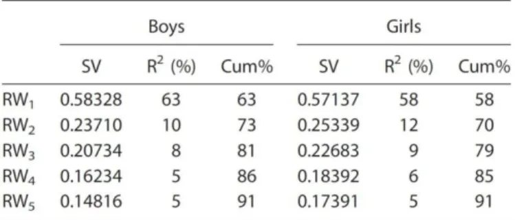
Figure 2 Relative warp (RW) plots for RW1 and RW2 in boys and girls 63
Figure 3 Femoral neck Ward’s area position changes in boys and girls
depicted by RW1 64
Pelvis width associated with bone mass distribution at the proximal femur in 10-11 yr old children
Figure 1 Geometric measures of the pelvic bone 101
Figure 2 Measurements of the Geometric measures of the proximal femur
geometry 102
Influence of physical activity and skeleton geometry on bone mass at the proximal femur in 10-12 year old children – a longitudinal study
Figure 1 Hip image from Hologic DXA scanner showing the region of interest of the superolateral femoral neck (A) and the inferomedial
femoral neck (B) 126
Figure 2 Geometric measures of the pelvic bone 127
Ward’s area location, Physical Activity and body composition in 8 and 9 years old
boys and girls
Table 1 Subject Characteristics 62
Table 2 Relative Warps Analysis 63
Table 3 Comparison of Age, Body Composition, Calcium Intake, Physical
Activity, and Bone Variables Between Group 1 and Group 2 66
Table 4 Regression Analysis: prediction of proximal shape variation 67
Sex specific association of physical activity on proximal femur BMD in 9 to 10 year-old children
Table 1 Characteristics of participants 83
Table 2 Standardized regression coefficients (β), level of significance (p) and coefficient of determination (R2) for proximal femur
sub-region models, with data for boys and girls pooled together 84
Table 3 Standardized regression coefficients (β), level of significance (p) and coefficient of determination (R2) for proximal femur
sub-region models, with data for boys and girls treated separately 86
Table 4 Effects of 10 minutes per day of additional physical activity on
femoral neck, trochanter, and intertrochanter BMD 87
Pelvis width associated with bone mass distribution at the proximal femur in 10-11 year-old children
Table 1 Participants characteristics: age, maturity, body composition and
physical activity 106
Table 2 Participants characteristics: BMD and bone geometric measures 107 Table 3 Regression coefficients (β), level of significance (p) and coefficient
of determination (R2) of the associations between physical activity, geometric measures of pelvis and proximal femur, and proximal
Table 4 Regression coefficients (β), level of significance (p) and coefficient of determination (R2) of the associations between physical activity, geometric measures of pelvis and proximal femur, and proximal
femur BMD ratios 109
Table 5 Main and interaction effects of inter-acetabular distance and
physical activity on proximal femur BMD ratios in girls 110
Table 6 Main and interaction effects of inter-acetabular distance and
physical activity on proximal femur BMD ratios in girls 111
Influence of physical activity and skeleton geometry on bone mass at the proximal femur in 10-12 year old children – a longitudinal study
Table 1 Descriptive characteristics of the participants at base line and one
year follow-up 133
Table 2 Random-effects GLS regression models for femoral neck (total, superolateral, and inferomedial) and trochanter BMDs testing for the effects of physical activity and geometric measures of the pelvis and hip, controlling for maturity, body height and body lean
mass 134
Table 3 Random-effects GLS regression models, using panel data, for proximal femur BMD ratios, controlling for maturity, body height and body lean mass and testing for the effects of physical activity
Abstract
In the context of bone health promotion, the aim of this Ph.D dissertation was to analyze potential explanatory factors of the effects of physical activity and of bone geometry on bone mass distribution at the proximal femur in 8-12 year old children. Four studies were undertaken to compare the bone mineral density (BMD) between: (a) the sub-regions of the proximal femur – the neck and its superolateral and inferomedial aspects, the trochanter and the intertrochanter; (b) sexes, concerning the associations/effects of non-targeted physical activity and bone geometry. Sex and regional specific effects of non-targeted physical activity on bone mass distribution at the proximal femur in children were observed. The geometry of the pelvis and the proximal femur, namely the pelvis width and the abductor lever arm, emerged as predictors of bone mass distribution at the proximal femur, therefore as explanatory factors of both the regional and the sex specific patterns. These geometric features might mediate the physical activity effects on bone mineralization at the proximal femur, as long as, when they are considered, the power of physical activity to explain the distribution of bone mass at this skeletal site seems limited.
Resumo
No contexto da promoção da saúde óssea, o objetivo desta dissertação de doutoramento foi analisar potenciais fatores explicativos dos efeitos da atividade física habitual e da geometria óssea na distribuição da massa óssea do fémur proximal, em crianças de 8-12 anos de idade. Para o efeito foram realizados quatro estudos comparando a densidade mineral óssea (DMO) entre: (a) as diversas sub-regiões do fémur proximal - o colo do fémur e os seus aspetos supero-lateral e infero-medial, o grande trocanter e a sub-região intertrocantérica; (b) os sexos, relativamente às associações/efeitos da atividade física habitual e da geometria óssea. Foram observadas associações/efeitos da atividade física habitual na massa óssea do fémur proximal diferenciados quanto ao sexo e sub-região. A geometria da pélvis e do femur proximal, nomeadamente a largura da pélvis e o braço de momento de força dos abdutores, surgiram como preditores da distribuição de massa óssea no fémur proximal e consequentemente como fatores explicativos de diferenciação da distribuição de massa óssea de acordo com o sexo e sub-região. Estas caraterísticas geométricas poderão mediar os efeitos da atividade física na mineralização do femur proximal uma vez que quando consideradas parecem limitar a capacidade explicativa da atividade física na distribuição de massa óssea no fémur proximal
Acknowledgments / Agradecimentos
Os contributos para esta tese foram muito diversificados, quer pela forma como se manifestaram, quer pela intensidade e frequência com que ocorreram.
O meu percurso como doutoranda foi marcado por um outro, paralelo, de grande sofrimento, motivado por questões familiares, que infelizmente, ou não, duraram quase a totalidade do tempo que tive para concluir este processo.
Os condicionalismos que esta situação provocou afetaram fortemente a minha capacidade para assumir compromissos e aceitar desafios, bem como as minhas vivências dentro da faculdade, o trabalho em equipa, a participação em projetos complementares e até a partilha de experiências com os meus colegas.
É neste contexto que os meus primeiros agradecimentos vão para a Professora Doutora Fátima Baptista, pelo seu valioso apoio ao longo deste processo. Agradeço também, para além da sua orientação científica que me proporcionou novos conhecimentos e importantes aprendizagens, a sua constante disponibilidade para compreender os meus atrasos e as minhas restrições, a sua capacidade em me valorizar e incentivar, e, principalmente, por nunca me ter deixado desistir. Sem estas suas qualidades humanas não teria conseguido chegar até aqui. Por tudo isto, o meu profundo agradecimento.
Agradeço também a toda a equipa do Laboratório de Exercício e Saúde, com quem partilhei experiencias, ideias, projetos, avaliações e conhecimentos, muitos deles estruturais neste meu percurso.
I would like to extend my gratitude to Professor Kathleen Janz from the Department of Health and Human Physiology and from Department of Epidemiology, University of Iowa (USA) for her suggestions and for her contribution in the articles.
À Professora Doutora Filomena Carnide agradeço o empenho e o carinho com que sempre se disponibilizou para me ajudar a refletir e ultrapassar problemas de estatística, deixando sempre a “suaporta aberta”.
Agradeço também ao Professor Doutor António Veloso pelas conversas, sempre interessantes, e pelos diversos momentos de reflexão, que para lá da biomecânica, me ensinaram a ver mais além.
Agradeço aos meus colegas de gabinete que sempre comigo trocaram experiências e conhecimentos, e em especial à Vera Zymbal e à Lurdes Rebocho, companheiras de muitos outros momentos… A ambas agradeço a oportunidade de partilhar e de trabalhar em conjunto, de dividir preocupações e alegrias e, mais do que isso, o afeto que sempre demonstraram.
Aos professores Carlos Ferreira, Paula Bruno e Nicoletta Rosati, que em momentos diferentes me ajudaram a resolver questões relacionadas com o tratamento estatístico e informática, o meu igual agradecimento.
A todos os meus amigos pela “presença viva” que têm na minha vida, por se
alegres destes últimos 4 anos. Em especial às minhas grandes amigas Margarida, João e Teresa, que me fazem acreditar cada vez mais no poder mágico da amizade.
Por fim, e sem dúvida, o maior agradecimento vai para a minha família, suporte precioso neste processo.
Aos meus pais pelo seu amor, pelo constante incentivo e em especial à minha mãe pelo extraordinário exemplo de coragem e de perseverança.
Ao Diogo, à Catarina e ao Francisco por encherem o meu coração de amor todos os dias e por me fazerem sentir tão feliz! Vocês foram e são, sem dúvida
nenhuma, uma das minhas maiores fontes de energia…
Ao Eduardo, que esteve sempre ao meu lado, para partilhar, para refletir, para
ajudar, para me ouvir, para me amparar… Que foi o amigo, o companheiro, o
marido e também cúmplice de todo este caminho…, que com a sua calma
1
Introduction
This dissertation presents my Ph.D research project’s main outputs. It’s core is the collection of the four research articles developed during this investigation period. This research is broadly motivated by a public health issue – the risk of bone fracture in the elderly phase of life.
It is now widely accepted that the risk of fracture in that phase, particularly at the spine and proximal femur, which is strongly associated to osteoporosis disease, constitutes a public health problem due to: its widespread incidence among adults; its dramatic consequences for the quality of life of those sustaining a fracture and due to the challenges it brings to national health care systems, namely those concerning resource allocation.
Being that there is no cure to osteoporosis and given the widespread belief that some of its origins may lay in childhood and adolescence, a large emphasis has been put on prevention strategies in these earlier phases of life, to such an extent that some authors consider that osteoporosis is a pediatric issue [1]. In fact, the development of bone mass during growth years is extensively described and documented in the literature [2], as are the effects of loads induced by physical activity, the main modifiable lifestyle factor.
Nevertheless, the effects of physical activity on bone mass vary considerably. The benefits of high-impact activities via specially designed intervention programs is a consolidated result in the literature, but increasing evidence of skeletal development benefits from lifestyle physical activity during childhood [5, 6, 7] should support the promotion of regular physical activity in youth - specially weight-bearing activity (as body weight increases the magnitude of loading) - as a strategy to reduce the risk of bone fracture later in life.
The understanding of the factors determining the specific effects on bone mass caused by physical activity induced loads is essential for the appropriate design of those prevention policies. But whereas the effects of intervention programs, or even very high intensity (and frequency) activities that some elite athletes engage in, are rather consolidated in the literature, the understanding of the effects of the habitual volunteer physical activity of non-athletes is less developed. Whether there is a distinct bone responsiveness to mechanical loading induced by non-targeted physical activity in boys and girls, or whether there are skeletal sex differentiating aspects that may influence the impact of physical activity on bone mineralization are among the questions that have arisen in this research branch.
In this context, the general aim of the present Ph.D dissertation was to analyze and explore the factors that explain the effects of physical activity induced mechanical loads on bone mass distribution at the proximal femur in children. To pursuit this general objective, several specific objectives were defined:
To analyze the potentially differentiated effects of physical activity at the proximal femur neck, trochanter and intertrochanter sub-regions;
To analyze the role of the pelvis and hip geometry on physical activity induced effects on the mineralization of the proximal femur.
To accomplish these tasks, body composition, bone mineral parameters and bone geometry features obtained from DXA scanning, and physical activity data from accelerometry and questionnaires were statistically treated in several regression models. To analyze the bone mass distribution at all three sub-regions of the proximal femur, intra-individual bone mineral density ratios of proximal femur sub-regions were defined.
This dissertation's structure is composed of an initial introductory chapter (chapter 2) where a literature review and theoretical background is presented, not as a substitute for the specific literature review that is part of each of the articles that constitute the core of the document, but solely with a unifying purpose of all the work developed. The methodological aspects are not treated in this chapter as they are instrumental for the research project objectives and are described in detail in each of the four studies presented in chapter 3. The fourth chapter, in a brief general discussion, attempts to integrate the main results of the studies produced, highlighting those that seem to be the most interesting aspects of the overall research project. Once more, they must not be considered without the studies' specific discussion sections presented in the precedent chapter.
Chapter 5 outlines the main conclusions and reflections for further research on the issues dealt with in this dissertation, and that the candidate believes deserve additional research.
References
1. Heaney, R. P., Abrams, S., Dawson-Hughes, B., Looker, A., Looker, A., . Marcus, R. et al. (2000). Peak bone mass. Osteoporos Int, 11(12):985-1009.
2. Gunter, K. B., Almstedt, H. C., Janz, K. F. (2012). Physical activity in childhood may be the key to optimizing lifespan skeletal health. Exerc Sport Sci Rev, 40(1):13.
3. Hind, K., Burrows, M. (2007). Weight-bearing exercise and bone mineral accrual in children and
adolescents: a review of controlled trials. Bone, 40(1):14-27.
4. Rizzoli R, Bianchi ML, Garabedian M, McKay HA, Moreno LA. Maximizing bone mineral mass gain during growth for the prevention of fractures in the adolescents and the elderly. Bone 2010;46:294–305.
5. Janz, K. F., Burns, T. L., Levy, S. M., Torner, J. C., Willing, M. C., Beck, T. J., et al. (2004). Everyday activity predicts bone geometry in children: the Iowa bone development study. Med Sci sports Exerc, 36(7):1124-1131.
6. Janz, K. F., Gilmore, J. M., Burns, T. L., Levy, S. M., Torner, J. C., Willing, M. C., et al. (2006). Physical activity augments bone mineral accrual in young children: The Iowa Bone Development study. The J Pediatr, 148(6):793-799.
2
Literature overview and theoretical background
Bone fracture in old age, usually associated to osteoporosis and often caused by falls, represents a serious public health problem, as it broadly affects the population and can significantly reduce individual wellbeing or even anticipate death [1, 2]. Among the adult population the risk of suffering any major fracture at the hip, the distal forearm, the proximal humerus and the spine after the age of 50 is estimated to be up to 20% for men and up to 50% for women [3- 7]. Given the population aging trend, an increase of the incidence of this type of fracture on the society as a whole is expected [8, 9]. This is also a relevant challenge for actual and future health-care systems, namely via the corresponding burden on their budgets [10, 11]. Bone fracture is also experienced in younger ages but its implications are not as dramatic from a public health policy perspective.
Bone fracture occurs whenever a bone is subjected to stress and it is not strong enough to hold it, originating a structure failure [12]. Stresses on bone are usually caused by the impact on an external object - in the event of a fall, a car accident or alike – or, in some
more extreme cases, by the action of the individual’s muscles. Therefore the study of bone strength – its ability to resist fracture –and the circumstances in which forces exerted over bones become abnormal are at the core of the prevention of osteoporosis-related fractures. The prevention approach may include both interventions to enhance bone strength and to reduce falls.
bone material, that despite being non negligible in the overall perspective, were assumed as exogenous in the present dissertation.
The mechanical properties of bones, as any other object, are also dependent on the object structure. This is why bone shape and geometric attributes, bone mass distribution and the microarchitecture of bone matter [12, 13]. Consequently bones with different bone structure may not be equally strong, given the same bone mass. But, conversely to other objects, bone structure cannot easily be studied in a lab, unless in rare postmortem situations, requiring, instead, sophisticated techniques that have not been available for long and are very expensive. This may justify the relatively lower maturity in the research of bone structure when compared to bone mass.
Bone mass is the most studied of the bone strength factors and it probably is the single most important determinant as it may explain over 50% of the mechanical strength [14-17]; the higher the bone mass, ceteris paribus, the strongest the bone. Bone mass
accrual is directly associated to the net effect of modeling and remodeling activity that naturally occurs on the bone during the first two decades of live, and determines the peak bone mass (PBM) - maximum bone mass. –– After the growth period, a slow and steady net loss is observed during the remaining lifetime [18]. With aging less new bone is formed than resorbed in each bone site remodeled, resulting in bone mass loss and structural damage, particularly in postmenopausal women. In cases of more intense loss in old ages or of malfunctioning of the modeling and remodeling processes, the risk of bone fracture may be exceptionally increased due to bone fragility, configuring a process of osteoporosis disease [19].
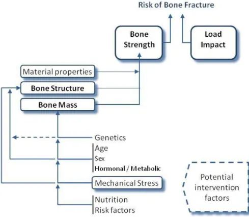
interactions. Beyond age and sex, a great variety of factors have been indicated in the literature [20-24] which, irrespective of alternative aggregation proposals, can be grouped as follows: genetic, hormonal and metabolic, nutritional; mechanical; and risk factors (Figure 1).
Figure 1 - Main known factors influencing bone mass, in the context of the risk of bone fracture problem.
Genetics has been reported as explaining 60-80% of the variance in PBM [21, 22, 25],
but unfortunately it is not influenced by any public health policy’s decision variable. As
appropriate medication to mitigate and prevent the impact of osteoporosis disease. All the remaining factors constitute potential vehicles to influence the bone mass through appropriate external intervention. Adequate diet may provide the necessary diary intake of calcium, proteins and vitamin D for a healthy bone development, whereas smoking and excessive alcohol intake are risk factors among the environmental factors [20, 23, 24]. The mechanical stress on bone, given its stimulus of the (re)modeling activity, is the intervention vehicle that allows the most powerful preventive strategies in promoting bone mass accrual and fighting osteoporosis, namely through appropriate physical activity in childhood and adolescence [27-35].
Despite de importance of the partial or direct relationship of each of the above described determining factors with the bone mass, it must not be disregarded their interrelation, given the complexity of the human biological system. Additionally, keeping in mind that bone mass accrual and loss do not necessarily happen homogeneously in the existing bone structure, those determining factors of bone mass are also, indirectly, determining factors of the bone structure [27].
Summing up, bone strength is at the core of the public health issue of bone fracture and is mainly determined by bone mass and structure, which are also the result of exogenous factors. Particularly relevant to bone mass and structure seems to be the mechanical stress stimulus that is singled out in the present dissertation.
research maturity stage. Assuming that it is due to the corresponding mechanical loads that physical activity benefits bone mass and bone structure, the role of the skeletal site specific biomechanics cannot be disregarded, as it might provide a better understanding of susceptibility to bone fracture.
The number of studies that integrates biomechanical models and physical activity are scarce, and we know none also adding the association/effect on bone mass distribution. Though with respect to the focus of this research project – the proximal femur –two biomechanical models were identified [27]: the biomechanics of the proximal femur, especially on the profile of forces observed at the femoral neck; and the biomechanics of the pelvis-hip system [36, 37]. The addition of the physical activity and the biomechanics topics to the previously identified ones, closes the setting up for the theoretical macro framework of this Ph.D dissertation (Figure 2).
The next sections present a literature overview of the four main topics that were linked and highlighted by the above framework.
2.1.1 The risk of bone fracture
Bone fractures are more frequent in the adolescence and in the older age periods, but may occur throughout the whole lifetime, with a site- and sex-specific profile, as suggested by several epidemiological studies [38-41]. The fracture of the distal forearm, hand and foot are among the most common during the adolescence, a period at which the incidence of a fracture is 27–40% in girls and 42–64% in boys [42, 43]. The reason for this predominance in boys is probably a combination of biological factors and social, sex-related differences associated to activity and risk taking, namely with boys showing a greater sports participation [32]. Sports are responsible for almost 40% of fractures at this age period, and soccer - mostly played by boys - contributes with the greatest number of cases [44]. Maturational factors also provide important explanations to understand fractures in adolescents, given that it is precisely during the pubertal growth spurt - when there is a relative decrease in bone mineral density due to bone expansion and insufficient mineralization - that their incidence is maximum [45].
Despite around one in two children in the adolescence experiencing a bone fracture, this is not considered a public health issue such as fractures in old age, since its consequences do not affect the individuals wellbeing so deeply [46]. During the old age period, low bone mineral density, low physical activity, low muscle mass and overweight are the principal risk factors for fractures [47, 48], that occur mainly at the hip, the distal forearm, the proximal humerus and the spine, with an epidemiologic pattern quite different between sexes [49, 50]. Women’s fracture risk more than doubles men’s, a disadvantage that starts as soon in life as the prepubertal period of skeletal
size, geometry and strength, benefiting boys [51-53]. But also during the last decades of life, women are also at a disadvantage position because of the accelerated bone mass loss caused by the increased bone remodeling as a consequence of the menopause-related estrogene deficiency. Menopause is a phenomena that is women specific and reduces the ability of bones to adapt to ageing by the natural periosteal apposition [51] through which bone size increases and partially compensates (concerning strength) bone mass loss [54, 55]. As a result, the worsening in bone strength parameters – such as size, bone mineral density, cross-sectional area, etc. – to levels that endanger bone structural ability to hold expected loads affects a higher proportion of elderly women than elderly men [56, 57].
Nevertheless, the difference in fracture incidence between sex described above has been narrowing, namely in hip fractures, probably as a combined consequence of women biased timely diagnosis programs, preventive measures, and therapeutic interventions [58-61].
Among all types of fracture at old age, the fractures of the proximal femur are the most serious ones; not only they are around ten times more frequent than any other femur fracture, but, in particular, their consequences are much more severe for the deterioration of patients quality of life, higher morbility, mortality and disability, as well as for their subsequent health care system costs [62-65]. Proximal femur fractures can be divided in two main groups – cervical (or neck) fractures, and trochanteric fractures
– and alike fractures in other skeletal, the incidence of femur fractures is higher in women, in particular at the femoral neck, where it is over 2.5 higher than in men, while
the literature suggests that both the fracture mechanisms and the risk factors are different between the two types of fracture [69-72] and that the relative incidence of
trochanteric fractures compared to cervicals’ increases moderately with aging [63-66].
The geometry of the proximal femur - the hip axis length, the neck-shaft angle and the neck width, included - and of the pelvis seem to be specially associated to cervical fractures, whereas trochanteric fractures particularly affect subjects with low bone density and osteoporosis mainly in the trabecular region [69-72, 74-79].
However the relative importance of proximal femur fracture risk factors is not consensual among researchers. Some support that the low level of bone mineral density (BMD), namely, areal density obtained by dual x-ray absorptiometry (DXA) scanning, is one of the best fracture predictors as the bone mass and its spatial distribution are strong determinants of bone strength [80-83]. Others question this predictive ability of BMD on the grounds that around half of supposedly osteoporotic fractures occur in individuals who are not diagnosed as suffering from osteoporosis [82, 84-88]. The fact that both bone mass and bone structure interact to simultaneously determine bone strength is probably the basis for this disagreement. Irrespective of this debate, research has focused on factors that influence bone mass, either its loss during aging or its accrual during growth.
2.1.2 Peak Bone Mass and bone health
Despite the fact bone consolidation can take place during the third decade of life,
total young adult bone mass is obtained [26, 90, 91]. Furthermore, the normal bone mass lifetime evolution shows that the bone mass gain in the two years around the time of peak bone gain approximately equals all the amount of bone mass loss in the three decades from 50 years on [92, 93].
A 10% increase above average (1 standard deviation) in bone mineral density during growth, delays postmenopausal osteoporosis in 13 years and reduces the fracture risk in 50% while a ~6% lower PBM seems to double the risk of fracture; [47, 94, 95].The pattern of bone mass gains has been reported to be site specific, with the peak bone mass occurring in the hip and the spine earlier than in the whole body, for example [96]. Concerning the bone mass accrual at the hip, in the 3 to 5 period around the peak height velocity girls and boys gain 25%-46% and 28%-43%, respectively, of the PBM observable at this site. At the femoral neck region and in the same period, the corresponding figures (here for both sexes) are 22% to 33% [90].
All this evidence supports the importance of promoting the maximization of the PBM as a prevention strategy to reduce the risk of bone fractures related to bone mass loss in old age [20, 94, 97-100], and drive the notion that osteoporosis is a pediatric issue. This
widely accepted policy “prescription” is based on the assumption that the intervention
during the growing age might contribute to reduce the fracture risk until the early adulthood.
Several equipments and technologies have been used to assess bone parameters particularly the bone mineral as areal or volumetric BMD. Despite the 2D nature of its measurements disregarding the 3D nature of bones, DXA is the widely used equipment for assessing areal BMD. DXA is easily available, has internationally accepted standardized measures and analysis protocols, has short scanning times, and relatively low radiation exposure when compared to alternative methods [4, 53, 103]. These two latter features are of paramount importance in pediatric applications, not only because of the side effects of x-ray radiation but also because children might find it difficult to keep still even for short periods. The computed tomography (CT) is another x-ray based technique, but with the advantage of returning volumetric BMD and 3D measurements.
However it is not as widely used due to its higher cost and complexity. It isn’t either
recommended for pediatric purposes. Radiography is also x-ray based but, even though it provides greater accuracy and precision than DXA or CT concerning geometric 2D measurements, it is not recommended for osteoporosis diagnosis due to its low sensitivity for bone mass. Using nonionizing radiation the magnetic resonation imaging (MRI) techniques return accurate 3D measurements and has a great potential for use in osteoporosis research, but it high cost has limited its large scale application and the long scanning periods are not compatible with studies involving children. Finally, the quantitative ultrasound (QUS) is a non-radiographic method to assess bone , but due to the low quality of its measurements it is not used in research work, despite its safety and low cost.
do not participate in vigorous activity three days per week for at least 20 minutes per session, the population-attributable risk of inactivity as a factor in adult fracture risk is likely to be considerable [105].
2.1.3 Effects of Physical Activity on bone mass in children
The mechanical stress or load exerted over bone is one of the main factors determining bone mass and structure. The underlying reason for this causality effect is the fact that bone responds to mechanical stress by getting stronger, just up to the required level (within certain limits), and to disuse by losing strength capabilities [106], as first suggested by Julius Wolff in the last quarter of the XIX century. According to the
Wolff’s Law, as it became known, when mechanical loads reach a certain magnitude, rate, and frequency they stimulate bone osteogenic activity [107, 108]. Apparently this osteogenic potential is particularly induced by the peak force reached during impact loading. Physical activity – comprising all movements from light leisure activities to the more vigorous ones like practicing organized sports or participating in target exercise –
is the main source of mechanical loads on the skeleton and has been considered to be the best intervention tool to efficiently and safely reduce osteoporosis and the associated fractures risk in old ages [109].
attention has been given by researchers on determining the type of physical activity with the best osteogenic effect. The evidence suggests that the participation in weight-bearing activities, such as jumping, gymnastics, football or handball, that impose high strains on bones, delivered quickly, convey the greatest benefit in the promotion of bone acquisition [110-113].
Concerning the responsiveness of bone to mechanical loads, there is a wide consensus that it is in the period prior to puberty, up to five years around the peak height velocity, that bone is especially adaptable to physical activity induced loads [3, 114]. Studies comparing athletes that initiated sports training prior to and after the puberty, support
the idea of that “golden period”, as well as a vast number of trials in youth whose
results for the percentage of bone mass gains due to physical activity range, between 1% and 6% prior to puberty but only 0.3% to 2% afterwards [91, 115]. However, looking for the pattern of physical activity participation through life, it is possible to fully understand its importance when fracture risk prevention is at stake. In fact pre-pubertal children are among the most active groups of the population and activity levels are reported to be lower in successive age groups, declining significantly during the adolescence [116]. This combination of high bone responsiveness to loads and of natural predisposition to engage in physical activity during the pre-pubertal period is the basis for considering this period to be an opportunity window for bone health prevention programs and for the belief that physical activity in childhood is its most powerful tool [3, 35, 114],
importance of physical activity promotion among children in the context of public health policy strategies.
The literature results strongly support this policy recommendation as several intervention studies with children, typically applying vigorous activities, including some sports, dancing aerobics or high impact exercises, three or more times per week during school days, for seven to twenty four months. In some cases, very low time intervention per session (12 minutes), have reported significant increases in bone mass parameters [3, 28, 67, 114, 117-121, 122-124]. Physical activity induced bone gains appear to last for several years, irrespectively of resulting from interventions programs, from sports participation or non-targeted physical activity. In fact it was possible to find bone mineral benefits at the hip 8 years after a 7 month intervention controlled trial [35] as well as in young adults [23-30 year old) that were more active at the 8-15 year old period (after controlling for their adult physical activity levels) [30]. Other longitudinal and retrospective studies involving subjects with sport participation in their peripubertal and pubertal ages have also showed sustained bone content and density benefits up to 10 years after sports retirement, namely at the lumbar spine and the femoral neck [101, 125-128].
bone response appears to be site (region) specific, as different results have been obtained for the impact on the three proximal femur sub-regions (neck, trochanter and intertrochanter), the neck and the trochanter being reported as the most positively affected – with up to 14% higher bone mineral content or density scores than less active peers – by children physical activity [31, 32, 132]. These results were observed in transversal, longitudinal, and observational studies, with targeted intervention in non-athletes, but also with young athletes increasing their robustness. However, in the perspective of policy intervention to improve population bone health, especially in children, particular attention shall be paid to the observational studies of habitual physical activity that children voluntarily engage in. The high loads imposed during an intervention program or experienced by (competition) athletes are not possible to generalize to a large population, irrespective of eventually configuring the best possible conditions for bone health promotion through physical activity. Here, the use of objective measures of physical activity, namely accelerometry based approaches, is a most valuable tool [31], without disregarding the contribution of questionnaire based approaches [133].
Another issue concerning the effects of physical activity on bone health is whether there is a sex specific bone response to mechanical loading. Objectively, a variety of studies
Girls lower participation is sports and vigorous activities, or physical activity intensities below some threshold level to effectively stimulate the osteogenic bone activity, constitutes one of the possible reasons behind those results, as in almost all of them girls were observed to be less active then boys [134, 135, 116, 143-146]. In fact the evidence shows that girls are consistently less active than boys at the same age group, a difference that widens during the adolescence because the observed decrease in activity level is lower in boys [146]. A second important explanation lies on the differences between sexes concerning the skeletal morphology and, consequently, the biomechanical and kinematic aspects of the whole loads transmitting mechanisms, as both the physical activity pattern (for the same type of activity) and the way the correspondent weight and muscle associated forces are transmitted through bone structures differ, as described in the following section.
Whatever the reasons for the observed sex differences in relation to the effective effects of physical activity, this issue shall be taken in consideration when designing any policy initiative intended to prevent fractures or osteoporosis among the population, eventually justifying different approaches for both sexes, concerning the type, timing, intensity and duration of proposed physical activity [147].
2.1.4 Geometry and biomechanics of the proximal femur and pelvis
In the case of hip fracture, a great variety of studies have identified geometric properties of the proximal femur as predictors of the most common types of fracture in this skeletal site. It has been reported that longer hip axis length (HAL) [150-153], wider femoral neck width [73, 74] and larger neck-shaft angles [74, 154] are associated to increased risk of proximal femur fractures. These proximal femur variables contribute to determining the way forces are exerted on bone (for example, a longer HAL result in a longer lever arm between the hip joint center and the femur shaft), but to better understanding the hip fractures geometry-related variables of the pelvis, such as inner and outer pelvis diameter or the pelvis width, have to be also included in the analysis [14, 155, 72-79].
Bone size is positively associated to bone strength as it improves bone mechanical properties to support loads, as has been suggested [156-162]. From a biomechanical perspective, the loads over a bone are a combination of compressive and tension forces as well as bending or torsional moments that are influenced by the geometric properties of the bone itself and of the system to which it belongs [12].
Concerning the focus of this dissertation, two biomechanical models seem to help understanding the bone mass distribution on the proximal femur and, consequently its structure and strength, given that, consistent with Wolff’s law, the trabecular architecture of the proximal femur is aligned with stress trajectories facilitating the transmission of loads from joints to diaphyseal cortical bone [163].
contraction of the abductor muscles, in order to balance the pelvis during gait, will cause a compression force over both parts of the neck, compensating partially the previous distinction of the nature of forces on the superior and inferior parts of the femoral neck.
Figure 3 Forces exerted on the femoral neck through the action of body weight and abductor muscle forces (adapted from Lovejoy, 1988 [164]).
It is based on this simple model that researchers have explained the fact that natural femoral neck bone loss with aging occurs mainly at the superior part, causing the thinning of this cortical region and resulting on a biased bone mass distribution towards the region most subjected to compression forces, the inferior part of the neck [165]. In fact these mechanical effects are particularly relevant during walking, the main physical activity of elderly people [35, 105].
This biomechanical contribute to the explanation of the neck’s bone mass distribution
Figure 4 - Stress and tension forces on the femoral neck (adapted from Tunner, 2005 [167])
The second biomechanical model adds some pelvis features in an integrated model for the pelvis-hip system. It was first proposed by Pauwels [36] and has been extensively used in the field of hip arthroplasty.
This model explains the intensity of the force exerted by the abductor muscles during the moments of single leg stance when walking or running, as a balanced system between the torque associated to this muscle force, given its lever arm, and the torque resulting from the bodyweight over the center of rotation of the femoral head (Figure 5).
In fact, during locomotion each hip alternatively carries the bodyweight without the
opposite leg’s weight. That bodyweight-related force (K, in Figure 5) acts on the hip joint multiplied by the lever arm h’ resulting in a force that is counterbalanced by the abductor muscles force M times it’s lever arm h [207]. In the analytical general form the model goes as follows:
M . h = K . h’
Figure 6 - “Disadvantaged” positioning of the abductor muscles in the
pelvis-hip system (adapted from Traiana et al., 2009 [77])
Based on this model, a study designed to simulate the load stresses on the proximal femur reported the relevance of the pelvis width on the load pattern [171].
The two models presented, added to the empirical evidence suggesting that the anatomy of the hip and pelvis may play a role in the risk factor patterns of hip fracture, give ground to the relevance of exploring the effects of the complex loads and forces exerted in the different regions of the proximal femur on the mineralization of those specific regions, as it has already been done for the neck region (as described above).
The detailed analysis of the distribution of bone mass in the proximal femur (even using DXA images) has revealed to be an interesting way of studying and understanding the risk of fracture as the identification of small spots of lower bone density (statistically associated to some type fracture) corresponds to those regions that are less mechanically stimulated during normal load bearing [172].
neck-shaft angles, lower hip offsets and wider pelvis [77]. These geometric differences may directly affect the forces and loading pattern on the proximal femur for biomechanical reasons, but also because they influence the pattern of locomotion and muscle activity, therefore the consequent weight loading forces on the femur [142, 173-175]. Consequently, these sex specific forces and loads may also help to understand the different proximal femur mineralization profile between sexes.
References
1. Salkeld G, Cameron I, Cumming R, Easter S, Seymour J, Kurrle S (2000). Quality of life related to fear of falling and hip fracture in older women: a time trade off study Commentary:
Older people's perspectives on life after hip fractures. Bmj, 320:341-346.
2. Kanis J, Burlet N, Cooper C, Delmas P, Reginster J, Borgstrom F, Rizzoli R (2008). European
guidance for the diagnosis and management of osteoporosis in postmenopausal women. Osteoporosis International, 19(4):399-428.
3. MacKelvie K, McKay H, Khan K, Crocker P (2001). A school-based exercise intervention augments bone mineral accrual in early pubertal girls. The Journal of pediatrics, 139 (4):501-508.
4. MacKelvie K, McKay A, Petit M, Moran O, Khan K (2002). Bone Mineral Response to a 7‐Month Randomized Controlled, School‐Based Jumping Intervention in 121 Prepubertal Boys: Associations with Ethnicity and Body Mass Index. Journal of Bone and Mineral Research, 17(5):834-844.
5. US Department of Health and Human Services (2004). Bone Health and Osteoporosis: A Report of the Surgeon General. Rockville, MD: Office of the Surgeon General.
6. Johnell O, Kanis J (2005). Epidemiology of osteoporotic fractures. Osteoporosis international, 16(2):S3-S7.
7. Nguyen N , Ahlborg H, Center J, Eisman J, Nguyen T (2007). Residual lifetime risk of fractures in women and men. Journal of Bone and Mineral Research, 22(6):781-788.
8. Cummings S, Melton L (2002). Epidemiology and outcomes of osteoporotic fractures. The Lancet, 359(9319):1761-1767.
9. Kannus P, Niemi S, Parkkari J, Palvanen M, Vuori I, Järvinen M (2006). Nationwide decline in
incidence of hip fracture. Journal of Bone and Mineral Research, 21(12):1836-1838.
10. National Osteoporosis Foundation. Fast Facts. http://www.nof. org/node/40. Accessed February 22, 2011.
12. Bouxsein M (2005). Determinants of skeletal fragility. Best Practice & Research Clinical Rheumatology, 19(6):897-911.
13. Griffith JF, Genant HK (2008). Bone mass and architecture determination: state of the art. Best Pract Res Clin Endocrinol Metab, 22:737-764.
14. Fardellone P (2008). Predicting the fracture risk in 2008. Joint Bone Spine, 75:661–664.
15. Bousson V, Le Bras A, Roqueplan F, Kang Y, Mitton D, Kolta S, Bergot C, Skalli W, Vicaut E, Kalender W, Engelke K, Laredo J (2006). Volumetric quantitative computed tomography of the proximal femur: relationships linking geometric and densitometric variables to bone strength. Role for compact bone. Osteoporos Int, 17:855–64.
16. Lang TF, Keyak JH, Heitz MW, Augat P, Lu Y, Mathur A, Genant HK (1997). Volumetric quantitative computed tomography of the proximal femur: precision and relation to bone strength. Bone, 21:101–8.
17. Lochmuller EM, Muller R, Kuhn V, Lill CA, Eckstein F (2003). Can novel clinical densitometric techniques replace or improve DXA in predicting bone strength in osteoporosis at the hip and other skeletal sites? J Bone Miner Res, 18:906–12.
18. Heaney R, Abrams S, Dawson-Hughes B, Looker A, Looker A, Weaver C (2000). Peak bone mass. Osteoporos Int, 11(12):985-1009.
19. Osteoporosis Prevention, Diagnosis, and Therapy (2001). NIH Consensus Development Panel on osteoporosis Prevention, Diagnosis, and Therapy. JAMA 285:785-95.
20. Bonjour J, Theintz G, Law F, Slosman D, Rizzoli R (1994). Peak bone mass. Osteoporos Int,
4:7–13.
21. Bonjour J, Chevalley T, Rizzoli R, Ferrari S (2007). Gene–environment interactions in the skeletal response to nutrition and exercise during growth. Med. Sport Sci, 51:64–80.
22. Davies J, Evans B, Gregory J (2005). Bone mass acquisition in healthy children. Arch. Dis. Child, 90:373–378.
23. Eisman J, Kelly P, Morrison N, Pocock N, Yeoman R, Birmingham J, Sambrook P (1993). Peak bone mass and osteoporosis prevention. Osteoporos Int, 3(1):56-60.
24. Seeman, E, Tsalamandri C, Formica C (1993). Peak bone mass, a growing problem? Int J Fertil Menopausal Stud, 38 (2):77–82.
25. Hopper J, Green R, Nowson C, Young D, Sherwin A, Kaymakci B, Larkins R, Wark J (1998). Genetic, common environment, and individual specific components of variance for bone mineral density in 10- to 26-year-old females: a twin study. Am J Epidemio, 147:17–29.
26. Bailey DA, McKay HA, Mirwald RL (1999). A six-year longitudinal study of the relationship of physical activity to bone mineral accrual in growing children: the university of Saskatchewan bone mineral accrual study. J Bone Miner Res, 14:1672–9.
27. Gunter K, Almstedt H, Janz F (2010). Physical Activity in Childhood May Be the Key to Optimizing Lifespan Skeletal Health. Exerc Sport Sci Rev, 40(1):13–21.
28. MacKelvie KJ, Petit MA, Khan KM, Beck TJ, McKay HA (2004). Bone mass and structure are enhanced following a 2-year randomized controlled trial of exercise in prepubertal boys. Bone,
34(4):755–764.
29. Scerpella TA, Dowthwaite JN, Rosenbaum PF (2011). Sustained skeletal benefit from childhood mechanical loading. Osteoporos Int, 22, 2205–2210.
31. Janz KF, Letuchy EM, Eichenberger Gilmore JM, Burns TL, Torner JC, Willing MC, Levy SM (2010). Early physical activity provides sustained bone health benefits later in childhood. Med. Sci. Sports Exerc, 42:1072–1078.
32. Gunter K, Kasianchuk A (2011). Examining the influence of participation in a community-based running program on skeletal health in growing girls. Osteoporosis Int, 22:417-439.
33. Janz KF, Gilmore JM, Burns TL, Levy SM, Torner JC, Willing MC, Marshall TA (2006). Physical activity augments bone mineral accrual in young children: The Iowa Bone Development study. J Pediatr, 148:793–799.
34. Hui SL, Slemenda CW, Johnston CC (1988). Age and bone mass as predictors of fracture in a prospective study. J Clin Invest, 81:1804–1809.
35. Gunter K, Baxter-Jones AD, Mirwald RL, Almstedt H, Fuchs R, Durski S, Snow C (2008). Impact exercise increases BMC during growth: an 8-year longitudinal study. J Bone Miner Res, 23:986–93.
36. Pauwels F 1980 Biomechanics of the locomotor apparatus. Berlin, Springer-Verlag
37. Pauwels F (1976). Biomechanics of the normal and diseased hip: theoretical foundation, technique, and results of treatment: an atlas. Springer-Verlag, Berlin
38. Alffram P, Bauer G (1962). Epidemiology of fractures of the forearm. A biomechanical investigation of bone strength. J Bone Joint Surg, 44:105–114.
39. Court-Brown C, Rimmer S, Prakash U, McQueen M (1998). The epidemiology of open long bone fractures. Injury, 29(7):529–534
40. Garraway W, Stauffer R, Kurland L, O'Fallon W (1979). Limb fractures in a defined population: I. Frequency and distribution. In Mayo Clinic proceedings. Mayo Clin, 54:701–
707.
41. Rennie L, Court-Brown C, Mok J, Beattie T (2007). The epidemiology of fractures in children.
Injury, 38:913–922.
42. Moustaki M, Lariou M, Petridou E (2001). Cross country variation of fractures in the childhood population. Is the origin biological or “accidental”? Inj Prev, 7:77-77.
43. Laudin LA (1983). Fracture patterns in children. Acta Orthop Scand, 54:1–109.
44. Hedström E, Svensson O, Bergström U, Michno P (2010). Epidemiology of fractures in children and adolescents: Increased incidence over the past decade: a population-based study from northern Sweden. Acta orthopaedica, 81(1):148-153.
45. Faulkner R A, Davison K S, Bailey D A, Mirwald R L, Baxter-Jones A D (2006). Size-corrected BMD decreases during peak linear growth: implications for fracture incidence during adolescence. J Bone Miner Res, 21 (12):1864-70.
46. Jones IE, Williams SM, Dow N, Goulding A (2002). How many children remain fracture-free during growth? A longitudinal study of children and adolescents participating in the Dunedin Multidisciplinary Health and Development Study. Osteoporos Int, 13:990–995.
47. Goulding A. Risk factors for fractures in normally active children and adolescents. In: Daly R, Petit M, eds. Optimising Bone Mass and Strength. The Role of Physical Activity and Nutrition during Growth. Med Sport Sci Basel: Karger 2007:51:102–20.
48. Kontulainen SA, Hughes JM, MacDonald HM, et al. The biomechanical basis of bone strength development during growth. In: Daly R, Petit M, eds. Optimising Bone Mass and Strength. The Role of Physical Activity and Nutrition during Growth. Med Sport Sci Basel: Karger
2007:51:13–32.
50. Cheng S, Levy A,. Lefaivre A, Guy P, Kuramoto L, Sobolev B (2011). Geographic trends in incidence of hip fractures: a comprehensive literature review. Osteoporos Int 22:2575–2586.
51. Ego Seeman (2002). Pathogenesis of bone fragility in women and men. Lancet, 359:1841–850
52. Tanner JM. Foetus into man: physical growth from Conception to maturity. Rev. ed. Cambridge (MA): Harvard University Press, 1990
53. Seeman E (2001). Clinical review 137: sexual dimorphism in skeletal size, density, and strength. J Clin Endocrinol Metab, 86:4576-84
54. Riggs BL, Melton LJ, Robb RA, Camp JJ, Atkinson EJ, Peterson JM, Rouleau PA, McCollough CH, Bouxsein ML, Khosla S (2004). Population-based study of age and sex differences in bone volumetric density, size, geometry, and structure at different skeletal sites. J Bone Miner Res, 19:1945–1954.
55. Sigurdsson G, Aspelund T, Chang M, Jonsdottir B, Sigurdsson S, Eiriksdottir G, Gudmundsson A, Harris TB, Gudnason V, Lang TF (2006). Increasing sex difference in bone strength in old age: The Age, Gene/Environment Susceptibility-Reykjavik study (AGESREYKJAVIK). Bone, 39:644–651.
56. Duan Y, Turner CH, Kim BT, Seeman E (2001). Sexual dimorphism in vertebral fragility is more the result of gender differences in age-related bone gain than bone loss. J Bone Miner Res, 16: 2267–75.
57. Duan Y, Parfitt M, Seeman E (1999). Vertebral bone mass, size and volumetric bone mineral density in premenopausal women, and postmenopausal women with and without spine fractures. J Bone Miner Res, 14:1796–1802.
58. Cummings SR, Black DM, Thompson DE, Applegate WB, Barrett-Connor E, Musliner TA, Palermo L, Prineas R, Rubin SM, Scott JC, Vogt T, Wallace R, Yates AJ, LaCroix AZ (1998). Effect of alendronate on risk of fracture in women with low bone density but without vertebral fractures: results from the Fracture Intervention Trial. JAMA, 280:2077–2082.
59. Cummings SR, Ensrud K, Delmas PD, LaCroix AZ, Vukicevic S, Reid DM, Goldstein S, Sriram U, Lee A, Thompson J, Armstrong RA, Thompson DD, Powles T, Zanchetta J, Kendler D, Neven P, Eastell R (2010). Lasofoxifene in postmenopausal women with osteoporosis. N Engl J Med, 362:686–696.
60. Cummings SR, San Martin J, McClung MR, Siris ES, Eastell R, Reid IR, Delmas P, Zoog HB, Austin M, Wang A, Kutilek S, Adami S, Zanchetta J, Libanati C, Siddhanti S, Christiansen C (2009). Denosumab for prevention of fractures in postmenopausal women with osteoporosis. N Engl J Med, 361:756–765.
61. Adams AL, Shi J, Takayanagi M, Dell RM, Funahashi TT, Jacobsen SJ (2012). Ten-year hip fracture incidence rate trends in a large California population, 1997–2006. Osteporos Int, doi: 10.1007/s00198-012-1938-5
62. Martinet O, Cordey J, Harder Y, Maier A (2000). The epidemiology of fractures of the distal femur. Injury, 31:C62–C63
63. Elmerson S, Zetterberg C, Andersson GB (1988). Ten-year survival after fractures of the proximal end of the femur. Gerontology, 34:186–91.
64. Cooper C (1997). The crippling consequences of fractures and their impact on quality of life.
Am J Med, 103:12–17.
65. Melton L J (1996). Epidemiology of hip fractures: implications of the exponential increase with age. Bone, 18:121–5.
67. Fuchs RK, Bauer JJ, Snow CM (2001). Jumping improves hip and lumbar spine bone mass in prepubescent children: a randomized controlled trial. J. Bone Miner. Res, 16:148–156.
68. Arinzon Z, Shabat S, Peisakh A, Gepstein R, Berner Y (2010). Gender differences influence the outcome of geriatric rehabilitation following hip fracture. Archives of gerontology and geriatrics, 50(1):86-91.
69. Mautalen CA, Vega E, Einhorn TA (1996). Are the etiologies of cervical and trochanteric hip fractures different? Bone. 1996;18(suppl): 133S–137S.
70. Pulkkinen P, Partanen J, Jalovaara P, Jamsa T (2004) Combination of bone mineral density and upper femur geometry improves the prediction of hip fracture. Osteoporos Int, 15:274–280.
71. Pulkkinen P, Eckstein F, Lochmuller EM, Kuhn V, Jamsa T (2006) Association of geometric factors and failure load level with the distribution of cervical vs. trochanteric hip fractures. J Bone Miner Res, 21:895–901.
72. Vega E, Mautalen C, Gomez H, Garrido A, Melo L, Sahores AO (1991) Bone mineral density in patients with cervical and trochanteric fractures of the proximal femur. Osteoporos Int,
1:81–86.
73. Gnudi S, Malavolta N, Testi D, Viceconti M (2004). Differences in proximal femur geometry distinguish vertebral from femoral neck fractures in osteoporotic women. Br J Radiol, 77:219–
223.
74. Gnudi S, Ripamonti C, Lisi L, Fini M, Giardino R, Giavaresi G (2002). Proximal femur geometry to detect and distinguish femoral neck fractures from trochanteric fractures in postmenopausal woman. Osteoporos Int, 13:69–73.
75. Nakamura N, Kyou T, Takaoka K, Ohzono K, Ono K (1992). Bone mineral density in the proximal femur and hip fracture type in the elderly. J Bone Miner Res, 7:755–759.
76. Uitewaal PJ, Lips P, Netelenbos JC (1987). An analysis of bone structure in patients with hip fracture. Bone Miner, 3:63–73.
77. Pulkkinen P, Partanen J, Jalovaara P, Jämsä T (2010). BMD T-score discriminates trochanteric fractures from unfractured controls, whereas geometry discriminates cervical fracture cases from unfractured controls of similar BMD. Osteoporos Int, 21:1269–1276.
78. Schott AM, Hans D, Duboeuf F, Dargent-Molina P, Hajri T, Breart G, Meunier PJ, EPIDOS Study Group (2005). Quantitative ultrasound parameters as well as bone mineral density are better predictors of trochanteric than cervical hip fractures in elderly women. Results from the EPIDOS Study. Bone, 37:858–863.
79. Yuki Maeda MD, Nobuhiko Sugano MD, Masanobu Saito MD, Kazuo Yonenobu MD (2011). Comparison of Femoral Morphology and Bone Mineral Density between Femoral Neck Fractures and Trochanteric Fractures. Clin Orthop Relat Res, 469:884–889.
80. Johnell O, Kanis JA, Oden A, Johansson H, De Laet C, Delmas P, Eisman JA, Fujiwara S,
Kroger H, Mellstrom D, Meunier PJ, Melton LJ 3rd, O’Neill T, Pols H, Reeve J, Silman A,
Tenenhouse A (2005). Predictive value of BMD for hip and other fractures. J Bone Miner Res,
20:1185–1194.
81. Cummings SR, Black DM, Nevitt MC, Browner W, Cauley J, Ensrud K, Genant HK, Palermo L, Scott J, Vogt TM (1993). Bone density at various sites for prediction of hip fractures. The study of osteoporotic fractures research group. Lancet, 341:72–75
82. Marshall D, Johnell O, Wedel H (1996). Meta-analysis of how well measures of bone mineral density predict occurrence of osteoporotic fractures. BMJ, 312:1254–9.
84. Wilkin TJ, Devendra D (2001). Bone densitometry is not a good predictor of hip fracture. BMJ,
323:795–797.
85. Kanis JA (2002). Diagnosis of osteoporosis and assessment of fracture risk. Lancet, 359:1929–
1936.
86. Robbins JA, Schott AM, Garnero P, Delmas PD, Hans D, Meunier PJ (2005). Risk factors for hip fracture in women with high BMD: EPIDOS study. Osteoporos Int, 16:149–154.
87. Stone KL, Seeley DG, Lui LY, Cauley JA, Ensrud K, Browner WS, Nevitt MC, Cummings SR, Osteoporotic Fractures Research Group (2003). BMD at multiple sites and risk of fracture of multiple types: long-term results from the Study of Osteoporotic Fractures. J Bone Miner Res, 18:1947–1954.
88. Schuit SC, van der KliftM,Weel AE, de Laet CE, Burger H, Seeman E, Hofman A, Uitterlinden AG, van Leeuwen JP, Pols HA (2004). Fracture incidence and association with bone mineral density in elderly men and women: the Rotterdam Study. Bone, 34:195–202.
89. Harel Z, Gold M, Cromer B, Bruner A, Stager M, Bachrach L (2007). Bone mineral density in postmenarchal adolescent girls in the United States: associated biopsychosocial variables and bone turnover markers J. Adolesc. Health, 40:44–53.
90. Baxter-Jones AD, Faulkner RA, Forwood M, Mirwald RL, Bailey DA (2011). Bone mineral accrual from 8 to 30 years of age: An estimation of peak bone mass. J. Bone Miner. Res,
26(8):1729-394.
91. Kontulainen S, Sievanen H, Kannus P, Pasanen M, Vuori I (2003). Effect of long-term impact-loading on mass, size, and estimated strength of humerus and radius of female racquet-sports players: a peripheral quantitative computed tomography study between young and old starters and controls. J. Bone Miner. Res, 18:352–359.
92. Arlot M, Sornay-Rendu E, Garnero P, VeyMarty B, Delmas PD (1997). Apparent pre- and postmenopausal bone loss evaluated by DXA at different skeletal sites in women: The OFELY cohort. J Bone Miner Res, 12:683–690.
93. Faulkner RA, Bailey DA. Osteoporosis: a pediatric concern? In: Daly R, Petit M, eds. Optimising Bone Mass and Strength. The Role of Physical Activity and Nutrition during Growth. Med Sport Sci Basel: Karger 2007:51:1–12.
94. Hernandez CJ, Beaupré GS, Carter DR (2003). A theoretical analysis of the relative influences of peak BMD, age-related bone loss and menopause on the development of osteoporosis.
Osteoporos Int, 14:843–7.
95. Bonjour JP, Chevalley T, Ferrari S, Rizzoli R (2009). The importance and relevance of peak bone mass in the prevalence of osteoporosis. Salud Publ Mex, 51:S5–S17.
96. Theintz G, Buchs B, Rizzoli R, Slosman D, Clavien H, Sizonenko P, Bonjour J (1992). Longitudinal monitoring of bone mass accumulation in healthy adolescents: Evidence for a marked reduction after 16 years of age at the levels of the lumbar spine and femoral neck in female subjects. J Clin Endocrinol Metab, 75:1060–1106.
97. Karlsson MK (2007). Does exercise during growth prevent fractures in later life? Med Sport Sci, 51:121–36.
98. Goulding A, Jones IE, Taylor RW, Williams SM, Manning PJ (2001). Bonemineral density and body composition in boys with distal forearm fractures: a dual-energy x-ray absorptiometry study. J Pediatr, 139:509–15.

![Figure 3 Forces exerted on the femoral neck through the action of body weight and abductor muscle forces (adapted from Lovejoy, 1988 [164])](https://thumb-eu.123doks.com/thumbv2/123dok_br/16927521.759540/41.892.307.568.312.520/figure-forces-exerted-femoral-action-abductor-adapted-lovejoy.webp)
![Figure 5 - Biomechanical model of the pelvis-hip system (adapted from Maquet, 1999 [169])](https://thumb-eu.123doks.com/thumbv2/123dok_br/16927521.759540/42.892.319.570.789.1058/figure-biomechanical-model-pelvis-hip-adapted-maquet.webp)
![Figure 6 - “Disadvantaged” positioning of the abductor muscles in the pelvis-hip system (adapted from Traiana et al., 2009 [77])](https://thumb-eu.123doks.com/thumbv2/123dok_br/16927521.759540/44.892.311.574.154.399/figure-disadvantaged-positioning-abductor-muscles-pelvis-adapted-traiana.webp)