From the Department of Dermatology and Immunodermatopathology Laboratory, Hospital das Clínicas, Faculty of Medicine, University of São Paulo – São Paulo/SP, Brazil.
E-mail: [email protected] Received for publication on
February 02, 2004.
ORIGINAL RESEARCH
ENDEMIC PEMPHIGUS FOLIACEUS (FOGO
SELVAGEM) AND PEMPHIGUS VULGARIS:
IMMUNOGLOBULIN G HETEROGENEITY DETECTED
BY INDIRECT IMMUNOFLUORESCENCE
Valéria Aoki, Milian H. T. Huang, Alexandre M. Périgo, Lígia M. I. Fukumori, Celina W. Maruta, Claudia G. Santi, Zilda N. P. Oliveira and Evandro Rivitti
AOKI V et al. Endemic pemphigus foliaceus (fogo selvagem) and pemphigus vulgaris: immunoglobulin G heterogeneity detected by indirect immunofluorescence. Rev. Hosp. Clín. Fac. Med. S. Paulo 59(5):251-256, 2004.
Pemphigus are autoimmune intraepidermal blistering diseases in which immunoglobulin G (IgG) autoantibodies are directed against desmosomal glycoproteins. The aim of this study was to determine the IgG subclass profile of endemic pemphigus foliaceus (fogo selvagem) and pemphigus vulgaris utilizing indirect immunofluorescence.
PATIENTS AND METHODS: Twenty-five patients with pemphigus vulgaris, 25 with endemic pemphigus foliaceus (fogo selvagem), and 25 healthy controls were analyzed by indirect immunofluorescence for circulating autoantibodies (total IgG and its subclasses).
RESULTS: Our data revealed a significant correlation (P <.05) of disease activity and autoantibody levels in both forms of pemphigus, i.e., negative titers related to clinical remission, whereas positive results related to active disease. Immunoglobulin G subclass analysis in fogo selvagem demonstrated that in patients in remission, 56% showed positive immunoglobulin G4; in active disease, immunoglobulin G4 was the predominant subclass (100% positive in all cases). The IgG subclass profile in pemphigus vulgaris showed that in patients in remission, only 10% were positive for immunoglobulin G4; in active disease, positivity for immunoglobulin G4 was present in 78% to 88% of the cases.
CONCLUSION: Subclass characterization of immunoglobulin G autoantibodies is a useful tool for pemphigus follow-up, since immunoglobulin G4 (IgG4) is the subclass that is closely related to recognition of pathogenic epitopes, and consequently with disease activity. Careful monitoring should be performed for fogo selvagem in clinical remission with a homogeneous IgG4 response, since this may indicate more frequent relapses.
KEY WORDS: Pemphigus vulgaris. Pemphigus foliaceus. Fogo selvagem. Autoimmunity. Immunofluorescence.
Pemphigus are autoimmune blister-ing diseases in which immunoglobu-lin G (IgG) antibodies are directed against desmogleins (desmosomal glycoproteins), leading to cell-cell de-tachment and acantholysis.1 They are
classified as 6 different types2,3: 1)
pemphigus foliaceus (erythematosus, classic and endemic or fogo selvagem); 2) pemphigus vulgaris (variant: vegetans); 3) drug-induced pemphigus; 4) paraneoplastic pemphi-gus; 5) IgA pemphipemphi-gus; 6) herpetiform
(clinical variant of either pemphigus vulgaris or foliaceus). Endemic pem-phigus foliaceus (EPF) or fogo selvagem (FS) shares the same clinical, histologic, and immunological profile
of the classic form, showing, however, unique epidemiological features, such as presence of familial cases and a high prevalence in South America, es-pecially in Brazil.4-7
Humoral response in pemphigus vulgaris (PV) and fogo selvagem (FS) is mediated by IgG autoantibodies.7,8
subclass autoimmune response in FS is similar to PV, showing an IgG4 re-sponse in active disease and IgG1 in preclinical stages or in disease remis-sion.11-15
Recent studies13 have shown that
due to an intramolecular epitope spread-ing of desmoglein 1, the autoantigen in-volved in FS, there is a heterogeneous IgG subclass response in this disease. In preclinical stages and in clinical remis-sion of FS, IgG1 autoantibodies are di-rected to the carboxy terminal EC-5 of desmoglein 1. Disease onset correlates with an IgG subclass switch (from IgG1 to IgG4), and IgG4 autoantibodies rec-ognize other epitopes of the desmoglein molecule, the EC1-2 amino-terminal do-mains.
The aim of our study was to ana-lyze, utilizing the indirect immun-ofluorescence method, the IgG sub-class profile of patients diagnosed with pemphigus vulgaris and fogo selvagem who were followed up at the Hospital das Clínicas, Department of Dermatology, University of São Paulo, São Paulo, Brazil.
MATERIAL AND METHODS
Source of Sera
Twenty-five patients with pemphi-gus vulgaris, 25 with fogo selvagem, and 25 healthy controls were selected to enter this study. The diagnosis of pemphigus was made according to clinical examination, histopathologic, and direct immunofluorescence analy-sis. All patients included were followed up at the outpatient clinic, Hospital das Clínicas, Department of Dermatol-ogy, University of São Paulo, from Au-gust 2002 to October 2003.
In the PV group (n = 25), 18 (72%) were female and 7 (28%) were male. Age varied from 23 to 83 years, with a mean age of 51 years. Clinical forms were distributed as follows: 10 (40%)
were in remission, 9 (36%) had loca-lized lesions (oral mucosa and/ or scalp), and 6 (24%) had generalized disease.
In the FS group (n = 25), 15 (60%) were female and 10 (40%) were male. Age varied from 15 to 79 years, with a mean age of 38 years. Clinical forms were distributed as follows: 9 (36%) were in remission, 4 (16%) had local-ized lesions, and 12 (48%) had gener-alized disease.
Blood drawing was performed af-ter patients’ consent, and serum was
separated and stored at -200C until
testing.
Indirect Immunofluorescence (Total IgG)
All sera were tested by indirect im-munofluorescence (IIF) using normal human foreskin as the substrate. Skin
cryosections of 4 µ on albuminized
slides were incubated for 30 minutes with serial serum dilutions (in Trizma acetate-buffered saline (TBS), pH 7.5,
with calcium added, or TBS-Ca++),
starting at 1:20, room temperature. Two 10-minute washes with the same buffer, followed by incubation with the secondary antibody (goat anti-human IgG-FITC, Sigma, USA) at a 1:130 di-lution, were performed. Two more
washes with TBS-Ca++ were made
be-fore mounting slides with buffered glycerin (pH 9.0/0.5M). Slides were then analyzed using the epilumines-cence microscope (Zeiss, 160 and 400X).
Indirect Immunofluorescence (IgG Subclasses)
All sera were tested by indirect im-munofluorescence using normal hu-man foreskin as the substrate. Skin
cryosections of 4 µ on albuminized
slides were incubated for 30 minutes with serial serum dilutions (in Trizma acetate-buffered saline (TBS), pH 7.5,
with calcium added, or TBS-Ca++),
starting at 1:20, room temperature. Two 10-minute washes with the same buffer was followed by an incubation with the secondary antibody (mouse anti-human IgG1,2,3,4 Sigma, USA), at a 1:500 dilution for IgG1, 2, and 3, and 1:1000 for IgG4. Two more washes with TBS-Ca++ were made, followed by
the last 30-minute incubation with the conjugate rabbit antimouse-FITC (Dako, Denmark) at a 1:30 dilution. Final steps included two 10-minute
washes with TBS-Ca++ and mounting
slides with buffered glycerin (pH 9.0/ 0.5M). Results were analyzed using an epiluminescence microscope (Zeiss, 160 and 400X).
Statistical Analysis
Category variables were presented in descriptive form in tables with ab-solute and relative frequencies. Propor-tions were compared utilizing Pearson’s χ2 testor Fisher’s test, when
the former could not be applied. Val-ues below P =.05 were considered sta-tistically significant.
RESULTS
Total IgG Autoantibodies in Pemphi-gus Vulgaris (PV)
In PV, IgG autoantibodies by IIF demonstrated the following pattern (Table 1):
1) PV in remission (n = 10): 70% negative, 30% positive, titers rang-ing from 1:80 to 1:320).
2) PV, localized form (n = 9): 11.1% negative, 88.9% positive, titers ranging from 1:160 to 1:1280). 3) PV, generalized form (n = 6):
corre-late with disease in remission, whereas positive results correlate with disease activity.
Total IgG Autoantibodies in Fogo Selvagem (FS)
In FS, IgG autoantibodies by IIF demonstrated the following pattern (Table 2):
1) FS in remission (n = 9): 67% nega-tive, 33% posinega-tive, titers ranging from 1:80 to 1:320).
2) FS, localized form (n = 4): 0% negative, 100% positive, titers ranging from 1:160 to 1:2560). 3) FS, generalized form (n = 12): 0%
negative, 100% positive, titers ranging from 1:80 to 1:5120). There was a clear correlation be-tween autoantibody titers and clinical presentation, i.e., negative titers corre-late with disease in remission, whereas positive results correlate with disease activity.
IgG Subclasses in Pemphigus Vul-garis (PV)
In PV, IgG subclass analysis by IIF revealed the following findings: 1) PV in remission (n = 10): 50%
showed negative results for all
sub-Table 1 - Statistical analysis of the different clinical forms of pemphigus vulgaris (PV) and indirect immunofluorescence (IIF) with total IgG (Fisher’s exact test; significant results when P <.05).
Localized, Generalized, PV in
IIF active Frequency active Frequency remission Frequency
PV PV
Negative 1 11.1% 1 16.7% 7 70%
Positive 8 88.9% 5 83.3% 3 30%
Total 9 100.0% 6 100.0% 1 0 100.0%
P = .0214
Table 2 - Statistical analysis of the different clinical forms of fogo selvagem (FS) and indirect immunofluorescence (IIF) with total IgG (Fisher’s exact test; significant results when P <.05).
Localized, Generalized, FS in
IIF active Frequency active Frequency remission Frequency
FS FS
Negative 0 0.0% 0 0.0% 6 67%
Positive 4 100.0% 1 2 100.0% 3 33%
Total 4 100.0% 1 2 100.0% 9 100.0%
P = .0011
Table 3 - Statistical analysis of pemphigus vulgaris under remission and indirect immunofluorescence (IIF) with IgG subclasses (Fisher’s exact test; significant results when P <.05).
IIF IgG1 Frequency IgG2 Frequency IgG3 Frequency IgG4 Frequency
Negative 6 60.0% 1 0 100.0% 1 0 100.0% 7 70.0%
Positive 4 40.0% 0 0.0% 0 0.0% 3 30.0%
Total 1 0 100.0% 1 0 100.0% 1 0 100.0% 1 0 100.0%
P = .0411
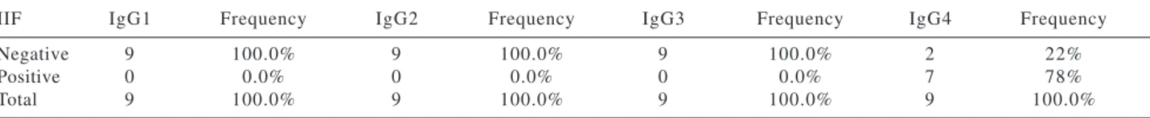
Table 4 - Statistical analysis of active, localized pemphigus vulgaris and indirect immunofluorescence (IIF) with IgG subclasses (Fisher’s exact test; significant results when P <.05).
IIF IgG1 Frequency IgG2 Frequency IgG3 Frequency IgG4 Frequency
Negative 9 100.0% 9 100.0% 9 100.0% 2 22%
Positive 0 0.0% 0 0.0% 0 0.0% 7 78%
Total 9 100.0% 9 100.0% 9 100.0% 9 100.0%
P <.001
classes, 20% were IgG1-positive, 20% had IgG1 and IgG4 antibod-ies, and only 10% were exclusively IgG4-positive (Figure 3); in Table 3, the analysis of each IgG subclass shows that 70% were IgG4-nega-tive, and 40% were IgG1-positive. 2) PV, localized form (n = 9): IgG4 was the only subclass present and was positive in 78% of the cases. (Table 4)
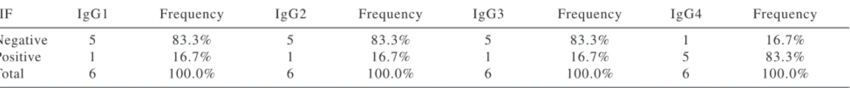
3) PV, generalized form (n = 6): IgG4 was present in 83% of the patients, and IgG1 and IgG2 were present only in 17% of the patients. (Table 5).
IgG Subclasses in Fogo Selvagem (FS)
1) FS in remission (n = 9): 56% showed positive results, all of them with an exclusive IgG4 response. (Table 6)
2) FS, localized form (n = 4): IgG4 was present in all patients (100%), contrasting with IgG1, which was present in only 25% of the samples. (Table 7)
3) FS, generalized form (n = 12): IgG4 was also present in 100% of the pa-tients, IgG1 and IgG2 were present in 33% of the patients, and IgG3 was positive in 17% of the pa-tients. (Table 8)
Total IgG and Subclasses in Healthy Controls
All sera showed negative results.
DISCUSSION
Autoimmunity is the result of a loss of self-tolerance, and depends on multiple interacting factors, such as genetics, infections, mechanical inju-ries, lymphocyte alterations, and hor-mones.11
Intercellular adhesion of the
epi-dermis is a vital function. When the self-tolerance mechanism is broken down, cell-cell detachment occurs due to the binding of autoantibodies to epidermal autoantigens, resulting in blister formation.
Pemphigus are considered auto-im-mune blistering diseases in which the target antigens are glycoproteins lo-cated on the desmosomal core, the so-called desmogleins,16-19 which belong
to the cadherins superfamily and are calcium-dependent adhesion mol-ecules.17,20,21 IgG autoantibodies in the
epidermis and in the sera of
pemphi-Table 5 - Statistical analysis of active, generalized pemphigus vulgaris and indirect immunofluorescence (IIF) with IgG subclasses (Fisher’s exact test; significant results when P <.05).
IIF IgG1 Frequency IgG2 Frequency IgG3 Frequency IgG4 Frequency
Negative 5 83.3% 5 83.3% 5 83.3% 1 16.7%
Positive 1 16.7% 1 16.7% 1 16.7% 5 83.3%
Total 6 100.0% 6 100.0% 6 100.0% 6 100.0%
P = .0293
Table 6 - Statistical analysis of fogo selvagem in remission, and indirect immunofluorescence (IIF) with IgG subclasses (Fisher’s exact test; significant results when P <.05).
IIF IgG1 Frequency IgG2 Frequency IgG3 Frequency IgG4 Frequency
Negative 9 100.0% 9 100.0% 9 100.0% 4 44%
Positive 0 0.0% 0 0.0% 0 0.0% 5 56%
Total 9 100.0% 9 100.0% 9 100.0% 9 100.0%
P = .0013
Table 7 - Statistical analysis of fogo selvagem in remission, and indirect immunofluorescence (IIF) with IgG subclasses (Fisher’s exact test; significant results when P <.05).
IIF IgG1 Frequency IgG2 Frequency IgG3 Frequency IgG4 Frequency
Negative 4 100% 4 100.0% 4 100.0% 0 0.0%
Positive 0 0.0% 0 0.0% 0 0.0% 4 100.0%
Total 4 100.0% 4 100.0% 4 100.0% 4 100.0%
P = .0110
Table 8 - Statistical analysis of generalized fogo selvagem, and indirect immunofluorescence (IIF) with IgG subclasses (Fisher’s exact test; significant results when P <.05).
IIF IgG1 Frequency IgG2 Frequency IgG3 Frequency IgG4 Frequency
Negative 8 67% 8 67% 1 0 83% 0 0.0%
Positive 4 33% 4 33% 2 17% 1 2 100.0%
Total 1 2 100.0% 1 2 100.0% 1 2 100.0% 1 2 100.0%
gus vulgaris and foliaceus patients were demonstrated by Beutner &
Jordon decades ago.18-19 The
patho-genicity of these autoantibodies was confirmed years later in animal mod-els,5,19 in which these diseases were
re-produced when injected with IgG from patients with PV or FS.
A clear correlation of IgG auto-antibody titers and pemphigus activ-ity has been reported by other au-thors.9,15,19,22 Our study corroborated
these data, i.e., the presence of circu-lating IgG is usually related to disease activity. In PV, 70% had a negative IIF with total IgG in cases in remission, and IIF was positive in 87% of active cases. In FS, 67% of the patients in re-mission had a negative IIF with total IgG, and all active cases showed total IgG deposits. Higher titers were ob-served in FS patients in comparison to PV patients: 16% of FS patients showed IIF titers above 1:1280, espe-cially when patients presented a gen-eralized form.
The determination of IgG sub-classes in pemphigus is a very inter-esting point. Studies demonstrate the predominance of IgG4 and IgG1 isotypes9,10,12,15 in both forms of
pem-phigus, with a major role of IgG4 in disease activity.9-10,12-13,15
Recent data focusing on epitope
mapping13 of desmoglein 1 (FS
auto-antigen) show that intramolecular epitope spreading may be responsible for this IgG subclass switch: in the preclinical or remission phase of the disease, there is a production of IgG1 autoantibodies directed against the EC-5 carboxyl portion of desmoglein 1. This condition changes when the disease becomes active, and an IgG4-oriented response starts, targeting other portions of the molecule (EC1-2), which are located on the amino-termi-nal side of desmoglein 1. Factors that may lead to this IgG switch include genetics and environmental aspects, which may play a crucial role in FS.5,13
Our findings in IgG subclass distri-bution by IIF in active PV and FS are also in accordance with other reports 13-15,19; IgG4 is the predominant subclass
in active disease, showing an 80%-sensitivity in PV, and a 100%-sensitiv-ity in FS.
Interesting findings in our data are related to those PV and FS patients in remission, since they show different patterns of IgG isotypes. In FS, our findings revealed that these patients had anti-IgG4 autoantibodies, despite the absence of cutaneous lesions. Our findings demonstrate a different profile from that shown by Warren et al.,12 who
found a negative IgG4 anti-Dsg1 re-sponse in 6 out of 8 FS patients, and a positive IgG1 anti-Dsg1 response in 5
out of 8 FS patients in clinical remis-sion by ELISA.
We propose a closer follow-up of our FS patients, since they are more prone to experiencing reactivation of the disease, according to their IgG subclass pattern. In contrast, our PV patients in clinical remission show a distinct IgG subclass profile, with only 10% showing exclusive IgG4 re-sponse. This may indicate that PV pa-tients, once in clinical remission, will tend to have less disease reactivation.
FS is the only auto-immune blister-ing disease with endemic features; the finding of an exclusive IgG4-based re-sponse in patients in clinical remission suggests that these patients may be un-der a constant environmental trig-ger,5,12,-3 which, in association with a
genetic predisposition, may lead to the production of pathogenic autoanti-bodies.
Our analysis indicates that moni-toring IgG autoantibodies in pemphi-gus vulgaris or foliaceus (fogo selvagem) of patients in clinical remis-sion is a very important matter, espe-cially if subclass characterization is performed. Further studies with an ELISA technique utilizing recom-binant desmogleins will be necessary to improve the determination of IgG isotypes in the humoral response of our pemphigus patients.
RESUMO
AOKI V e col. Pênfigo foliáceo endê-mico (fogo selvagem) e pênfigo vulgar: hetregeneidade da imuno-globulina G detectada através da imunofluorescência indireta. Rev. Hosp. Clín. Fac. Med. S. Paulo 59(5):251-256, 2004.
Pênfigos são enfermidades auto-imunes bolhosas intraepidérmicas, onde auto-anticorpos IgG se dirigem
contra glicoproteínas desmossomais. O objetivo deste estudo foi determinar o perfil de subclasses de imunoglu-bulina G no pênfigo foliáceo endê-mico (fogo selvagem) e no pênfigo vulgar através da imunofluorescência indireta.
MÉTODOS: Vinte e cinco doentes de pênfigo foliáceo endêmico (fogo selvagem), 25 de pênfigo vulgar e 25 controles sadios foram analisados
atra-vés da imunofluorescência indireta, com respeito aos auto-anticorpos circulantes (imunoglobulina G total e subclasses).
clínica, enquanto resultados positivos correlacionaram-se com doença em atividade. A análise de subclasses de IgG mostrou que 56% dos doentes de fogo selvagem em remissão apresenta-ram apenas IgG4 positiva; na doença ativa, IgG4 foi a subclasse predominan-te, sendo positiva em 100% dos casos. Nos doentes de pênfigo vulgar, apenas 10% dos doentes em remissão apresen-taram positividade exclusiva para
IgG4; na doença em atividade, IgG4 esteve presente em 78-83,3% dos ca-sos.
CONCLUSÕES: A caracterização de subclasses de imunoglobulina G consiste em um instrumento de gran-de valia no seguimento gran-de doentes gran-de pênfigo, uma vez que a IgG4 é a subclasse intimamente relacionada com o reconhecimento de epítopos patogênicos, e consequentemente com
atividade da enfermidade. No fogo sel-vagem em remissão com uma resposta homogênea ‘as custas de IgG4, uma monitoração cuidadosa deve ser reali-zada, uma vez que isto pode signifi-car uma maior chance de reativação.
UNITERMOS: Pênfigo vulgar.
Pênfigo Foliáceo. Fogo Selvagem. Auto-imunidade. Imunofluores-cência.
REFERENCES
1 . Stanley JR. Pemphigus. In: Fitzpatrick´s Dermatology in General Medicine. 6th edition. USA: Ed. Mc Graw Hill; 2003. p. 558-74.
2 . Santi CG, Maruta CW, Aoki V, Sotto MN, Rivitti EA, Diaz LA. Pemphigus herpetiformis is a rare, clinical expression of nonendemic pemphigus foliaceus, fogo selvagem and pemphigus vulgaris. J Am Acad Dermatol 1996;34:40-6. 3 . Amagai M. Adhesion molecules. I: Keratinocyte-keratinocyte
interactions: cadherins and pemphigus. Progress in Dermatology. J Invest Dermatol 1995;104:146-52.
4 . Warren SJP, Lin MS, Giudice GJ, Hoffmann RG, Hans-Filho G, Aoki V, et al. The prevalence of antibodies against desmoglein 1 in endemic pemphigus foliaceus in Brazil. N Engl J Med 2000;343:23-30.
5 . Aoki V, Millikan RC, Rivitti EA, Hans-Filho G, Eaton DP, Warren SJP, et al.Environmental risk factors of Fogo Selvagem. J Investig Dermatol Symp Proc 2004; 9:34-40.
6 . Lombardi C, Borges PC, Chaul A, Sampaio SAP, Rivitti EA, Friedman H, et al. Environmental risk factors in endemic pemphigus foliaceus (fogo selvagem). J Invest Dermatol 1992;98:847-50.
7 . Sampaio SAP, Rivitti EA, Aoki V, Diaz LA. Brazilian pemphigus foliaceus, endemic pemphigus foliaceus or fogo selvagem (wild fire). Contemporary Tropical Dermatology 1994;12(4):765-76.
8 . Diaz LA, Sampaio SAP, Rivitti EA, Martins CR, Cunha PR, Lombardi C, et al. Endemic pemphigus foliaceus (fogo selvagem): I. Clinical features and immunopathology. J Am Acad Dermatol 1989;20:657-9.
9 . Bhol K, Ahmed RA. Correlation of subclasses of IgG with disease activity in pemphigus vulgaris. Dermatology 1994; 189(suppl 1):85-89.
10. Bhol K, Natarajan K, Nagarwalla N, Mohimen A, Aoki V, Ahmed RA. Correlation of peptide and IgG subclass with pathogenic and nonpathogenic autoantibodies in pemphigus vulgaris: A model for autoimmunity. Proc Natl Acad Sci USA 1995;92:5239-43.
11. Hacker MK, Janson M, Fairley JA, Lin MS. Isotypes and antigenic profiles of pemphigus foliaceus and pemphigus vulgaris autoantibodies. Clin Immunol2002;105(1):64-74.
12. Warren SJP, Arteaga L, Rivitti EA, Aoki V, Hans-Filho G, Qaqish BF, et al. The role of IgG subclass switch in the pathogenesis of fogo selvagem. J Invest Dermatol 2003;120:104-8. 13. Li N, Aoki V, Hans-Filho G, Rivitti EA, Diaz LA. The role of
intramolecular epitope spreading in the pathogenesis of endemic pemphigus foliaceus (fogo selvagem). J Exp Med 2003;197(11):1501-10.
14. Santos SNMB, Patrus AO, Filgueira AL, Diaz LA. Perfil evolutivo das subclasses de imunoglobulinas gama em pacientes de pênfigo foliáceo endêmico. An Bras Dermatol 2001;76(5):561-74.
15. Rock B, Martins CR, Theophilopoulos AN, Diaz LA. The pathogenic effect of IgG autoantibodies in endemic pemphigus foliaceus (fogo selvagem). N Engl J Med 1989;320:1463-9. 16. Amagai M, Klaus-Kotvun V, Stanley JR. Autoantibodies against a novel epithelial cadherin in pemphigus vulgaris, a disease of cell adhesion. Cell 1991;67:869-77.
17. Buxton RS, Magee AI. Structure and Interactions of Desmosomal and Other Cadherins. Cell1992;3:157-167.
18. Fine, DJ. Pemphigus vulgaris, foliaceus, and paraneoplastic. Topics in Clinical Dermatology: Bullous Diseases. New York- Tokyo: IGAKU-SHOIN; 1993. p. 52-74.
19. Aoki V. Avaliação da técnica de imunoprecipitação com a desmogleína 1 recombinante em população de risco para o pênfigo foliáceo endêmico (fogo selvagem). 80p. Doctoral Thesis (Doutorado em Medicina) 1999 – Faculdade de Medicina, Universidade de São Paulo, São Paulo. Brasil. 20. Goodwin L, Hill JE, Raynor K, Raszi L, Manabe M, Cowin P.
Desmoglein shows extensive homology to the Cadherin Family of Cell Adhesion Molecules. Bioch Biophys Res Commun1990; 173: 1224-1230.