www.bjournal.com.br
www.bjournal.com.br
Institutional Sponsors
The Brazilian Journal of Medical and Biological Research is partially financed by
Volume 43 (5) 409-521 May 2010
Braz J Med Biol Res, May 2010, Volume 43(5) 492-499
Acute lead-induced vasoconstriction in the vascular beds of
isolated perfused rat tails is endothelium-dependent
Acute lead-induced vasoconstriction in
the vascular beds of isolated perfused rat
tails is endothelium-dependent
E.A. Silveira
1, J.H.F. Lizardo
1, L.P. Souza
1, I. Stefanon
1and D.V. Vassallo
1,21Departamento de Ciências Fisiológicas, Universidade Federal do Espírito Santo, Vitória, ES, Brasil 2Centro de Ciências da Saúde de Vitória, EMESCAM, Vitória, ES, Brasil
Abstract
Chronic lead exposure induces hypertension in humans and animals, affecting endothelial function. However, studies concern-ing acute cardiovascular effects are lackconcern-ing. We investigated the effects of acute administration of a high concentration of lead
acetate (100 µΜ) on the pressor response to phenylephrine (PHE) in the tail vascular bed of male Wistar rats. Animals were
anesthetized with sodium pentobarbital and heparinized. The tail artery was dissected and cannulated for drug infusion and mean perfusion pressure measurements. Endothelium and vascular smooth muscle relaxation were tested with acetylcholine (5 µg/100 µL) and sodium nitroprusside (0.1 µg/100 µL), respectively, in arteries precontracted with 0.1 µM PHE.
Concentration-response curves to PHE (0.001-300 µg/100 µL) were constructed before and after perfusion for 1 h with 100 µΜ lead acetate.
In the presence of endothelium (E+), lead acetate increased maximal response (E
max) (control: 364.4 ± 36, Pb2+:480.0 ± 27
mmHg; P < 0.05) and the sensitivity (pD2; control: 1.98 ± 0.07, 2.38 ± 0.14 log mM) to PHE. In the absence of endothelium (E-)
lead had no effect but increased baseline perfusion pressure (E+: 79.5 ± 2.4, E-: 118 ± 2.2 mmHg; P < 0.05). To investigate the
underlying mechanisms, this protocol was repeated after treatment with 100 µM L-NAME, 10 µM indomethacin and 1 µM tempol in the presence of lead. Lead actions on Emax and pD2 were abolished in the presence of indomethacin, and partially abolished
with L-NAME and tempol. Results suggest that acute lead administration affects the endothelium, releasing cyclooxygenase-derived vasoconstrictors and involving reactive oxygen species.
Key words: Lead acetate; Vascular reactivity; Endothelium; Hypertension
Introduction
Correspondence: E.A. Silveira, Departamento de Ciências Fisiológicas, PPGCF/CBM, UFES, Av. Marechal Campos, 1468, 29040-091 Vitória, ES, Brasil. Fax: +55-27-2122-7330. E-mail: [email protected]
Received September 26, 2009. Accepted March 16, 2010. Available online April 9, 2010. Published May 14, 2010. Environmental exposure to pollutant metals, including
lead, contributes to cardiovascular disease (1). Moreover, this metal is recognized as a common environmental and occupational health hazard (2). Lead is an important toxic agent that can exert adverse effects in humans given
suf-ficient exposure and/or accumulation in the body (3). Ex -perimental studies in animals and epidemiological reports suggest a close relationship between lead exposure and hypertension (4,5).
Lead is one of the metals most extensively used in the industrial sector in several countries including Brazil, a fact that contributes to its wide environmental distribution. Therefore, usually all humans have lead in their organism as a result of exposure to exogenous sources (6). Some workers are exposed to high levels of lead in their activities. This exposure occurs during the manufacture of
ammuni-tion, batteries, sheet lead, solder, ceramic glazes, caulking, some brass and bronze plumbing, circuit boards, military equipment, some surgical equipment, and some medicines (herbal remedies from China, India) (6). Barbosa Jr. et al. (7), studying 62 lead-exposed subjects, demonstrated a positive correlation between the increased inhibition of nitric oxide (NO) with increasing blood lead or plasma lead
concentrations. These investigators suggested a significant
inhibitory effect of lead on NO and provided clinical evidence for a mechanism involving the association between lead exposure and increased cardiovascular risk.
Lead-induced vasoconstriction in isolated rat tail artery 493
including endothelial dysfunction (10) and inhibition of the sarcolemmal sodium potassium ATPase (11). These actions also include direct effects on the excitability and contractility of the heart (8,9), vascular actions on the compliance and contractility of smooth muscle (12-14), and tissue damage by free radicals (15). This metal also promotes reductions
in vascular β-adrenoceptor density and cAMP levels, fac -tors that contribute to increased blood pressure (16). In addition, harmful effects on other possible sites of action as within the central nervous system might occur affecting blood pressure regulation (17,18).
The actions of lead on the endothelium seem to involve
the release of vasoactive factors, although the findings ob
-tained to date remain controversial. Previous findings have
shown that lead exposure might increase (19) or decrease (20) plasma endothelin levels, as well as increase or de-crease urine NO concentration or endothelial NO synthase (eNOS) overexpression (21).
Although the chronic and acute effects of lead on liv-ing animals are well known, there are comparatively few studies concerning the acute effects of lead on vascular reactivity. Therefore, the aim of the present study was to investigate the acute effects of continuous perfusion with a
high concentration of lead acetate (100 µΜ) on the pressor
reactivity of the perfused rat tail artery.
Material and Methods
Animals
Studies were performed on male Wistar rats (250-300 g). All experiments were conducted in accordance with the guidelines for biomedical research as stated by the Brazilian Societies of Experimental Biology. The experimental proto-col was approved by the Ethics Committee of the Institute of Biomedical Sciences, EMESCAM (Protocol #003/2007). All rats had free access to water and rat chow ad libitum.
Isolated rat tail vascular bed preparation
Isolated rat tail arteries were perfused as previously
reported (22). Rats were first anesthetized with sodium
pentobarbital (65 mg/kg, ip), and heparin (500 IU, ip) was administered 10 min later. At the base of the tail 2 cm of the tail artery were dissected free and cannulated with an intracath (Nipro 24G ¾, Brazil). The whole tail (average length of 18 to 20 cm) was then severed from the body,
placed in a tissue bath and perfused at a constant flow of 2.5
mL/min with a peristaltic pump (Milan, Brazil), with Tyrode solution (120 mM NaCl, 5.4 mM KCl, 1.2 mM MgCl2, 1.25
mM CaCl2, 20 mM HEPES buffer, 11 mM glucose, and 3
µM EDTA), bubbled with 100% O2, at 36 ± 0.5°C).
The HEPES buffer was carefully chosen to prevent lead acetate precipitation. After a 45-min equilibration period, the experimental protocol was initiated. The mean perfusion pressure (MPP) was measured using a pressure transducer (TSD104A, BIOPAC Systems, Inc., USA), and data were
recorded using an interface and software for computer data acquisition (model MP100A, BIOPAC Systems, Inc.). As a
constant flow was maintained, changes in MPP represent
changes in vascular resistance.
Effects of the acute administration of lead on the actions of phenylephrine in the presence and absence of endothelium
After a 45-min stabilization period, endothelial integrity was evaluated using acetylcholine (Ach) administration (5 µg/100 µL) in the tail artery precontracted with 0.1 µM phenylephrine (PHE). Endothelial integrity was considered to be adequate when relaxation attained more than 50% of maximum contraction induced by a 0.1 µM PHE. Smooth muscle viability was tested using sodium nitroprusside (SNP: 0.1 mg/mL) in the tail artery, which had been previously contracted with PHE. This test was performed before and after each complete experiment.
The vasoconstrictor response to the stimulation of an α1
-adrenoceptor agonist was evaluated by administering PHE (0.001-300 µg/100 µL) to preparations (N = 10) with intact
endothelium (control group). After the first
concentration-response curve to PHE the preparations were perfused for
1 h with a buffer solution containing 100 µΜ lead acetate
and a second concentration-response curve of PHE was
constructed in the presence of 100 µΜ lead acetate. To
ensure that the effects were not dependent on time, an-other group of rats was used under the same conditions in a time control experiment performed 1 h later, after iv administration of 0.9% saline. This lead concentration was selected because it is known to produce a clear negative inotropic effect and to reduce myosin ATPase activity in the rat myocardium (23).
The same protocol was performed as described above in preparations following endothelium damage (E-: N = 10)
induced by {3-[(3-cholamidopropyl) dimethylammonio]-1-propane sulfonate} (CHAPS, 8 mg in 80 µL), as previously described (24). The absence of functional endothelium
was confirmed by the inability of ACh (5 µg in 100 µL) to
produce relaxation. The endothelium-independent vasodi-lator response to SNP (0.1 mg/mL) was also determined. Concentration-response curves to PHE were constructed at 30 min after endothelial damage and repeated after
perfusion with a solution containing 100 µΜ lead acetate
for a period of 1 h (E- Pb2+, N = 10).
Time-control concentration-response curves to PHE (0.001-300 µg in 100 µL) were constructed before and after an infusion period of 1 h with Tyrode solution in preparations with (N = 10) and without endothelium (N = 10).
The role of prostanoids and nitric oxide in the effects of lead on the phenylephrine-induced pressure response
of lead acetate on the pressure responses to PHE, two protocols were performed. Preparations were perfused for 1 h with 100 µM lead acetate in both situations: with the NO synthase inhibitor NG-nitro-L-arginine methyl ester (L-NAME
100 µM, Pb2+ L-NAME, N = 13) and with indomethacin (10
µM, Pb2+ indomethacin, N = 9), respectively. Basal NO release was evaluated by comparing the areas under the concentration-response curves for PHE, obtained in the absence and presence of L-NAME (25). The same protocol was also performed without lead acetate in the absence and in the presence of 10 µM indomethacin (N = 9) or 100 µM L-NAME (N = 8).
The role of free radicals in the effects of lead on the phenylephrine-induced pressure response
To investigate whether free radicals play a role in the effects of lead acetate on the pressure response to PHE, preparations were perfused for 1 h with 1 µM tempol (4-hydroxy-2,2,6,6-tetramethylpiperidine 1-oxyl), an agent that mimics superoxide dismutase (SOD), in the absence and in the presence of lead acetate.
Drugs and reagents
Lead acetate, PHE, L-NAME, indomethacin, tempol, ACh hydrochloride, SNP, CHAPS, HEPES, EDTA, and sodium pentobarbital were purchased from Sigma (USA); and heparin was purchased from Roche (Brazil). NaCl, KCl, MgCl2, CaCl2, and glucose were purchased from
Merck (Germany).
The drugs were dissolved in distilled water and all solutions were freshly prepared before use and protected from light. Indomethacin was dissolved in 0.1 M Tris buf-fer, pH 7.4.
Data analysis
Results regarding perfusion pressure measurements are presented as changes in MPP by subtracting peak pressure
from baseline pressure (ΔMPP, mmHg). The relaxation re -sponses to ACh and SNP are reported as percent relaxation in preparations precontracted with 0.1 µM PHE.
For each PHE concentration-response curve, the maximum effect and the bolus dose (µg) that produced one-half Emax (-log EC50) were estimated using non-linear
regression analyses (Graph Pad Prism Software, USA). The sensitivity of the agonist is reported as pD2 (-log EC50)
and the maximum effect as maximal response (Emax). To
compare the effects of different drugs on the response to PHE, results were reported as “differences of area under the concentration-response curves” (dAUC) in control and experimental situations. AUCs were calculated from the individual dose-response curves and the differences were reported as a percent of AUC (%dAUC) of the correspond-ing control situation (26).
Data are reported as means ± SEM and were analyzed by the Student t-test (paired or unpaired) and by one-way
analysis of variance (ANOVA). When ANOVA showed a
significant effect of treatment, the Tukey post hoc test was used to compare means. A P value <0.05 was considered
to be significant.
Results
Effects of the acute administration of lead on the actions of phenylephrine in the presence and absence of endothelium
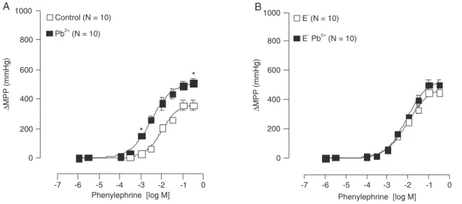
To evaluate the interference of the duration of the experi-ments with the stability of the preparations, concentration-response curves to PHE were constructed before and after 1 h of continuous perfusion with Tyrode solution. This protocol showed that the duration of the experiments did not interfere with the Emax (before: 522 ± 19.6, after: 506 ± 13.2 mmHg)
or the pD2 (before: 1.6 ± 0.04, after: 1.7 ± 0.03 log mM).
On the other hand, 1 h of perfusion with lead acetate (100
µΜ) increased Emax and pD2 (Figure 1A).
Figure 1B demonstrates that, in the absence of en-dothelium, lead acetate increased the baseline perfusion pressure (E+: 79.5 ± 2.4 vs E-: 118 ± 2.2 mmHg; P < 0.05,
t-test) but did not change Emax or pD2 (Figure 1B).
To investigate how acute lead acetate treatment af-fected the endothelium or smooth muscle integrity, single doses of ACh, an endothelial-dependent vasodilator, and SNP, an endothelial-independent vasodilator, were used. Treatment with lead damaged the endothelium since the
relaxation produced by ACh was significantly decreased
(control: 60%; Pb2+: 20%; P < 0.05). The integrity of smooth
muscle was preserved since SNP-induced vasorelaxation was unchanged (control: 81%; Pb2+: 91%).
Role of endothelial factors in the effects of lead
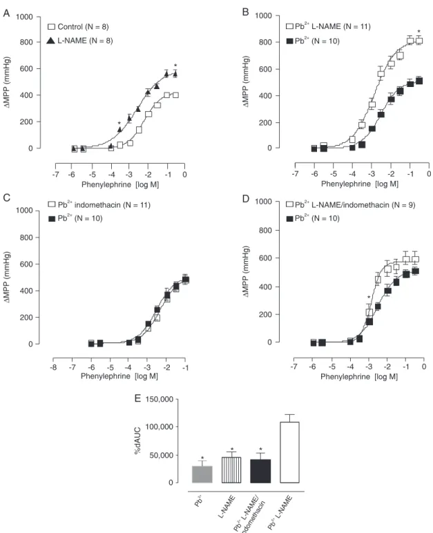
L-NAME was used to investigate the putative role of NO in the effects of lead acetate on the pressor response to PHE. In control groups, L-NAME produced the expected increase in Emax and pD2 (Figure 2A). However, after perfusion with
lead acetate plus L-NAME, a significant increase in Emax
was observed when compared with the separate effect of L-NAME on the untreated preparations (Figure 2B).
When the COX pathway-derived prostanoids were in-vestigated with the use of indomethacin, just a small increase of Emax to PHE was observed before lead administration
Lead-induced vasoconstriction in isolated rat tail artery 495
procedure, it was observed that the Pb2+
L-NAME/indo-methacin treatment showed a leftward displacement of the concentration-response curve to PHE (Figure 2D); however, the effects of lead on the Emax to PHE were abolished. The
magnitude of the response of these groups was evaluated using the areas under the curves (Figure 2E). This analysis suggested the involvement of a COX-derived vasoconstrictor in the effect of lead.
The role of free radicals on the effects of lead acetate
We also investigated the role of free radicals in the effect of lead. Tempol, an SOD mimetic, was used to investigate
whether free radicals play a role in the action of 100 µΜ
lead acetate on the pressor responses to PHE. Tempol promoted a small increase in the sensitivity to PHE (Figure 3B). Moreover, in the presence of tempol, the effects of lead acetate were partially abolished (Figure 3C). Figure 3D shows the magnitude of these responses.
Discussion
The main finding of the present study shows that acute exposure to 100 µΜ lead acetate increases the reactiv -ity of the tail artery to PHE. This increased reactiv-ity is endothelium-dependent, since the effects of lead were abolished in the absence of endothelium. Additionally, this study demonstrated that COX inhibition by indomethacin prevented vasoconstriction, suggesting that the increased reactivity of the tail artery depends on the participation of a COX-derived vasoconstrictor. Results also showed that the blockade of the superoxide anion by tempol partially
prevented the effects of lead, suggesting the involvement of free radicals in the effects of this metal.
It is well established that chronic lead exposure in-duces the production or release of endothelin, a powerful vasoconstrictor (19), inhibits the production or release of a hyperpolarizing factor (26) and reduces the release or production of NO (21,27). The combination of these lead-induced effects might increase vascular tonus and consequently blood pressure.
These effects appeared to be related to an imbalance of endothelial-derived vasoconstrictor and vasodilator compounds (19). In addition, other investigators have demonstrated that chronic exposure to moderate levels of lead increases blood pressure (14,28), probably due to
an increased vascular reactivity to α-adrenergic agonists.
Previous studies have shown that chronic administration of this metal can act directly on vascular smooth muscle (29), also increasing the pressor response to norepinephrine in isolated arteries (20). In addition, Heydari et al. (30) reported that lead was able to increase Emax and pD2 in the
pres-ence of PHE in thoracic aortic rings of rats treated with lead for 8 to 12 weeks. Furthermore, it is known that this metal increases vascular responses to endogenous compounds such as norepinephrine (14) and enhances sympathetic
activity, modifying the baroreflex sensitivity and inducing
alterations in the signal transduction system present in cell membranes, such as cAMP, Ca2+ and NO (31).
To further understand the vasoconstrictor effect promoted by lead acetate and its capacity to harm the vascular en-dothelium and the smooth muscle cells we investigated the actions of lead on the vascular reactivity of the tail artery.
Figure 1. Changes in mean perfusion pressure (∆MPP; mmHg) of the tail artery in response to increasing doses of
phenylephrine in Wistar rats. A, With endothelium, before (control, open squares) and after (filled squares, Pb2+) infusion
of 100 µΜ lead acetate for 1 h. B, Without endothelium before (open squares, E-) and after (filled squares, Pb2+) infusion
of 100 µΜ lead acetate for 1 h. Data are reported as means ± SEM. *P < 0.05 for maximal response (Emax) and sensitivity
Figure 2. Changes in mean perfusion pressure (∆MPP; mmHg) in response to increasing doses of phenylephrine in the
tail artery of Wistar rats. A, Before (open squares, control) and after (filled triangles, L-NAME) perfusion with L-NAME. B,
Effect of lead acetate in the absence (filled squares, Pb2+) and in the presence of L-NAME (open squares, Pb2+ L-NAME). C, Effect of lead acetate in the absence (filled squares, Pb2+) and in the presence of indomethacin (open squares, Pb2+
indomethacin). D, Effect of lead acetate in the absence (filled squares, Pb2+) and in the presence of L-NAME and
indo-methacin (open squares, Pb2+ L-NAME/indomethacin). E, Difference of areas under the concentration-response curves
(dAUC) to phenylephrine for the following groups: Pb2+vs control, L-NAME vs control, and Pb2+ L-NAME/indomethacin vs Pb2+. Data are reported as means ± SEM. *P < 0.05 for maximal response (Emax) and sensitivity (pD2): L-NAME vs
Lead-induced vasoconstriction in isolated rat tail artery 497
Our findings showed that the action of lead in the tail
artery with intact endothelium increased the vasoconstrictor response to PHE, the maximal response (Emax) and the
sen-sitivity (pD2). These actions were endothelium-dependent,
since lead effects were abolished after endothelium removal.
Considering that a significant impairment of endothelial
function was suggested, lead could be inhibiting the release of endothelium-derived vasodilators or stimulating the
re-lease of endothelial vasoconstrictors. Our findings seem to
corroborate those of Marques et al. (32), as they reported that lead-induced hypertension damaged both
endothelium-dependent and -inendothelium-dependent relaxation. This response was accompanied by increased eNOS protein expression and down-regulation of soluble guanylate cyclase (sGC). Marques et al. (32) also suggested that these responses seem to involve reactive oxygen species (ROS) acting on the relaxation mechanisms of NO/cyclic guanosine mono-phosphate in the vascular wall.
We observed a significant increase of the Emax promoted
by lead acetate in the presence of NAME. However, L-NAME was not able to completely abolish the response promoted by lead acetate. This result suggests that the
Figure 3. Changes in mean perfusion pressure (∆MPP; mmHg) in response to increasing doses of phenylephrine
in the tail artery of Wistar rats. A, Before (control, open squares) and after (Pb2+,filled squares) infusion of 100 µΜ
lead acetate for 1 h. B, Before (control, open squares) and after (filled triangles) infusion of tempol. C, Effect of lead acetate in the absence (Pb2+, open squares) and presence of Tempol (Pb2+ Tempol, filled squares). D, Difference of
areas under the concentration-response curves (dAUC) to phenylephrine between: (A: Control vs Pb2+; B: Control vs Tempol; C: Pb2+ Tempol vs Pb2+). Data are reported as means ± SEM. *P < 0.05 for maximal response (E
max)
and sensitivity (pD2): Pb2+ vs control; #P < 0.05 for Emax: Pb2+vs Pb2+ Tempol; +P < 0.05 for %dAUC: Pb2+vs Pb2+
effects on the pressor response to PHE cannot be totally attributed to NO. To better clarify this issue, our next step was to evaluate the pathway of COX-derived prostanoids. For this, preparations were treated with indomethacin, a COX inhibitor. We observed that the lead-induced vaso-constrictor effects on the tail arterial bed were abolished by indomethacin. These results suggest that lead acetate might stimulate the release of COX-derived vasoconstric-tor prostanoids. Although suggesting the production of a
vasoconstrictor prostanoid, our findings did not enable us to
distinguish which compound caused this. However, Courtois et al. (33) found an increase in the expression of COX-2 protein in aortic rings perfused with lead and suggested that this response was associated with a deregulation of
the β1-sGC subunit and an increased production of ROS
as the vasoconstrictor mechanism.
Given that NO production was still present after inhibi-tion by indomethacin because of the intact endothelium, the production of another endothelium-derived vasoconstrictor had to be taken into account. To better clarify this issue we also carried out a protocol associating two drugs, L-NAME and indomethacin, in the presence of lead. In this protocol, we observed a leftward displacement of the
concentration-response curve to PHE, which confirms that the effects of
lead on the vascular tail bed are probably mediated by the release of a COX-derived vasoconstrictor plus another constrictor component.
There is growing evidence that the chronic effects of lead are also associated with oxidative stress (15,19,21). In contrast, previous studies have shown that ROS up-regulates the expression of COX-2 isoforms, which is primarily responsible for the synthesis of prostaglandins involved in pathological processes, including hypertension (34). Therefore, the ability of lead acetate to generate ROS,
specifically the superoxide anion (O2•-), was evaluated.
For this, the preparations were perfused with tempol. This treatment partially abolished the actions of lead acetate on the pressor response to phenylephrine, suggesting that
acute treatment with lead probably involved the pathway of O2•- production and/or liberation. Our findings show
-ing that COX is also involved reinforce the importance of ROS-induced generation by lead. However, this response does not rule out the possibility that the chronic action of this metal can induce the production of other kinds of free radicals, such as the radical hydroxyl (OH•-) and hydrogen peroxide (H2O2), as previously suggested (35,36).
It is important to emphasize that the present findings reinforce the biological significance of lead as an environ -mental contaminant that damages the human organism, producing harmful effects on the cardiovascular system.
Although significant progress has occurred regarding
environmental contamination, there are still serious prob-lems produced by heavy metals. The positive correlation between the concentration of plasma lead and hyperten-sion (37,38), one of the most prominent cardiovascular problems in many countries, and the increase in all causes of lead-related deaths, including circulatory and cancer
mortality (39) reinforce the biological significance of lead
as an important hazard.
The results obtained thus far might be interpreted
considering two limitations. The first limitation regards the
lead concentration we used in this study, which is clearly toxic and far above safety values for human beings. We used a high lead concentration to begin to understand its effects. In the future, we will use smaller concentrations. The second limitation is that it is not possible to compare the present results with those for other vascular beds such as resistance vessels or the greater conductance vessels such as the aorta. However, the results of the present study provide guidance for further studies using much lower lead concentrations to better elucidate the cardiovascular effects of the metal.
In conclusion, the present study raises the possibility of a new mechanism of action of lead, i.e., an endothelial COX-derived vasoconstrictor mechanism, reinforcing the importance of ROS-induced generation by lead.
References
1. Bhatnagar A. Cardiovascular pathophysiology of environ-mental pollutants. Am J Physiol Heart Circ Physiol 2004; 286: H479-H485.
2. Agency for toxic substances and disease registry (ATSDR):
toxicological profile for lead. US Department of Health and
Human Services, Public Health Service, 1993, Atlanta.
3. Sharifi MA, Darabi R, Akbarloo N, Larijani B, Khoshbaten
A. Investigation of circulatory and tissue ACE activity dur-ing development of lead-induced hypertension. Toxicol Lett
2004; 153: 233-238.
4. Moller L, Kristensen TS. Blood lead as a cardiovascular risk factor. Am J Epidemiol 1992; 136: 1091-1100.
5. Maheswaran R, Gill JS, Beevers DG. Blood pressure and industrial lead exposure. Am J Epidemiol 1993; 137: 645-653.
6. Levin SM, Goldberg M. Clinical evaluation and management of lead-exposure construction workers. Am J Ind Med 2000; 37: 23-43.
7. Barbosa F Jr, Sertorio JTC, Gerlach RF, Tanus-Santos JE. Clinical evidence for lead-induced inhibition of nitric oxide formation. Arch Toxicol 2006; 80: 811-816.
8. Kopp SJ, Baker JC, D’Agrosa LS, Hawley PL. Simultaneous recording of His bundle electrogram, electrocardiogram, and
systolic tension from intact modified Langendorff rat heart
preparations, I: Effects of perfusion time, cadmium, and lead. Environ Health Perspect 1978; 46: 475-487.
9. Prentice RC, Kopp SJ. Cardiotoxicity of lead at various perfusate calcium concentrations: Functional and metabolic responses of the perfused rat heart. Toxicol Appl Pharmacol
Lead-induced vasoconstriction in isolated rat tail artery 499
10. Vaziri ND, Liang K, Ding Y. Increased nitric oxide inactivation by reactive oxygen species in lead-induced hypertension.
Kidney Int 1999; 56: 1492-1498.
11. Weiler E, Khalil-Manesh F, Gonick H. Effects of lead and na-triuretic hormone on kinetics of sodium-potassium-activated adenosine triphosphatase: Possible relevance to hyperten-sion. Environ Health Perspect 1988; 78: 113-115.
12. Rosenblum WI. Effects of lead and other cations on pial arteries of the mouse. Acta Neuropathol 1965; 5: 54-60. 13. Piccinini F, Favalli L, Chiari MC. Experimental investigations
on the contraction induced by lead in arterial smooth muscle.
Toxicology 1977; 8: 43-51.
14. Webb RC, Winquist RJ, Victery W, Vander AJ. In vivo and
in vitro effects of lead on vascular reactivity in rats. Am J Physiol 1981; 241: H211-H216.
15. Lyn Patrick ND. Lead toxicity part II: The role of free radical damage and the use of antioxidants in the pathology and treatment of lead toxicity. Altern Med Rev 2006; 11: 114-127.
16. Tsao D-A, Yu H-S, Cheng J-T, Ho C-K, Chang H-R. The
change of β-adrenergic system in lead-induced hyperten -sion. Toxicol Appl Pharmacol 2000; 163: 127-133. 17. Stöfen D. Environmental lead and the heart. J Mol Cell
Car-diol 1974; 6: 285-290.
18. Hejtmancik MR, Williams BJ. Effects of chronic lead expo-sure on the direct and indirect components of the cardiac response to norepinephrine. Toxicol Appl Pharmacol 1979; 51: 239-245.
19. Khalil-Manesh F, Gonick HC, Weiler EW, Prins B, Weber MA, Purdy RE. Lead-induced hypertension: possible role of endothelial factors. Am J Hypertens 1993; 6: 723-729. 20. Skoczynska A, Stojek E, Gorecka H, Wojakowska A. Serum
vasoactive agents in lead-treated rats. Int J Occup Med Environ Health 2003; 16: 169-177.
21. Vaziri ND, Ding Y, Gonick HC. Altered nitric oxide me-tabolism and increased oxygen free radical activity in lead-induced hypertension: effects of lazaroid therapy. Kidney Int
1997; 52: 1042-1046.
22. França AS, Rossoni LV, Amaral SMC, Vassallo DV. Reactiv-ity of the isolated perfused rat tail vascular bed. Braz J Med Biol Res 1997; 30: 891-895.
23. Vassallo DV, Lebarch EC, Moreira CM, Wiggers GA, Ste-fanon I. Lead reduces tension development and the myosin ATPase activity on the right ventricular myocardium. Braz J Med Biol Res 2008; 41: 789-795.
24. Rossoni LV, Cunha V, França A, Vassallo DV. The influence
of nanomolar ouabain on vascular pressor responses is modulated by the endothelium. J Cardiovasc Pharmacol
1999; 34: 887-892.
25. Rossoni LV, Salaices M, Miguel M, Briones AM, Barker LA, Vassallo DV, et al. Ouabain-induced hypertension is
accom-panied by increases in endothelial vasodilator factors. Am J Physiol Heart Circ 2002; 283: H2110-H2118.
26. Oishi H, Nakashima M, Totoki T, Tomokuni K. Chronic lead exposure may inhibit endothelium-dependent hyperpolar-izing factor in rats. J Cardiovasc Pharmacol 1996; 28: 558-563.
27. Mittal CK, Harrell WB, Mehta CS. Interaction of heavy metal toxicants with brain constitutive nitric oxide synthase. Mol Cell Biochem 1995; 149/150: 263-265.
28. Chai S, Webb RC. Effects of lead on vascular reactivity.
Environ Health Perspect 1988; 78: 85-89.
29. Watts SW, Chai S, Webb RC. Lead acetate-induced contrac-tion in rabbit mesenteric artery: interaccontrac-tion with calcium and protein kinase C. Toxicology 1995; 99: 55-65.
30. Heydari A, Norouzzadeh A, Khoshbaten A, Asgari A,
Ghasemi A, Najafi S, et al. Effects of short term and sub -chronic lead poisoning on nitric oxide metabolites and vas-cular responsiveness in rat. Toxicol Lett 2006; 6: 180-189. 31. Carmignani M, Volpe AR, Boscollo P, Qiao N, Di Gioacchino
M, Grilli A, et al. Catecholamine and nitric oxide systems as targets of chronic lead exposure in inducing selective functional impairment. Life Sci 2000; 68: 401-415. 32. Marques M, Millás I, Jiménez A, García-Colis E,
Rodriguez-Feo J, Velasco S, et al. Alteration of the soluble guanylate cyclase system in the vascular wall of lead-induced hyper-tension in rats. J Am Soc Nephrol 2001; 12: 2594-2600. 33. Courtois E, Marques M, Barrientos A, Casado S,
López-Farré A. Lead-induced downregulation of soluble guanylate cyclase in isolated rat aortic segments mediated by reactive oxygen species and cyclooxygenase-2. J Am Soc Nephrol
2003; 14: 1464-1470.
34. Nakamura T, Sakamoto K. Reactive oxygen species up-regulates cyclooxygenase-2, p53 and Bax mRNA expres-sion in bovine luteal cells. Biochem Biophys Res Commun
2001; 284: 203-210.
35. Gonick HC, Ding Y, Bondy SC, Ni Z, Vaziri ND. Lead-induced hypertension: interplay of nitric oxide and reactive oxygen species. Hypertension 1997; 30: 1487-1492.
36. Ni Z, Hou S, Barton CH, Vaziri ND. Lead exposure raises superoxide and hydrogen peroxide in human endothelial and vascular smooth muscle cells. Kidney Int 2004; 66: 2329-2336.
37. Harlan WR. The relationship of blood lead levels to blood pressure in the U.S. population. Environ Health Perspect
1988; 78: 9-13.
38. Navas-Ancien A, Guallar E, Silbergeld EK, Rothenberg SJ. Lead exposure and cardiovascular disease - A systematic review. Environ Health Perspect 2007; 115: 472-482. 39. Lustberg M, Silbergeld E. Blood lead levels and mortality.