The e rythro cyte cyto ske le to n
pro te in 4 .2 is no t de m o nstrable
in se ve ral m am m alian spe cie s
1Seção de Hematologia, Divisão de Patologia, Instituto Adolfo Lutz,
São Paulo, SP, Brasil
2LIM-23 and LIM-27, Departamento de Psiquiatria, Faculdade de Medicina,
Universidade de São Paulo, São Paulo, SP, Brasil E.M. Guerra-Shinohara1
and O .C. de O . Barretto2
Abstract
Erythrocyte membrane proteins from 44 representative mammals
were studied. Protein 4.2 was not detected in guinea pigs (
Cavia
porcellus
) (N = 14), Southern Brazilian swamp large rats (
Myocastor
coypus
) (N = 2), cutias (
Dasyprocta
sp) (N = 4), and horses (
Equus
caballus
) (N = 13). These animals also presented high ankyrin
concen-trations except for the horse which did not exhibit a sharp band,
although minor components located between proteins 2 and 3 could
account for the ankyrin family. The rodents studied did present band 6,
which was not detectable in other common rodents such as white rats
(
Rattus norvegicus
) (N = 9) and mice (
Mus musculus
) (N = 12). Since
the absence of protein 4.2 does not disrupt the cytoskeleton
mem-brane, we suggest that it is not an essential protein. Its absence may be
compensated physiologically by the higher ankyrin concentration
observed.
Co rre spo nde nce
E.M. Guerra-Shinohara Av. Dr. Arnaldo, 355 01246-092 São Paulo, SP Brasil
Fax: + 55-11-853-3505
Presented at the XIII Annual Meeting of the Federação de Sociedades de Biologia Experimental, Caxambu, MG, Brasil, August 26-29, 1998. Research supported by CNPq. Publication supported by FAPESP. Part of a doctoral thesispresented by E.M. Guerra-Shinohara to the Departamento de Análises Clínicas e Toxicológicas, Faculdade de Ciências Farmacêuticas da Universidade de São Paulo.
Received April 14, 1998 Accepted March 1, 1999
Ke y wo rds ·Protein 4.2
·Erythrocyte membrane cytoskeleton
·Mammals
Human erythrocyte membrane proteins
have been extensively studied, and band 3
and glycophorins have been classified as
integral proteins, and the spectrins, band 2.1,
band 4.1, band 4.2, band 4.5, band 4.9, band
5 (actin), band 6
(glyceraldehyde-3-phos-phate dehydrogenase), and band 7 as
periph-eral proteins. The periphperiph-eral proteins which
form the cytoskeleton are very important for
maintaining the protein network just below
the membrane lipid bilayer (1). In general,
mammals present small variations in
eryth-rocyte membrane protein concentration as
well as in molecular weight (2). Variable
spectrin and ankyrin concentrations have been
reported in mice (3-8), as well as a molecular
weight variation of band 3 in non-human
primates (9), its enrichment in llamas (10),
and the absence of band 6 in rats and mice
(11,12).
Table 1 - M ammals w hose erythrocyte membrane proteins w ere studied and their relative amount of protein 4.2.
* The densitometer w as not able to discriminate betw een band 4.1 and band 4.2, although both bands could be observed visually.
Scientific name N Family Order band 4.2 percent (SD)
Homo sapiens 20 Hominidae Primates 7.5 (1.7)
Cebus apella 15 Cebidae Primates 9.4 (1.5)
Alouatta sp 6 Cebidae Primates 8.0 (2.2)
Ateles paniscus chamek 4 Cebidae Primates 6.5 (1.2)
Gorilla gorilla 1 Pongidae Primates 7.1
Pongo pygmaeus 2 Pongidae Primates 5.4 (1.0)
Erythrocebus pata 2 Cercopithecidae Primates 9.5 (1.2)
Papio cynocephalus 1 Cercopithecidae Primates 5.9
Arctocephalus tropicalis 2 Otariidae Pinnipedia 7.7 (0.7)
Arctocephalus australis 1 Otariidae Pinnipedia 7.2
Acinonyx jubatus 1 Felidae Carnivora 5.8
Felis concolor 1 Felidae Carnivora 7.3
Panthera onca 3 Felidae Carnivora 8.9 (1.0)
Panthera leo 6 Felidae Carnivora 8.9 (1.8)
Panthera tigris 2 Felidae Carnivora 8.0 (2.2)
Panthera pardus 2 Felidae Carnivora 7.0 (1.5)
Procyon cancrivorus 1 Procyonidae Carnivora 4.3
Nasua nasua 6 Procyonidae Carnivora 8.1 (0.6)
Chrysocyon brachyurus 6 Canidae Carnivora 6.7 (0.4)
Cerdocyon thous 6 Canidae Carnivora 8.6 (1.8)
Canis familiaris 13 Canidae Carnivora 7.0 (1.3)
Cavia porcellus 14 Caviidae Rodentia 0
Rattus novergicus 9 M uridae Rodentia 10.2* (0.6)
M us musculus 12 M uridae Rodentia 5.9 (1.3)
M yocastor coypus 2 M yocastoridae Rodentia 0
Dasyprocta sp 4 Dasyproctidae Rodentia 0
M esocricetus auratus 8 Cricetidae Rodentia 12.9* (0.9)
Oryctolagus cuniculus 15 Leporidae Lagomorpha 7.4* (1.2)
Bradypus tridactylus 2 Bradypodidae Edentata 10.2* (0.9)
Elephas maximus 1 Elephantidae Proboscidea 5.1
Camelus bactrianus 3 Camelidae Artiodactyla 6.3 (0.9)
Giraffa camelopardalis 1 Giraffidae Artiodactyla 5.2
Cervus elaphus 2 Cervidae Artiodactyla 6.0 (0.7)
Ozotoceros bezoarticus 1 Cervidae Artiodactyla 5.7
M azama gouazoubira 3 Cervidae Artiodactyla 7.5 (1.4)
Ovis aries 15 Bovidae Artiodactyla 5.4 (1.1)
Bos taurus 6 Bovidae Artiodactyla 10.1* (1.1)
Bos indicus 15 Bovidae Artiodactyla 10.6* (1.9)
Capra hircus 19 Bovidae Artiodactyla 3.2 (0.2)
Tapirus terrestres 1 Tapiridae Perissodactyla 12.3*
Equus callus 13 Equidae Perissodactyla 0
Inia geoffrensis 6 Iniidae Cetacea 10.9* (0.9)
Tadarida brasiliensis 1 M olossidae Chiroptera 9.5*
Trichechus inunguis 4 Trichechidae Sirenia 11.1 (1.7)
mM sodium phosphate buffer, pH 8.0,
con-taining the following protease inhibitors: 0.2
mM phenylmethylsulfonyl fluoride, 0.2 mM
N-ethylmaleimide, 1.0 mM disodium
ethyl-enediaminetetraacetate, 0.1 mM
diisopro-pylfluorophosphate, 0.1 mM
Na-p-Tosyl-L-lysine chloromethyl ketone, 0.1 mM
p-hy-droxymercuribenzoic acid, and 1 µM
pepstatin A.
The ghosts were washed with the same
buffer, solubilized by standard methods and
submitted to SDS-polyacrylamide gel
elec-trophoresis (SDS-PAGE), using 10%
poly-acrylamide gel (13) as well as an exponential
gradient (3-17%, with piston) polyacrylamide
gel (14). A ghost sample without protease
inhibitors was also prepared.
Coomassie Blue (0.605 mM) in
isopropanol:acetic acid:water (5:2:6) was
used to stain the SDS gels. An Ultrascan XL
laser densitometer (Pharmacia) was used with
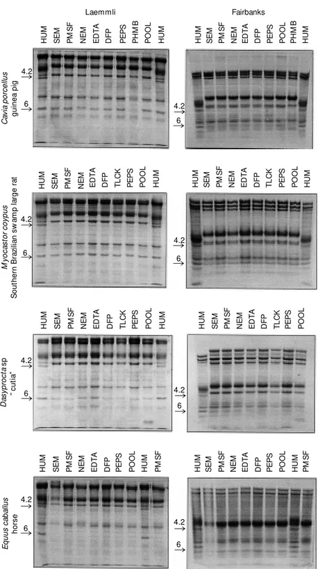
gelscan/92 software. Figure 1 illustrates the
SDS-PAGE of the erythrocyte proteins from
the animals which did not present protein 4.2
and shows that the patterns were constant in
the presence of individual inhibitors and the
pool of all inhibitors.
The red cell morphology of all animals
lacking band 4.2 is shown in Figure 2. In
comparison with human red cells, it can be
seen that Equus caballus erythrocytes are
microcytic and that Rodentia erythrocytes
are similar to human except for those of
Dasyprocta sp, which exhibit occasional
stomatocytes. However, it is difficult to
as-cribe these morphological differences to the
absence of protein 4.2.
The following Rodentia representatives
did not contain demonstrable protein 4.2:
guinea pig (Cavia porcellus) (N = 14),
South-ern Brazilian swamp large rats (Myocastor
coypus) (N = 2), and cutias (Dasyprocta sp)
(N = 4). Horses (Equus caballus) (N = 13)
also did not exhibit protein 4.2. These
ani-mals also presented high ankyrin
concentra-tions except for horses, which did not exhibit
a sharp band although they showed a group
Figure 1 - Absence of erythrocyte membrane protein 4.2 in Cavia porcellus, M yocastor coypus, Dasyprocta sp and Equus caballus in SDS-PAGE. HUM , Human erythrocyte mem-brane protein; SEM , w ithout any inhibitor; PM SF, phenylmethylsulfonyl fluoride; NEM , N-ethylmaleimide; EDTA, disodium ethylenediaminetetraacetate; DFP, diisopropylfluorophos-phate; PEPS, pepstatin A (isovaleryl-VaL-VaL--Sta-Ala-Sta); PHM B, p -hydroxymercuriben-zoic acid; POOL, mixture of all inhibitors; TLCK, Na-p-Tosyl-L-lysine chloromethyl ketone.
of minor components located between
pro-teins 2 and 3, which could account for an
ankyrin family. Protein 4.2 has been
consid-ered to play an important role in
cytoskel-eton anchorage to the integral protein 3,
together with ankyrin (band 2.1).
Inaba and Maede (15) reported the
find-ing of a putative membrane protein 4.2
be-tween 4.1a and 4.1b of horse erythrocyte
membrane. This, however, was not observed
in our study. Bands 4.1a and 4.1b always
remain together, and the hypothesis raised
by these investigators of an undefined band
between 4.1a and 4.1b may be due to an
artifact since an antibody against 4.2 was not
employed in their study. If a band 4.2 exists
in horse erythrocytes, it should be detected
as a protein of lower molecular weight than
proteins 4.1a and 4.1b.
The rodent species studied here did
pres-ent band 6, which does not occur in other
common Rodentia (11,12) such as white rats
(Rattus norvegicus) (N = 9) and mice (Mus
musculus) (N = 12) (see Figure 1).
Protease inhibitors were employed in
or-der to exclude the possibility that the
ab-sence of band 4.2 was due to proteolysis.
However, since antibodies to band 4.2 were
Equus caballus
horse
Cavia porcellus
guinea pig
Dasyprocta sp “ cutia”
Homo sapiens
man
M yocastor coypus
Southern Brazilian sw amp large rat Figure 2 - Red cell morphology
not used in the present study, the possibility
of a band immunologically similar to the
protein of band 4.2 but of different
molecu-lar weight cannot be ruled out.
Our data, however, do suggest that
pro-tein 4.2 is not an essential propro-tein since its
absence does not disrupt the cytoskeleton
membrane. Physiologically its absence may
be compensated for by the higher ankyrin
concentration observed in the mammals
lack-ing band 4.2.
Re fe re nce s
1. Palek J (1995). The red cell membrane. In: Beutler E, Lichtman M A, Coller BS & Kipps TJ (Editors), Williams Hematology. 5th edn. M cGraw -Hill, New York, 536-563. 2. Guerra-Shinohara EM (1996). Estudo das proteínas de membrana eritrocitária de mamíferos de treze ordens da Classe M ammalia. Doctoral thesis, Faculdade de Ciências Farmacêuticas da Universidade de São Paulo, São Paulo.
3. Greenquist AC & Shohet SB (1978). M arked reduction of spectrin in heredi-tary spherocytosis in the common house mouse. Blood,51: 1149-1155.
4. Whitfield CF, M ylin LM & Goodman SR (1983). Species-dependent variations in erythrocyte membrane skeletal proteins.
Blood, 61: 500-506.
5. Brookoff D, M aggio-Price L, Bernstein S & Weiss L (1982). Erythropoiesis in ha/ha
and sph/sph mice, mutants w hich pro-duce spect rin-def icient eryt hrocyt es.
Blood, 59: 646-651.
6. Reinhart W H, Sung LPA, Sung PKL,
Bernestein SE & Chien S (1988). Impaired echinocytic transformation of ankyrin and spectrin-deficient erythrocytes in mice.
American Journal of Hematology, 29: 195-200.
7. Bernestein SE (1980). Inherited hemolytic disease in mice: a review and update.
Laboratory Animal Science, 30: 197-205. 8. Bodine IV DM , Birkenmeier CS & Barker
JE (1984). Spectrin deficient inherited hemolytic anemias in the mouse: charac-terization by spectrin synthesis and mRNA activity in reticulocytes. Cell, 37: 721-729. 9. Palatnik M , Simões M LM S, Alves ZM S & Laranjeira NSM (1990). The 60 and 63 kDa proteolytic peptides of red cell mem-brane band-3 protein = their prevalence in human and non-human primates. Human Genetics, 86: 126-130.
10. Khodadad JK & Weintein RS (1983). The band 3-rich membrane of llama erythro-cytes: studies on cell shape and the or-ganization of membrane proteins. Journal of M embrane Biology, 72: 161-171.
11. Ballas SK, Kliman HJ & Smith ED (1985). Glyceraldehyde-3-phosphate dehydrogen-ase of rat erythrocytes has no membrane com ponent. Biochimica et Biophysica Acta, 831: 142-149.
12. Ballas SK (1987). Comparative distribution of glyceraldehyde-3-phosphate dehydro-genase activity in human, guinea-pig, rab-bit, and mouse erythrocytes. Compara-tive Biochemistry and Physiology, 87B: 837-842.
13. Laemmli UK (1970). Cleavage of struc-tural proteins during the assembly of head of bacteriophage T4. Nature, 227: 680-685.
14. Fairbanks G, Steck TL & Wallach DFH (1971). Electrophoretic analysis of the major polypeptides of the human erythro-cyte membrane. Biochemistry, 10: 2606-2617.