O R I G I N A L
A R T I C L E
Phytol, a diterpene alcohol, inhibits the
inflammatory response by reducing
cytokine production and oxidative stress
Renan O. Silva
a, Francisca Beatriz M. Sousa
a, Samara R.B. Damasceno
a,
Nathalia S. Carvalho
a, Valdela
̂
nia G. Silva
a, Francisco Rodrigo M.A.
Oliveira
b, Dami
~
ao P. Sousa
c, Karoline S. Araga
̃
o
d, Andr
e L.R. Barbosa
a,
Rivelilson M. Freitas
b, Jand Venes R. Medeiros
a*
a
Biotechnology and Biodiversity Center Research (BIOTEC), Federal University of Piauı, Av. S~ao Sebasti~ao, 2819, CEP: 64202-020, Parnaıba, PI, Brazil
b
Postgraduate Program in Pharmaceutical Sciences, Federal University of Piauı, Av. N S Fatima, CEP 64049-550, Teresina, PI, Brazil
c
Department of Physiology, Federal University of Paraıba, CP5009, CEP 58051-970, Jo~ao Pessoa, PB, Brazil
dDepartment of Physiology and Pharmacology, Federal University of Ceara
́, Rua CR Nunes de Melo 1315, CEP 60430-270, Fortaleza, CE, Brazil
Keywords
anti-inflammatory, cytokine, oxidative stress, phytol
Received 29 June 2013; revised 12 August 2013; accepted 20 August 2013
*Correspondence and reprints: [email protected]
A B S T R A C T
Studies have shown that diterpenes have anti-inflammatory and redox-protective pharmacological activities. The present study aimed to investigate the anti-matory properties of phytol, a diterpene alcohol, in a mouse model of acute inflam-mation, and phytol effect on leukocyte recruitment, cytokines levels, and oxidative stress. The anti-inflammatory activities of phytol were assessed by measuring paw edema induced by different inflammatory agents (e.g., k-carrageenan, compound 48/80, histamine, serotonin, bradykinin, and prostaglandin E2[PGE2]),
myeloperox-idase (MPO) activity, peritonitis model and cytokine levels. Further, oxidative stress was evaluated by determining glutathione (GSH) levels and malondialdehyde (MDA) concentration. The results showed that phytol (7.5, 25, 50, and 75 mg/kg) signifi-cantly reduced carrageenan-induced paw edema, in a dose-dependent manner. In addition, phytol (75 mg/kg) inhibited compound 48/80-, histamine-, serotonin-, bradykinin- and PGE2-induced paw edema. It also inhibited the recruitment of total
leukocytes and neutrophils; decreased MPO activity, tumor necrosis factor-a(TNF-a) and interleukin-1b(IL-1b) levels, and MDA concentration; and increased GSH levels during carrageenan-induced acute inflammation. These results suggest that phytol attenuates the inflammatory response by inhibiting neutrophil migration that is partly caused by reduction in IL-1band TNF-alevels and oxidative stress.
I N T R O D U C T I O N
Inflammation is a complex biological response of vascu-larized tissues to harmful stimuli, such as pathogens, damaged cells, or irritants [1]. It is well established that this process involves the local formation of kinins and cytokines that promote vascular endothelial cell activa-tion, followed by leukocyte migration into the inflamed site [2]. Another important component of he
inflamma-tory response is oxidative stress leading to the generation of molecules, such as hydrogen peroxide, superoxide anion, and peroxynitrite, which are produced in response to stimuli and can exacerbate this process [3].
The clinical signs and symptoms of inflammation include edema, fever, erythema, pain, and cell migra-tion (primarily neutrophil migramigra-tion) into the site of injury [4]. The drugs used to treat these symptoms, such as nonsteroidal anti-inflammatory drugs
(NSA-Fundam
e
ntal &
Clinical
P
h
a
rm
a
co
lo
IDs), are not only associated with major adverse effects, such as gastrointestinal ulcers, bleeding, and renal disorders, but also have low therapeutic efficacy [5]. Thus, the search for new products with therapeutic potential for the treatment for inflammation has increased in recent years [6].
Many studies have been conducted as a part of the search for new therapeutic options for inflammation, and classes of secondary metabolites from natural sources, such as lactones [7], alkaloids [8], and terpe-noids [9], have attracted the attention of many researchers because of their pharmacological activities.
Phytol (Figure1) is an acyclic diterpene alcohol found in the essential oils of some aromatic plants, such as Cleome serrata [10] and Lantana radula [11]. Various therapeutic activities of phytol have been reported in previous studies, including its activity against mycobacteria [12], and anticonvulsant [13], antispasmodic [14], and anticancer activities [15].
Some studies have demonstrated promising anti-inflammatory pharmacological activities of diterpenes [16,17], but few have focused on phytol. The aim of this study was to investigate the anti-inflammatory properties of phytol, a diterpene alcohol, in mouse models of acute inflammation. Furthermore, the study investigated the roles of leukocyte recruitment, cyto-kines, and oxidative stress in phytol-induced effects.
M A T E R I A L S A N D M E T H O D S
Drugs and chemicals
k-Carrageenan, compound 48/80, serotonin, histamine, bradykinin, prostaglandin E2 (PGE2), indomethacin, dimethyl sulfoxide (DMSO), and phytol (97% purity)
were purchased from Sigma Chemical (St. Louis, MO, USA). Heparin and morphine were provided by Merck (Brazil). All drugs were dissolved in sterile 0.9% (w/v) NaCl (saline). The phytol was dissolved in 2% DMSO. All other chemicals were of analytical grade and obtained from standard commercial suppliers.
Animals
Male Swiss mice weighing 25–30 g were randomly
housed in appropriate cages at 23 2°C under a
12/12-h light/dark cycle with free access to food (Puri-na, Brazil) and water. Experimental protocols were approved by the Ethics Committee in Research of the Federal University of Piauı (protocol No. 0066/10), and handling procedures were in accordance with the
Guide for Care and Use of Laboratory Animals (National Institute of Health, Bethesda, MD, USA).
Effect of phytol on carrageenan-induced paw edema
Initially, the animals were randomly divided into seven groups (n=5), and edema acute hind paw edema was produced by injecting of 50lL of a suspension of carrageenan (500lg/paw, prepared in 0.9% sterile saline) into the right hind paw (group I). In the other groups, mice were pretreated intraperitoneally (i.p.) with carrageenan + 2% DMSO (group II) or only 2% DMSO (group III); indomethacin 10 mg/kg (group IV, standard drug) or phytol 7.5, 25, 50, or 75 mg/kg (groups V, VI, VII and VIII, respectively). Right paw volume was measured by the dislocation of the water column of a plethysmometer (Panlab, Barcelona, Spain) before (V0; time zero) at 1, 2, 3, and 4 h after
carrageenan treatment (Vt) as previously described
[18]. The effect of pretreatment was calculated as per-cent inhibition of edema relative to the paw volume of the DMSO-treated controls using the following formula:
% inhibition of edma
¼ðVt V0ÞControl ðVt V0ÞTreated
ðVt V0ÞControl
100
Effect of phytol on paw edema induced by different agents
To induce edema with different agents, the animals received 50-lL injections of compound 48/80 (12 lg/ paw), serotonin (5-HT; 1% w/v), histamine (Hist; 100lg/paw) bradykinin (BK; 6.0 nmol/paw), or PGE2
(3 nmol/paw) into the right hind paw, as previously described [4,19]. The contralateral paw received 50lL of 2.0% DMSO and served as control untreated. Phytol (75 mg/kg) or indomethacin (10 mg/kg, reference con-trol) was injected i.p. Thirty minutes before intraplan-tar injections of phlogistic agents. Right paw volume was measured by the dislocation of the water column of a plethysmometer (Panlab, Barcelona, Spain) before (V0; time zero) at 30, 60, 90, and 120 min after (Vt)
phlogistic agents administration.
Myeloperoxidase (MPO) activity
Briefly, 50–100 mg of paw tissue was homogenized in potassium buffer containing 0.5% hexadecyltrimethy-lammonium bromide (HTAB). The homogenate was centrifuged at 4500 g for 15 min at 4 °C. The pellet
was resuspended, and MPO activity was assayed by measuring the change in absorbance at 450 nm using o-dianisidinedihydrochloride and 1% hydrogen perox-ide. Myeloperoxidase activity was reported as units/mg of tissue. A unit of MPO activity was defined as that converting 1lmol of hydrogen peroxide to water in 1 min at 22°C.
Evaluation of neutrophil migration
Mice were injected intraperitoneally with 2.0% DMSO, phytol 75 mg/kg, or indomethacin 10 mg/kg. Thirty minutes later, the animals were injected with carra-geenan (500lg/cavity; 250 ll). After 4 h, mice were sacrificed and peritoneal cavity was washed with 1.5 mL of heparinized phosphate-buffered saline (PBS). The volumes recovered were similar in all experimental groups and were equivalent to ~95% of the injected volume. Total cell counts were performed in a Neu-bauer chamber, and differential cell counts (100 cells total) were carried out on cytocentrifuge slides stained with hematoxylin and eosin. The results are presented as the number of neutrophils per milliliter of peritoneal exudate. Aliquots of the peritoneal exudates were stored at 70°C for later analysis of cytokine content, glutathione (GSH) levels, and malondialdehyde (MDA) concentration.
Cytokine measurements
The levels tumor necrosis factor (TNF)-a and interleu-kin (IL)-1b were evaluated using sandwich ELISA as described previously [20]. Briefly, microliter plates were coated overnight at 4°C with antibody against mice TNF-a or IL-1b (2lg/mL). Blocking of nonspecific binding sites was accomplished by incubating plates with PBS containing 2% BSA for 90 min at 37°C. After blocking the plates, the test samples and each standard at various dilutions were added in duplicate and incubated at 4°C for 24 h. The plates were washed three times with buffer. After washing the plates, 50lL of biotinylated sheep polyclonal anti-TNF-a and anti-IL-1b (diluted 1 : 1000 with assay buffer 1% BSA) was added to the wells. After further incubation at room temperature for 1 h, the plates were washed, and 50lL of streptavidin-HRP diluted 1 : 5000 was added to all wells. The reagent
o-phenylenediamine dihydrochloride (50 lL) was added 15 min later, and the plates were incubated in the dark at 37°C for 15–20 min. After color
develop-ment, the reaction was stopped with the addition of sulfuric acid (1M), and absorbance was measured at
490 nm. The results are expressed as pg/mg protein and reported as meanSD.
Glutathione (GSH) levels
The concentration of glutathione in the peritoneal exudates was estimated according to the method previ-ously described [21]. Aliquots (600lL) of the perito-neal exudates were centrifuged at 3000 rpm for 15 min at 4°C. Next, 400lL of each supernatant was mixed with 800lL of Tris buffer (0.4M, pH 8.9)
and 20lL of 0.01M 5,5-dithio-bis (2-nitrobenzoic
acid). Subsequently, the samples were stirred for 3 min and read on a spectrophotometer at 412 nm. Glutathione concentration was determined via a reduced GSH standard curve, which was generated in parallel. Glutathione levels are expressed as lg/ml of exudates.
Malondialdehyde (MDA) concentration
The MDA concentration in the peritoneal exudates from each group was measured according to the method previously described [22]. Briefly, aliquots (500lL) peritoneal exudates were centrifuged at 3000
g for 15 min at 4 °C, then 250lL each supernatant
was added to 1.5 mL of 1% phosphoric acid (H3PO4)
and 0.5 mL of 0.6% thiobarbituric acid (aqueous solu-tion). Then, this mixture was stirred and heated in a boiling water bath for 45 min. The mixture was then cooled immediately in an ice water bath followed by the addition of 4 mL of n-butanol. This mixture was shaken, and the butanol layer was separated by centri-fugation at 12009g for 10 min. Optical density was
determined to be 535 and 520 nm, and the optical density difference between the two determinations was calculated as the tert-butyl alcohol value. Malondialde-hyde concentrations are expressed as mmol/ml of exudates.
Statistical analysis
Results are expressed as meanSEM from at least five animals per group, and statistical analysis was per-formed using one-way analysis of variance (ANOVA)
followed by the Newman–Keuls post hoc test, when appropriate. Statistical significance was set at
R E S U L T S
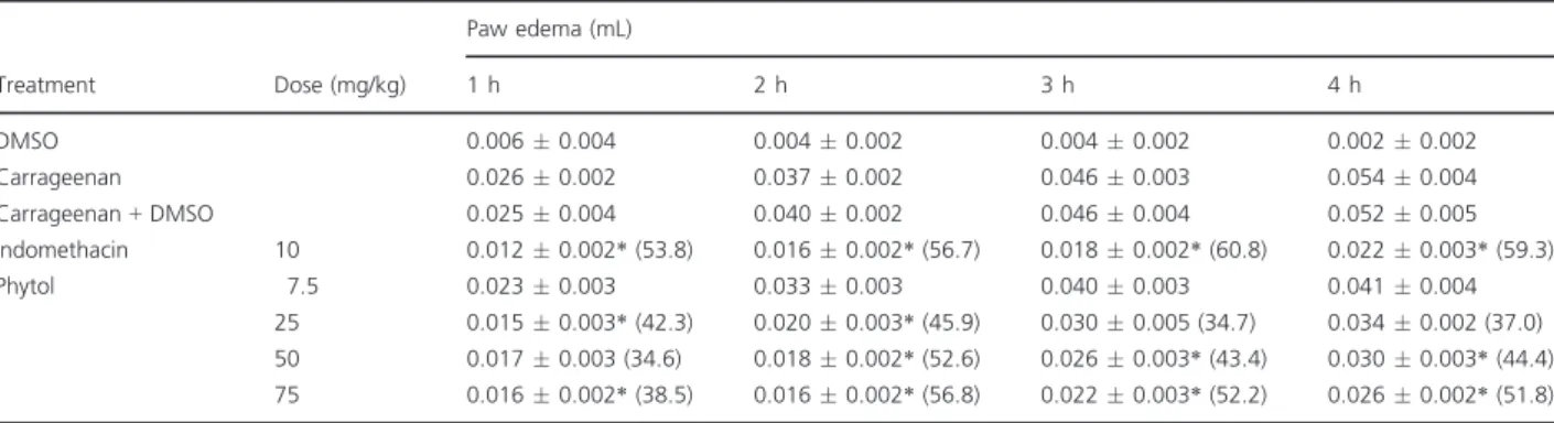
Phytol reduced carrageenan-induced paw edema As shown in TableI, carrageenan administration was effective in inducing time-dependent paw edema, which peaked after 4 h (0.0540.004 mL). Phytol (25, 50, and 75 mg/kg) significantly inhibited (P< 0.05) the development of carrageenan-induced paw edema in a dose-dependent manner, at all time points. The maximum inhibitory effect—51.8% inhibition—was
achieved 4 h after the administration of 75 mg/kg phytol (0.0260.002 mL). The reference drug indomethacin (10 mg/kg) also significantly decreased (P<0.05) paw edema throughout the experimental period, with the maximum inhibition being 59.3% 4 h after treatment with carrageenan (Table I). Because the 75 mg/kg phytol dose produced the maximum inhibi-tory effect against carrageenan-induced paw edema, it was used to study the possible mechanisms involved in the anti-inflammatory activity of phytol.
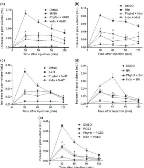
Phytol reduced paw edema induced by different phlogistic agents
As shown in Figure 2, the injection of compound 48/ 80 (0.1080.011 mL; Figure 2a), Hist (0.082 0.0 03 mL; Figure2b), 5-HT (0.084 0.008 mL; Fig-ure 2c), BK (0.0700.009 mL; Figure2d), or PGE2
(0.069 0.003 mL; Figure 2e) induced intense paw edema. Pretreatment with phytol (75 mg/kg) signifi-cantly inhibited (P< 0.05) paw edema induced by compound 48/80 (0.038 0.008 mL; 64.8% inhibition), Hist (0.0300.010 mL; 64.4% inhibi-tion), 5-HT (0.0280.005 mL; 64.4% inhibition), BK (0.018 0.06 mL; 74.3% inhibition), or PGE2
(0.030 0.007 mL; 56.5% inhibition). Indomethacin
(10 mg/kg) also significantly reduced the paw edema induced by different phlogistic agents.
Phytol reduced carrageenan-induced MPO activity
Figure 3 shows that compared with the group treated with carrageenan alone (9.850.96 U/mg of tissue), the groups pretreated with phytol (75 mg/kg) should significantly reduced (P<0.05) MPO activity (3.210.82 U/mg of tissue). Furthermore, compared with treatment with carrageenan alone, treatment with the reference drug, indomethacin, significantly reduced (P <0.05) MPO activity (4.371.45 U/mg of tissue).
Phytol inhibited cell migration in the carrageenan-induced peritonitis model
Carrageenan administration induced a significant increase in the total leukocyte (4.830.999103 cells/mL; Figure 4a) and neutrophil recruitment (3.550.329 103 cells/mL;Figure 4b) to the
perito-neal cavity. Compared with treatment with carra-geenan alone, pretreatment with phytol (75 mg/kg) significantly reduced (P<0.05) the leukocyte recruit-ment (2.39 0.629103 cells/mL) and neutrophil
migration (0.42 0.459 103 cells/mL) to the perito-neal cavity. Indomethacin (10 mg/kg) reduced leuko-cyte (1.980.329103 cells/mL) and neutrophil
counts (0.580.119103cells/mL) in the peritoneal cavity (Figure4).
Phytol decreased carrageenan-induced TNF-aand IL-1bproduction
Carrageenan significantly increased (P<0.05) TNF-a
(208.7 39.29 103 pg/mL; Figure 5a) and IL-1b
Table IEffect of phytol on carrageenan-induced paw edema.
Treatment Dose (mg/kg)
Paw edema (mL)
1 h 2 h 3 h 4 h
DMSO 0.0060.004 0.0040.002 0.0040.002 0.0020.002 Carrageenan 0.0260.002 0.0370.002 0.0460.003 0.0540.004 Carrageenan+DMSO 0.0250.004 0.0400.002 0.0460.004 0.0520.005 Indomethacin 10 0.0120.002*(53.8) 0.0160.002*(56.7) 0.0180.002*(60.8) 0.0220.003*(59.3) Phytol 7.5 0.0230.003 0.0330.003 0.0400.003 0.0410.004
25 0.0150.003*(42.3) 0.0200.003*(45.9) 0.0300.005 (34.7) 0.0340.002 (37.0) 50 0.0170.003 (34.6) 0.0180.002*(52.6) 0.0260.003*(43.4) 0.0300.003*(44.4) 75 0.0160.002*(38.5) 0.0160.002*(56.8) 0.0220.003*(52.2) 0.0260.002*(51.8) Values of paw edema are expressed in meanSEM of 5–6 animals per group.
% Inhibition of paw edema is indicated in parenthesis.
(1073.0180.19103pg/mL; Figure5b) production
in peritoneal exudates 4 h after stimulus injection, compared with the control treatment (100.7 15.59 103 pg/mL and 176.999.5 pg/mL, for
TNF-aand IL-1b, respectively). However, pretreatment with phytol (75 mg/kg) significantly decreased (P <0.05) TNF-a (106.8 32.81 pg/mL; Figure 5a) and IL-1b
(607.1147.6 pg/mL;
Figure5b) levels. Similarly, indomethacin produced significant inhibitory effects on both the parameters analyzed.
Phytol inhibited the carrageenan-induced increase in GSH levels
Figure6 shows that carrageenan treatment signifi-cantly increased (P<0.05) GSH levels (59.738.19lg/mL) in peritoneal exudates, 4 h after stimulus injection, compared with the control treatment(127.6020.12lg/mL). Compared with
treatment with carrageenan alone, pretreatment with phytol (75 mg/kg) significantly reduced (P< 0.05) GSH levels (94.0517.17lg/mL).
Phytol reduced the carrageenan-induced increase in MDA concentration
Malondialdehyde concentration increased significantly (25.174.47 nmol/mL) in peritoneal exudates 4 h after carrageenan injection (P<0.05), compared with the control treatment (14.12 1.71 nmol/mL) (Figure 7). However, compared with treatment with carrageenan alone, pretreatment with phytol (75 mg/kg) significantly decreased (P<0.05) MDA concentration (10.731.75 nmol/mL).
D I S C U S S I O N
Natural compounds with different mechanisms of action may be used to treat inflammatory diseases and
(a) (b)
(c) (d)
(e)
Figure 2 Effect of phytol on paw edema induced by different agents. Animals were pretreated with 2% DMSO, phytol (75 mg/kg, i.p.), or indomethacin (Indo; 10 mg/kg, i.p.). Edema was induced 30 min later by compound 48/ 80 (panel a), histamine (Hist; panel b), serotonin (5-HT; panel c), bradykinin (BK; panel d), and prostaglandin E2 (PGE2; panel e). Each column
the essential oils, and their constituents, especially the terpenes, have been shown to possess important prop-erties in several inflammatory processes [17]. The pharmacological results of the present study revealed that phytol, a diterpene alcohol, could reduce neutro-phil migration, cytokine levels, and oxidative stress in acute inflammation models.
The carrageenan-induced paw edema model was used to evaluate the anti-inflammatory effects of phy-tol. This inflammation model has been well established as a standard for screening the anti-inflammatory activity of natural product-derived compounds [23].
The initial phase (1–2 h) of edema is associated with alterations in the vascular permeability induced by the action of vasoactive amines, such as histamine and serotonin released from mast cells. The delayed phase (3–4 h) involves the overproduction and secretion of BK and prostaglandins in paw tissue, which is accom-panied by increased neutrophil migration and produc-tion of neutrophil-derived free radicals, such as hydrogen peroxide, superoxide, and hydroxyl radicals [24]. In this study, we found that phytol was equally effective in both phases of carrageenan-induced paw edema, suggesting that the anti-inflammatory effect observed is due, at least in part, to the inhibition of pro-inflammatory mediators, and reduction in neutro-phil migration and oxidative stress.
To elucidate this mechanism, different inflammatory agents, including compound 48/80, Hist, 5-HT, BK, and PGE2, were used in the paw edema study.
Com-pound 48/80 induces edema by Hist and 5-HT release from mast cell degranulation, leading to osmotic edema, characterized by increased vascular permeabil-ity [25]. Our data demonstrated that phytol inhibited the paw edema induced by compound 48/80, which could be because of the stabilization of mast cell mem-branes and prevention of degranulation. This was con-firmed by the fact that phytol inhibited paw edema induced by Hist and 5-HT.
Furthermore, phytol reduced BK- and PGE2-induced
paw edema. BK is a peptide with potent activity on vascular permeability and promotes arteriolar vasodila-tion and inflammatory cell chemotaxis [26]. Once released, BKs activate their receptors, resulting in the production and release of pro-inflammatory mediators derived from the arachidonic acid pathway, including
Figure 3 Effect of phytol on carrageenan-induced paw tissue myeloperoxidase (MPO) activity. Animals were pretreated with 2% DMSO, phytol (75 mg/kg, i.p.), or indomethacin (Indo; 10 mg/kg, i.p.) and injected with carrageenan (500lg/paw) 30 min later. Myeloperoxidase activity in the paw tissue was determined 4 h later. Results are expressed as the meanSEM of 5–6 animals per group.*P<0.05 vs. DMSO-treated group;
#P
<0.05 vs. carrageenan group.
(a) (b)
Figure 4 Effect of phytol on cell migration in carrageenan-induced peritonitis. Animals were pretreated with 2% DMSO, phytol (75 mg/kg, i.p.), or indomethacin (Indo; 10 mg/kg, i.p.) and injected with 250lL of carrageenan (500lg/cavity, i.p.) 30 min later. Neutrophil migration was evaluated 4 h later. Panel (a), leukocyte counts; panel (b), neutrophil counts. Each column represents the meanSEM of 5–6 animals per group.*P<0.05 vs. DMSO-treated group;
#
PGE2 [27], as well as cytokines, and histamine and
serotonin following mast cell degranulation [28]. Therefore, PGE2 is also considered being a key
pro-inflammatory mediator and causes an increase in paw volume by altering vascular permeability in synergy with the other mediators described earlier [29]. Thus, our findings suggest that the anti-inflammatory activi-ties of phytol are due to inhibition of the synthesis, release, or action of pro-inflammatory mediators. This notion is reinforced by the inhibitory effect of phytol in the initial phase of carrageenan-induced edema, as described previously.
Cell migration within the injured tissue is an impor-tant step of the inflammatory process. Thus, to evalu-ate whether the anti-inflammatory effect of phytol involved the inhibition of neutrophil migration, we measured MPO activity in paw tissue. Myeloperoxidase is an enzyme abundantly found in the azurophilic granules of neutrophils and is released after their acti-vation, within the phagosome or in the extracellular space [30]. Myeloperoxidase activity is directly propor-tional to neutrophil chemotaxis and infiltration into inflamed tissues [31]. Our results showed that phytol inhibited neutrophil infiltration, which was evident
(a) (b)
Figure 5 Effect of phytol on cytokine production in carrageenan-induced peritonitis. Animals were pretreated with 2% DMSO, phytol (75 mg/kg, i.p.), or indomethacin (Indo; 10 mg/kg, i.p.), and 250lL of carrageenan (500lg/cavity, i.p.) was injected 30 min later. The levels of tumor necrosis factor (TNF)-a(panel a) and interleukin (IL)-1b(panel b) in the peritoneal cavity were measured 4 h after carrageenan injection. Each point represents the meanSEM of 5–6 animals per group.*P<0.05 vs. DMSO-treated group;
#P
<0.05 vs. carrageenan group.
Figure 7Effect of phytol on malondialdehyde (MDA)
concentration in carrageenan-induced peritonitis. Animals were pretreated with 2% DMSO or phytol (75 mg/kg, i.p.), and 250lL of carrageenan (500lg/cavity, i.p.) was injected 30 min later. Malondialdehyde concentration in the peritoneal cavity was measured 4 h after carrageenan injection. The results are expressed as the meanSEM of 5–6 animals per group. *P<0.05 vs. DMSO-treated group;#P
<0.05 vs. carrageenan group.
from the reduced MPO activity. This suggests that phytol can suppress neutrophil recruitment to the site of inflammation.
The carrageenan-induced peritonitis model was used to confirm the role of cell migration in the anti-inflammatory effect of phytol. Literature shows that carrageenan induces an inflammatory response in the peritoneal cavity, which is characterized by intense exudation and migration of inflammatory cells, espe-cially neutrophils [32]. In the present study, we showed that phytol significantly reduced the migration of poly-morphonuclear cells to the peritoneal cavity, as demon-strated by neutrophil counts. This inhibitory effect corroborated the data analyzing MPO activity and con-firmed the importance of cell migration in the mainte-nance and exacerbation of the carrageenan-induced inflammatory response in the peritoneal cavity.
Next, we demonstrated that the carrageenan-induced inflammatory response in the peritoneal cavity led to a substantial increase in TNF-a and IL-1b levels; this finding was in agreement with those of previous studies [2,8,33]. These pro-inflammatory cytokines are impor-tant mediators associated with several inflammatory diseases, such as bacterial sepsis, rheumatoid arthritis, and skin inflammation [34,35]. Therefore, suppressing these mediators is believed to be an effective strategy for the treatment of various pathological conditions. In our study, pretreatment with phytol significantly reversed this significant increase, suggesting that phytol exerted an anti-inflammatory action and inhib-ited polymorphonuclear cell migration, probably by decreasing TNF-aand IL-1blevels.
Oxidative stress products, primarily generated by infiltrating neutrophils, are another important aspect of the inflammatory process [36,37]. It has been shown that cytokines, including TNF-aand IL-1b, can induce the production of H2O2 and superoxide, leading to
nuclear factorjB (NFjB) activation, which upregulates cytokine production [38]. Free radicals have been established as one of the major causes of damage from inflammation [39]. Reactive oxygen species are mole-cules with one or more unpaired outer shell electrons, which are generally highly unstable and extremely reactive [3].
Glutathione levels and MDA concentration were evaluated to explore the redox-protective action of phytol during acute inflammation. Under normal con-ditions, oxidative stress is kept under control by the endogenous antioxidant system, which includes enzymatic and nonenzymatic antioxidants, such as
superoxide dismutase (SOD) and reduced GSH [40,41]. However, during inflammation, excess free radicals lead to tissue damage and activate inflammatory mediators, leading to marked downregulation of endogenous defense mechanisms [42].
In this context, MDA is the end-product of lipid peroxidation and reflects an imbalance between the oxidative and antioxidant systems. Increased MDA con-centration, due to free radical-induced plasma mem-brane damage, has been reported in the carrageenan-induced inflammation model [43,44]. On the other hand, GSH is a free radical scavenger and has been suggested to play an important role against carra-geenan-induced local inflammation, by promoting hydrogen transference and acting as a cofactor for the enzyme GSH peroxidase [43,45].
Our results confirmed that carrageenan administra-tion caused an increase in MDA concentration, whereas GSH level decreased, indicating a role of oxi-dative stress in this model [46,47]. However, pretreat-ment with phytol reduced MDA formation and restored the depleted GSH content in peritoneal exudates. Therefore, we could infer that the redox-protective effect of phytol might be explained, at least in part, by an increase in GSH concentration. An alternative possibility is that the increase in GSH levels could be secondary to a decrease in free radical production.
In summary, our results suggest that phytol has an anti-inflammatory activity in acute inflammation mod-els. Although there are many mechanisms through which this effect can occur, our data support the hypoth-esis that the inhibition of neutrophil migration is of essential importance. This effect is due, at least in part, to reduce IL-1band TNF-alevels and oxidative stress.
A C K N O W L E D G E M E N T S
The authors gratefully acknowledge the financial support from National Counsel of Technological and Scientific Development – CNPq (Brazil) and Research
foundation for the State of Piauı–FAPEPI.
R E F E R E N C E S
1 Weiss U. Inflammation. Nature (2008)454427. 2 Chaves L.S., Nicolau L.A.D., Silva R.O. et al.
3 Wang Z.Q., Porreca F., Cuzzocrea S. et al. A newly identified role for superoxide in inflammatory pain. J. Pharmacol. Exp. Ther. (2004)309869–878.
4 Vasconcelos D.I.B., Leite J.A., Carneiro L.T. et al. Anti-inflammatory and antinociceptive activity of ouabain in mice. Mediators Inflamm. (2011)20111–11.
5 Quintans-Junior L.J., Guimar~aes A.G., Santana M.T. et al. Citral reduces nociceptive and inflammatory response in rodents. Braz. J. Pharmacog. (2011)21497–502. 6 Tirapelli C.R., Ambrosio S.R., de Oliveira A.M., Tostes R.C.
Hypotensive action of naturally occurring diterpenes: a therapeutic promise for the treatment of hypertension. Fitoterapia (2010)81690–702.
7 Valerio D.A., Cunha T.M., Arakawa N.S. et al. Anti-inflammatory and analgesic effects of the sesquiterpene lactone budlein A in mice: inhibition of cytokine production-dependent mechanism. Eur. J. Pharmacol. (2007)562 155–163.
8 Silva V.G., Silva R.O., Damasceno S.R.B. et al. Anti-inflammatory and antinociceptive activity of epiisopiloturine, an imidazole alkaloid isolated fromPilocarpus microphyllus. J. Nat. Prod. (2013)761071–1077.
9 Gershenzon J., Dudareva N. The function of terpene natural products in the natural world. Nat. Chem. Biol. (2007)3 408–414.
10 Mcneil M.J., Porter R.B., Williams L.A. Chemical composition and biological activity of the essential oil from Jamaican
Cleome serrata. Nat. Prod. Commun. (2012)71231–1232. 11 Passos J.L., Barbosa L.C., Demuner A.J., Alvarenga E.S., Silva
C.M., Barreto R.W. Chemical characterization of volatile compounds ofLantana camaraL. andL. radulaSw. and their antifungal activity. Molecules (2012)1711447–11455. 12 Saikia D., Parihar S., Chanda D. et al. Antitubercular
potential of some semisynthetic analogues of phytol. Bioorg. Med. Chem. Lett. (2010)20508–512.
13 Costa J.P., Ferreira P.B., Sousa D.P., Jordan J., Freitas R.M. Anticonvulsant effect of phytol in a pilocarpine model in mice. Neurosci. Lett. (2012)523115–118.
14 Pongprayoon U., Baeckstr€om P., Jacobsson U., Lindstr€om M., Bohlin L. Antispasmodic activity of beta-damascenone and e-phytol isolated fromIpomoea pes-caprae. Planta Med. (1992) 5819–21.
15 Lee K.L., Lee S.H., Park K.Y. Anticancer activity of phytol and eicosatrienoic acid identified from Perilla leaves. J. Korean Soc. Food Sci. Nutr. (1999)281107–1112.
16 Fernandez M.A., Tornos M.P., Garcıa M.D., Heras B., Villar A.M., Saenz M.T. Anti-inflammatory activity of abietic acid, a diterpene isolated fromPimenta racemosavar. grissea. J. Pharm. Pharmacol. (2001)53867–872.
17 Demetzos C., Dimas K., Hatziantoniou S., Anastasaki T., Angelopoulou D. Cytotoxic and anti-inflammatory activity of labdane and cis-clerodane type diterpenes. Planta Med. (2001)67614–618.
18 Winter C.A., Risley E.A., Nuss G.W. Carrageenin-induced edema in hind paw of the rat as an assay for antiiflammatory drugs. Proc. Soc. Exp. Biol. Med. (1962)111544–547.
19 Claudino R.F., Kassuya C.A., Ferreira J., Calixto J.B. Pharmacological and molecular characterization of the mechanisms involved in prostaglandin E2-induced mouse paw edema. J. Pharmacol. Exp. Ther. (2006)318611–618. 20 Cunha F.Q., Boukili M.A., Motta J.I.B., Vargaftig B.B.,
Ferreira S.H. Blockade by fenspiride of endotoxin-induced neutrophil migration in the rat. Eur. J. Pharmacol. (1993) 23847–52.
21 Sedlak J., Lindsay R.H. Estimation of total, protein-bound, and nonprotein sulfhydryl groups in tissue with Ellman’s reagent. Anal. Biochem. (1968)241992–2005.
22 Mihara M., Uchiyama M. Determination of malonaldehyde precursor in tissues by thiobarbituric acid test. Anal. Biochem. (1978)86271–278.
23 Kumar P.P., Kuttan G.Vernonia cinereaL. scavenges free radicals and regulates nitric oxide and proinflammatory cytokines profile in carrageenan induced paw edema model. Immunopharmacol. Immunotoxicol. (2009)3194–102. 24 Pereira L.P., da Silva R.O., Bringela P.H.S.F., da Silva K.E.S.,
Assreuya A.M.S., Pereira M.G. Polysaccharide fractions of
Caesalpinia ferreapods: potential anti-inflammatory usage. J. Ethnopharmacol. (2012)139642–648.
25 Sousa A.A., Benevides N.M., de Freitas P.A. et al. A report of a galactan from marine algaGelidium crinalewith in vivo anti-inflammatory and antinociceptive effects. Fundam. Clin. Pharmacol. (2013)27173–180.
26 Siqueira-Junior J.F., Dantas C.J.S. Mecanismos celulares e
moleculares da inflamacß~ao, MEDSI, Rio de Janeiro, Brazil, 2000.
27 Saleh T.S.F., Calixto J.B., Medeiros Y.S. Pro-inflammatory effects induced by bradykinin in a murine model of pleurisy. Eur. J. Pharmacol. (1997)33143–52.
28 Gaginella T.S., Kachur J.F. Kinin mediators of intestinal secretion. Am. J. Physiol. (1989)2561–15.
29Ozd€ €ol N.C., Melli M. Formation of 8-isoprostaglandin F2aand prostaglandin E2 in carrageenan-induced air pouch model in rats. Eur. J. Pharmacol. (2004)506189–197.
30 Van Der Veen B.S., de Winther M.P., Heeringa P.
Myeloperoxidase: molecular mechanisms of action and their relevance to human health and disease. Antioxid. Redox Signal. (2009)112899–2937.
31 Gaut J.P., Yeh G.C., Tran H.D. et al. Neutrophils employ the myeloperoxidase system to generate antimicrobial
brominating and chlorinating oxidants during sepsis. Proc. Natl Acad. Sci. USA (2001)9811961–11966.
32 Foster S.J., McCormick M.E., Howarth A., Aked D. Leukocyte recruitment in the subcutaneous sponge implant model of acute inflammation in the rat is not mediated by leukotriene B4. Biochem. Pharmacol. (1986)351709–1717.
33 Loram L.C., Fuller A., Fick L.G., Cartmell T., Poole S., Mitchell D. Cytokine profiles during carrageen-induced inflammatory hyperalgesia in rat muscle and hind paw. J. Pain (2007)8 127–136.
cytokines expressions via nuclear factor-kbinactivation. J. Agric. Food Chem. (2008)5610265–10272.
35 Feghali C.A., Wright T.M. Cytokines in acute and chronic inflammation. Front. Biosci. (1997)112–26.
36 Nagib M.M., Tadros M.G., ELSayed M.I., Khalifa A.E. Anti-inflammatory and anti-oxidant activities of olmesartan medoxomil ameliorate experimental colitis in rats. Toxicol. Appl. Pharmacol. (2013)271106–113.
37 Rao R.S., Medhi B., Khanduja K.L., Pandhi P. Correlation of seizures and biochemical parameters of oxidative stress in experimentally induced inflammatory rat models. Fundam. Clin. Pharmacol. (2010)24325–331.
38 Bowie A., O’Neill L.A. Oxidative stress and nuclear factor-kappaB activation: a reassessment of the evidence in the light of recent discoveries. Biochem. Pharmacol. (2000)59 13–23.
39 Young I.S., Woodside J.V. Antioxidants in health and disease. J. Clin. Pathol. (2001)54176–186.
40 Urso M.L., Clarkson P.M. Oxidative stress, exercise, and antioxidant supplementation. Toxicology (2003)18941–54. 41 Valko M., Rhodes C.J., Moncol J., Izakovic M., Mazur M. Free radicals, metals and antioxidants in oxidative stress-induced cancer. Chem. Biol. Interact. (2006)1601–40.
42 Valerio D.A., Georgetti S.R., Magro D.A. et al. Quercetin reduces inflammatory pain: inhibition of oxidative stress and cytokine production. J. Nat. Prod. (2009)721975–1979. 43 Chang H.Y., Sheu M.J., Yang C.H. et al. Analgesic effects and
the mechanisms of anti-inflammation of hispolon in mice. Evid. Based Complementary Altern. Med. (2011)20111–8. 44 El-Shitany N.A., El-Masry S.A., El-Ghareib M.A., El-Desoky K.
Thioctic acid protects against carrageenan-induced acute inflammation in rats by reduction in oxidative stress, downregulation of COX-2 mRNA and enhancement of IL-10 mRNA. Fundam. Clin. Pharmacol. (2010)2491–99. 45 Liao J.C., Deng J.S., Chiu C.S. et al. Anti-inflammatory
activities ofCinnamomum cassiaconstituentsin vitroandin vivo. Evid. Based Complementary Altern. Med. (2012)2012 1–12.
46 Cuzzocrea S., Costantino G., Zingarelli B., Mazzon E., Micali A., Caputi A.P. The protective role of endogenous glutathione in carrageenan-induced pleurisy in the rat. Eur. J. Pharmacol. (1999)372187–197.