87
Radiol Bras. 2012 Mar/Abr;45(2):87–92
Enhancement of radiological protection through an internal
quality assessment cycle
*
Melhoria da proteção radiológica mediante um ciclo de avaliação interna da qualidade
Filipe Morais de Figueiredo1, Zenewton André da Silva Gama2
Objective: To determine the level of quality in radiation protection of patients during radiological examination, evalu-ating the effectiveness of an intervention aimed at enhancing the quality of such a protection. Materials and Meth-ods: A quality improvement cycle was implemented in a radiology service of the Regional Health Administration, in Algarve, Portugal. Based on six quality criteria, an initial evaluation was performed and followed by an intervention fo-cused on the most problematic points (over an eight-month period) and a subsequent quality reassessment. A random sampling (n = 60) has allowed the authors to infer the point estimates and confidence intervals for each criterion, as well as calculating the statistical significance of the results by means of the Z-test. Results: Initially, deficiencies were observed in relation to all the quality criteria. After the intervention, a minimum relative improvement of 33% was ob-served in five of the six criteria, with statistical significance (p < 0.05) in two of them. The absolute frequency of non-compliance decreased from 38 (first evaluation) to 21 (second evaluation), corresponding to a 44.7% improvement. Conclusion: The first institutional evaluation cycle showed a seemingly incipient improvement margin. However, the implemented intervention was effective in stimulating good practices and improving the level of radiological protection of patients.
Keywords: Radiological protection; Quality enhancement; Institutional evaluation; Health care quality assurance.
Objetivo: Determinar o nível de qualidade da proteção radiológica para os pacientes durante a realização de exames radiológicos e avaliar a efetividade de uma intervenção dirigida a melhorar a qualidade. Materiais e Métodos: Rea-lizou-se um ciclo de melhoria em um serviço de radiologia da Administração Geral de Saúde do Algarve, Portugal. Utilizando seis critérios de qualidade, foram efetivadas uma avaliação, uma intervenção focada nos critérios mais pro-blemáticos (durante oito meses) e uma reavaliação. As amostras foram aleatórias (n = 60), possibilitando a inferência sobre as estimativas pontuais e intervalos de confiança do cumprimento de cada critério, assim como o cálculo da significância estatística da melhoria identificada, através do teste Z. Resultados: Na avaliação inicial, todos os crité-rios de qualidade apresentaram falhas. Após a intervenção, a melhoria relativa mínima foi de 33% em cinco dos seis critérios, sendo significativa (p < 0,05) em dois deles. A frequência absoluta de não conformidades diminuiu de 38 (primeira avaliação) para 21 (segunda avaliação), que corresponde a uma melhoria de 44,7%. Conclusão: O início do ciclo de avaliação institucional revelou uma margem de melhoria que antes parecia incipiente, porém, a intervenção implementada foi efetiva para estimular boas práticas e aumentar o nível de proteção radiológica para os pacientes. Unitermos: Proteção radiológica; Melhoria da qualidade; Avaliação institucional; Garantia da qualidade dos cuidados de saúde.
Abstract
Resumo
* Study developed at Universidad de Murcia, Spain. 1. Master of Quality Management, Universidad de Murcia, Spain, Radiology Technician at the Unit of Radiology, Centro de Saúde de Olhão, Algarve, Portugal.
2. PhD, Universidad de Murcia, Spain, Professor, Department of Collective Health, Universidade Federal do Rio Grande do Norte (UFRN), Natal, RN, Brazil, Tutor, Program of Mastership of Qual-ity Management in Health Services, Universidad de Murcia, Spain. Mailing Address: Dr. Zenewton André da Silva Gama. Depar-tamento de Saúde Coletiva, Universidade Federal do Rio Grande do Norte. Rua General Gustavo Cordeiro de Farias, s/nº, Petró-polis. Natal, RN, Brazil, 59010-180. E-mail: [email protected]
Figueiredo FM, Gama ZA. Enhancement of radiological protection through an internal quality assessment cycle. Radiol Bras. 2012 Mar/Abr;45(2):87–92.
gram can be started, resort to detected qual-ity problems to analyze and develop solu-tions for such problems(1,2).
A problem intrinsically associated with radiology services is related to the fact that the population is increasingly exposed to a greater amount of ionizing radiation origi-nated from medical diagnosis appara-tuses(3). According to the 2006 report of the
National Council on Radiation Protection and Measurements (NCRP), in 2006, the North American population was exposed to seven times more ionizing radiation origi-activities with the specific purpose of
pro-moting continuous improvement of qual-ity”(1,2). In what regards the three starting
points of the program, three areas of differ-ent activities may be iddiffer-entified in the man-agement program, namely, improvement cycles, monitoring and quality planning(1,2).
The improvement cycles, corresponding to one of the activities by which the imple-mentation of a quality management pro-INTRODUCTION
A quality management program can be defined as a “set of structural elements and
nating from medical procedures than in 1980(3). According to the report of the
United Nations Scientific Committee on the Effects of Atomic Radiation, in 2000, the patients were exposed to approximately 200 times more ionizing radiation than health workers, and in some countries such value may be almost 500 times higher. According to the World Health Organiza-tion, at least 3,000 patients were affected by incidents involving ionizing radiation during medical procedures over the previ-ous 30-year period(4).
Among all the diagnostic imaging tech-niques, conventional radiology exposes the patient to lower radiation doses for a shorter period of time, in comparison with techniques such as interventional radiology and computed tomography(5), but one
should not neglect any procedure which may minimize the ionizing radiation dose to which the patient is exposed(6), as
dur-ing the performance of such exams, the operator must always follow the ALARA (As Low As Reasonably Achievable) prin-ciple, i.e., utilize the lowest possible radia-tion dose to achieve the best diagnostic result(7).
In radiology services, there are basic principles of radiological protection in-tended to minimize the ionizing radiation dose to which patients are exposed(8), but
such principles are not always followed by the involved professionals. Thus, it is im-portant to optimize the work procedures, since they directly affect the quality and safety in patients care(9).
Some of the main radiological protec-tion measures which can/must be adopted in the pursuit of minimizing undesirable effects of ionizing radiation, and which are many times forgotten on account of vari-ous factors, are the following: a) always utilize gonad shields and lead skirt aprons on patients, except in cases where such shielding exclude or degrade important diagnostic information(10); b) always make
the best efforts to minimize the repetition of radiographic studies(4); c) utilize
appro-priate collimation for the area of interest in the study(9); d) optimize technical factors
(acquisition time, mA and kV) to reduce the radiation dose while maintaining radio-graphic quality(3); e) avoid studies during
pregnancy(11)
.
Based on the above considerations, the authors have implemented a cycle of im-provement of radiological protection for patients during examinations in a radiology service.
The general objective of the present study was the maximization of the radio-logical protection of patients while remind-ing radiologists of the importance of radio-logical protection. More specifically, the objectives were the following: to evaluate the quality of radiological protection with basis on appropriate criteria; to identify the most representative problems in order to guide their solution; and assess the effec-tiveness of an intervention aimed at im-proving baseline quality.
MATERIALS AND METHODS
Design and scope of the study
The present study approaches an inter-nal improvement cycle characterized by the identification and prioritization of a prob-lem of quality (radiological protection of patients during the performance of radio-logical examinations), its analysis, assess-ment of quality based on criteria, interven-tion to promote improvement and quality reevaluation to verify the effectiveness of the applied intervention.
Such cycle of improvement was imple-mented during the year of 2010 in the Ser-vice of Radiology at Unidade Funcional de Olhão, Unidade de Recursos Assistenciais Partilhados do Agrupamento de Centros de Saúde (ACES) Central, which together with the ACES do Barlavento and ACES do Sotavento comprises the Administração Regional de Saúde do Algarve (Algarve Regional Health Administration), located in the Algarve region, in Portugal. In this radiology service, where only conventional radiology studies are performed, there are three radiologists, although occasionally radiologists from other ACES Central ser-vices provide their assistance in the opera-tion of the service.
Development of the quality criteria
After a qualitative analysis on the causes of the inappropriate radiological protection by means of a cause-effect dia-gram(12), a task group comprising three
ra-diologists developed requirements or
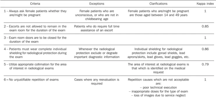
qual-ity criteria related to radiological protection of patients. Definitions, exceptions, and clarification of each one of those criteria are presented on Table 1.
All of the defined criteria are related to the assistance process, as they comprise the activities or procedures undertaken by the health professionals to transform re-sources into results. Additionally, the au-thors have taken the precaution of analyz-ing the validity and reliability of the crite-ria. Purpose, contents and foundations for each one of the criteria were considered appropriate and, in a pilot study by means of a test-retest design (n = 30), a satisfac-tory reliability (kappa index) was demon-strated.
Population and sample
The target population of the criteria comprised all patients seeking the service for their exams, except in the case of crite-rion 1, which applied only to female pa-tients. The temporal parameters for case ex-traction varied according to the criteria over one week (criteria 2, 3, 4 and 5), one month (criterion 1) and a quadrimester (criterion 6). The sampling of all criteria was system-atic and random, and the sample included 60 cases for each criteria.
Data collection
Several information sources were uti-lized in the collection of data related to compliance with the criteria, namely, re-view of the clinical process (criterion 1), patient questionnaire (criterion 2), proce-dural compliance (criteria 3 and 4) and analysis of the images on the image treat-ment console (criteria 5 and 6). As regards timing, the evaluation was concurrent for criteria 3 and 4, and retrospective for cri-teria 1, 2, 5 and 6.
The initiative of undertaking such evaluation came from the professionals themselves, that is, it was an internal pro-cess, with the professionals from the ser-vice being responsible for collecting the data and performing a cross-analysis where each professional evaluates the actions of another peer.
Improvement intervention
For the development of an improvement intervention plan, the authors have resorted to a participative planning method which included and comprised the radiologists related to the processes which are object of improvements. The set of interventions that originated from the generation of ideas within the group was distributed over an affinity diagram into three groups of ac-tions to be implemented, as follows:
1. Education of the radiologists on movement and transfer of patients, children immobilization, radiological protection and interaction with patients.
2. Changes in registration forms, add-ing YES and NO filladd-ing spaces on the re-quests for female patients asking whether they are/might be pregnant.
3. Disclosure of results: by means of a storyboard recording the progress of the activities located in a place where all pro-fessionals could see it, and awareness de-velopment actions for the follow-up of the study results.
Once the improvement action was de-fined, the authors decided to utilize two in-struments to assure and supervise the implementation of the action plan: the storyboard, utilized to record the progress of the activities at the sight of all involved professionals; and a Gantt chart, which is a graphic representing the scheduled time
required for the execution of the actions, as well as the names of the responsible agents for each one of them.
Data analysis
Both in initial evaluations and in the re-evaluation, calculations of point and inter-val estimates (95% confidence level) were performed on the compliance with criteria in random selected samples.
In order to estimate the improvement observed between the reevaluation and the initial evaluation, absolute and relative improvements were calculated for each one of the criteria. In order to prove the statis-tical significance of the detected improve-ment, a unilateral hypothesis test was per-formed by means of the calculation of the Z value, considering as the null hypothesis the absence of improvement, which was rejected whenever the p-value was lower than 0.05.
Additionally, the main quality defects identified at both evaluations were graphi-cally represented. For such a purpose a before-and-after Pareto chart was utilized, for being a complete and informative rep-resentation(2) which makes the
prioriti-zation of intervention strategies easier. In the early stage of the chart construction, a table of absolute and relative non-compli-ance frequencies was developed. Subse-quently, the before-and-after Pareto chart
was built on a three-axis Cartesian plane, where the central axis represents the abso-lute frequencies demonstrating the results from the two evaluations, and the left and right axes represent, respectively, the rela-tive percentage of non-conformities in the first evaluation and in the reevaluation. The lines drawn on the chart represent the ac-cumulated frequency of quality defects observed in each evaluation.
RESULTS
Basic level of quality in radiological protection
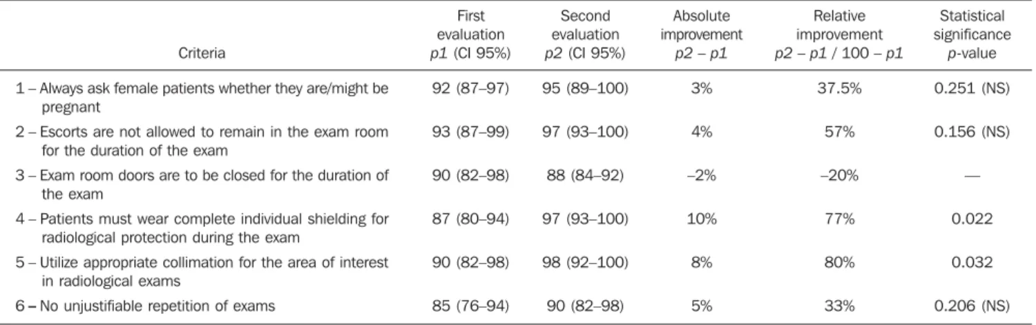
Table 2 demonstrates that all six crite-ria presented a high rate of compliance al-ready in the first evaluation (minimum = 85%; maximum = 93%). The highest com-pliance level was observed for the criterion “Escorts are not allowed to stay in the room during the performance of the exam” (cri-terion 2), with a compliance rate of 93% (CI 95%: 87–99), followed by the criteria “Always ask female patients whether they are/might be pregnant” (criterion 1), with a compliance rate of 92% (CI 95%: 87–97).
Analysis of identified quality defects and intervention priorities
On the before-and-after Pareto chart, one can observe and compare the values of the corresponding non-compliances for Table 1 Quality criteria developed to measure radiological protection quality level.
Criteria
1 – Always ask female patients whether they are/might be pregnant
2 – Escorts are not allowed to remain in the exam room for the duration of the exam 3 – Exam room doors are to be closed for the
duration of the exam
4 – Patients must wear complete individual shielding for radiological protection during the exam
5 – Utilize appropriate collimation for the area of interest in radiological exams
6–No unjustifiable repetition of exams
Exceptions Female patients who are unconscious, or who are not in
childbearing age Patients who do require full time
assistance of an escort
Whenever the radiological protection exclude or degrade important diagnostic information
Cases where any reevaluation is required
Clarifications
Female patients who are/might be pregnant are those aged between 14 and 49 years
Individual shielding for radiological protection include gonad shields, lead aprons/skirts, lead gloves, lead goggles, etc.
The area of interest at radiological exams is that which is identified on the medical
request
Repetition causes which are not acceptable are:
– poor technical execution – inappropriate doses for the type of exam
– loss of images due to service neglect
Kappa index 1
0.85
1
0.86
0.79
each one of the six criteria at the two evalu-ations. It is also possible to identify the most problematic criteria denominated as “vital few” according to the “Pareto’s prin-ciple”(3).
In the first evaluation, a pair of criteria (criteria 6 and 4) stood above the others for representing 44.7% of all defects identified amongst the 100% representing all defects under the six criteria included in the present study, which allows those two criteria to be considered as the “vital few” criteria which were prioritized in the effort to obtain im-provements (as previously indicated under the topic “Improvement Intervention”). In the second evaluation, criteria 3 and 6 rep-resented approximately 62% of the non-compliance cases, i.e., being considered as “vital few” criteria. Thus the planning for a new intervention should prioritize those two criteria, not neglecting the others, as in the second evaluation all criteria presented non-conformities.
Table 2 Compliance with the radiological protection quality criteria before and after improvement intervention.
Criteria
1 – Always ask female patients whether they are/might be pregnant
2 – Escorts are not allowed to remain in the exam room for the duration of the exam
3 – Exam room doors are to be closed for the duration of the exam
4 – Patients must wear complete individual shielding for radiological protection during the exam
5 – Utilize appropriate collimation for the area of interest in radiological exams
6–No unjustifiable repetition of exams
First evaluation p1 (CI 95%) 92 (87–97) 93 (87–99) 90 (82–98) 87 (80–94) 90 (82–98) 85 (76–94)
Second evaluation p2 (CI 95%) 95 (89–100) 97 (93–100) 88 (84–92) 97 (93–100) 98 (92–100) 90 (82–98)
Absolute improvement
p2 – p1 3% 4% –2% 10% 8% 5%
Relative improvement p2 – p1 / 100 – p1
37.5% 57% –20%
77% 80% 33%
Statistical significance
p-value 0.251 (NS) 0.156 (NS)
— 0.022 0.032 0.206 (NS)
p1, compliance rate at the first evaluation; p2, compliance rate at the second evaluation; NS, non statistically significant (p > 0.05). Reevaluation of the level of quality
and effectiveness of the improvement intervention
All of the six criteria presented a high compliance rate in the reevaluation (Table 2). Additionally, in absolute terms, im-provements were observed for every crite-ria, except for the criterion “examination room doors must be closed throughout the performance of the exam” (criterion 3), which presented a higher number of non-conformities in the reevaluation than in the first evaluation.
All of the criteria which demonstrated improvements (criteria 1, 2, 4, 5, and 6) presented relative improvement rates above 30% between the two evaluations. As re-gards statistical significance of such im-provement, the criteria “The patients must wear complete individual shielding for ra-diological protection during the exam” and “Utilize collimation appropriate for the area of interest in radiological
examina-tions” (criteria 4 and 5 respectively) pre-sented a p-value lower than 0.05, charac-terizing a statistically significant improve-ment in the level of quality regarding those criteria. Statistically significant improve-ments were not achieved in the remaining criteria. (Table 2).
According to Table 2 and Figure 1, one can observe that the set of the six criteria totals 21 non-conformities in the second evaluation, 17 less than in the first evalua-tion, corresponding to an absolute improve-ment of approximately 45% between the evaluations (corresponding to the area highlight on the chart of the second evalu-ation). However, in a negative result, cri-terion 3 (“exam room doors must be closed throughout the performance of the exam”) presented a higher rate of non-compliances in the reevaluation than in the first evalua-tion (absolute improvement = –2%). The statistical significance of the worsening was not calculated, as the sensitivity of the
test for the improvement hypothesis was prioritized with the method of unilateral analysis described in the methodology.
The two criteria considered as “vital few” in the first evaluation — criteria 6 and 4, on which more improvement activities were developed in the intervention — pre-sented a significant decrease in the number of non-conformities in the reevaluation, with a relative improvement of 77% for criterion 4, while criterion 6 presented a relative improvement of 33%. However, criterion 6 (“No repetition of exams”), al-though presenting a decrease in the num-ber of non-conformities between evalua-tions, remained as the second criteria with more non-conformities (second only to cri-terion 3).
DISCUSSION
The results obtained in this improve-ment cycle contribute to the understanding of the effectiveness of the cycles of insti-tutional quality assessments in radiology services. In general, the method based on the internal quality management scope was effective in changing the professionals’ at-titude and in improving the level of good practices in radiological protection. Al-though the improvement intervention planned and implemented by the profes-sionals in the center were not completely effective in solving all of the evaluated ra-diological protection deficiencies, the con-solidation of the philosophy and structure of quality management in this institution paved the way to the continuity of the same evaluation cycle and for the evaluation and improvement of other priority problems in the pursuit of excellence.
During the improvement cycle in the present study, actions which were within the capabilities of the professionals were utilized as quality criteria, aiming at the optimization of the radiological protection of the patients and which must always be adopted during the performance of a radio-logical exam and therefore, ideally, non-conformities should not exist with respect to the criteria utilized for the improvement cycle in the present study(10,13,14). However, the results of the present study demonstrate that non-conformities occurred in every criterion, even in the reevaluation after the
implementation of some improvement strategies.
The fact that the reevaluation revealed a criterion which presented a higher num-ber of non-conformities than in the first evaluation, and also other criteria which did not present statistically significant im-provements (criteria 1, 2, and 6), demon-strates that the improvement activities implemented during the intervention did not produce optimum effects for such cri-teria. The cause for such facts may reside in mistakes that may have been made in analyzing the causes of the problems or in the planning and/or implementation of the intervention.
Thus, the continuity of the assessment cycle is important, improving the analysis of causes and planning of the intervention, which are the key steps to achieve improve-ments. The continuity of the improvement cycle also allows the consolidation of the processes, methods and tools utilized in this type of activity on a theme with accu-mulated experience, as well as it strength-ens the improvements achieved in the first cycle, helping such improvements to be-come sustainable(2,15).
As regards the difficulties experienced during the improvement cycle, which can be similar to those in other institutions undertaking similar projects, the task group reported some difficulties in the implementation and in the form of utiliza-tion of tools and methods applied in the activities, causing some delays in the im-provement cycle. Possibly, this occurred because of the fact that this was the first time in which most of the involved radi-ologists had a contact with systematic quality improvement activities. One of the phases where more difficulty was encoun-tered was the implementation of improve-ment strategies adopted after the first evaluation, where delays occurred in rela-tion to the timeline initially established on the Gantt chart. A positive note, consider-ing that the improvement cycle was the first contact of all intervenients with quality improvement activities, refers to the fact that the analyzed problems were internally prioritized by the professionals themselves. This allowed them to work on a known field, where they found themselves directly involved, recognizing its relevance, thus
fa-cilitating the commitment with the quality improvement actions.
CONCLUSIONS
The results of the present study demon-strated that the radiological protection qual-ity level in the evaluated service, although reasonably high, presented a margin for improvement, particularly in the criteria concerning the non-repetition of exams and the utilization of appropriate individual shielding for radiological protection. The identification of such problems has moti-vated and guided an intervention based on the participative principle, allowing a sig-nificant improvement in two of the aspects with greater impact on quality, demonstrat-ing the effectiveness of the evaluation cycle in this context. The fact that an optimum quality level was not reached (absence of non-conformities with the criteria) only highlights the relevance of the continuity of the evaluation cycle with the purpose of further improving the processes and the currently prioritized criteria. Finally, it is possible to say that undertaking such an improvement cycle has been beneficial for the patients, as the optimization of their radiological protection means that they will be exposed to lower radiation doses, a ben-efit of utmost importance, even not being directly perceived by them.
REFERENCES
1. Saturno PJ. Gestión de la calidad. Concepto y componentes de un programa de gestión de la calidad. Manual del máster en gestión de la cali-dad en los servicios de salud. Módulo 1: Concep-tos básicos. Unidad temática 2. 2ª ed. Murcia: Universidad de Murcia; 2008.
2. Juran JM, Gryna FM, Binghan RS. Manual de control de la calidad. 2ª ed. Barcelona: Reverté; 1990.
3. National Council on Radiation Protection and Measurements. Medical radiation exposure of the U.S. population greatly increased since the early 1980s. 2009. [cited 2011 Feb 23]. Available from: h t t p : / / w w w. n c r p o n l i n e . o r g / P r e s s _ R e l / Rept_160_Press_Release.pdf
4. Henriques S. IAEA culture shift needed to achieve patient radiation safety. 2011. [cited 2011 May 17]. Available from: http://www.iaea.org/newscenter/ news/2010/cultureshift.html
5. Wall BF, Hart P. Revised radiation doses for typi-cal X-ray examinations. Report on a recent review of doses to patients from mrdical X-ray examina-tions in the UK by NRPB. Br J Radiol. 1977;70: 437–9.
May 23]. Available from: http://www.iaea.org/ newscenter/news/2010/guidanceprotect.html 7. International Atomic Energy Agency. Heavy
com-ponent replacement in nuclear power plants: ex-perience and guidelines. IAEA Nuclear Energy Series No. NP-T-3.2. Vienna: IAEA; 2008. 8. International Atomic Energy Agency.
Internatio-nal action plan for the radiological protection of patients. 46th IAEA General Conference (2002) Documents; GOV/2002/36-GC(46)/12; IAEA. 2002. [cited 2011 May 27]. Available from: h t t p : / / w w w. i a e a . o r g / A b o u t / P o l i c y / G C / GC46Documents/English/gc46-12_en.pdf.
9. Pisco JM. Imagiologia básica – texto e atlas. 2ª ed. Lisboa: Lidel Edições Técnicas; 2009. 10. Soares FAP, Pereira AG, Flôr RC. Utilização de
vestimentas de proteção radiológica para redução de dose absorvida: uma revisão integrativa da li-teratura. Radiol Bras. 2011;44:97-103. 11. D’Ippolito G, Medeiros RB. Exames radiológicos
na gestação. Radiol Bras. 2005;38:447-50. 12. Saturno PJ, Gascon JJ. Métodos de análisis de los
problemas de calidad. Manual del máster en ges-tión de la calidad en los servicios de salud. Mó-dulo 3: Actividades básicas para la mejora conti-nua: métodos y herramientas para la realización
de ciclos de mejora. Unidad temática 11. 1ª ed. Murcia: Universidad de Murcia; 2008. 13. Ministério da Saúde. Portugal. Despacho nº258/
2003 (2ª série). Diário da República nº 6, II Sé-rie, de 8 de janeiro de 2003.
14. Ministério da Saúde. Portugal. Decreto-Lei nº180/ 2002. Diário da República nº 182, Série I-A, de 8 de agosto de 2002.