Imaging diagnosis in snapping syndromes*
Diagnóstico por imagem nas síndromes do estalido ou do ressalto
Henrique Ribeiro da Silva1, Marcelo Novelino Simão2, Jorge Elias Júnior3, Valdair Francisco Muglia3, Marcello Henrique Nogueira-Barbosa3
Snapping syndromes occur during certain movements in several joints and may be accompanied by local pain and crepitation or audible snapping sensation. Snapping joints may eventually have no pathologic significance and in this case no treatment is required. There is an array of intra- and extra-articular causes of snapping syndrome and the lack of precise clinical findings impairs the definition of the most appropriate imaging method to confirm the diagnosis. The present study is aimed at discussing the most frequent causes of snapping syndrome in different joints, emphasizing the indications and limitations of each of the different diagnostic methods in specific situations of the clinical practice.
Keywords: Joints; Snapping; Ultrasonography; Computed tomography; Magnetic resonance imaging.
A síndrome do estalido ou do ressalto ocorre durante a movimentação de várias articulações e pode ser acompanhada de dor local e de crepitação ou de estalido audível. Nem sempre o estalido audível ou a cre-pitação articular à palpação têm significado patológico ou implicam necessidade de tratamento. Esta síndrome tem causas variadas intra-articulares e extra-articulares e os achados clínicos podem ser pouco precisos, com dificuldade para definir o melhor método de imagem que confirme o diagnóstico. O objetivo deste tra-balho é discutir as causas mais comuns da síndrome do estalido em cada articulação e enfatizar a indicação e a limitação de cada um dos diferentes métodos diagnósticos em situações específicas da prática clínica.
Unitermos: Articulações; Ressalto; Ultrassonografia; Tomografia computadorizada; Imagem por ressonância magnética.
Abstract
Resumo
* Study developed at Unit of Radiodiagnosis, Center of Imag-ing Sciences and Medical Physics – Hospital das Clínicas da Faculdade de Medicina de Ribeirão Preto da Universidade de São Paulo (CCIFM/HC-FMRPUSP), Ribeirão Preto, SP, Brazil.
1. MD, Resident, Hospital das Clínicas da Faculdade de Me-dicina de Ribeirão Preto da Universidade de São Paulo (HC-FMRPUSP), Ribeirão Preto, SP, Brazil.
2. Master, Physician Assistant at Unit of Radiodiagnosis, Center of Imaging Sciences and Medical Physics – Hospital das Clíni-cas da Faculdade de Medicina de Ribeirão Preto da Universidade de São Paulo (CCIFM/HC-FMRPUSP), Ribeirão Preto, SP, Brazil. 3. PhDs, Professors at Center of Imaging Sciences and Med-ical Physics – Hospital das Clínicas da Faculdade de Medicina de Ribeirão Preto da Universidade de São Paulo (CCIFM/HC-FMRPUSP), Ribeirão Preto, SP, Brazil.
Mailing address: Dr. Marcello Henrique Nogueira-Barbosa. Avenida Bandeirantes, 3900, Campus Universitário. Ribeirão Preto, SP, Brazil, 14048-900. E-mail: [email protected]
Received June 8, 2008. Accepted after revision September 16, 2008.
Treatment, if required, is generally con-servative. Eventually, surgery or arthroscopy may be required, and outcomes vary, depending on the etiology of the syn-drome(3–10).
The most frequent presentations of snapping syndromes in large limb joints will be discussed in the present review and the role played by the different imaging techniques in these cases is emphasized.
SNAPPING HIP
The most frequently reported form of snapping syndrome occurs in hip, generally with extra-articular causes(11–14). Many
times, snapping hip syndrome, or coxa saltans, presents as an audible snapping or
popping sound or palpable prominence associated with pain when the hip is flexed and extended.
Medial snapping hip is related to the sudden displacement of the iliopsoas ten-don over the iliopectineal eminence or ad-jacent bone structures. Lateral snapping hip is related to the sudden displacement of the
Silva HR, Simão MN, Elias Jr J, Muglia VF, Nogueira-Barbosa MH. Imaging diagnosis in snapping syndromes. Radiol Bras. 2009;42(1):49–55.
anatomic abnormalities, loose intra-articu-lar bodies, previous surgery or trauma, ar-ticular degeneration and inflammatory processes. These causes may be extra- or intra-articular. Snapping joints can be found with no association with other com-plaints and in these cases no treatment is required.
The diagnosis is primarily based on the clinical history and physical examination of the patient. At clinical examination an au-dible snapping can be identified or a pal-pable prominence is observed, generally during specific maneuvers or when there is an attempt to reproduce the movements that result in complaint by the patient. Imaging diagnosis methods are utilized for confirm-ing and documentconfirm-ing the anatomic struc-tures or lesions associated, and can be use-ful for the management of the case, allow-ing the plannallow-ing or choice of the an appro-priate treatment.
It is important to know the most fre-quent causes of this syndrome in the differ-ent joints to allow the choice of the appro-priate imaging diagnosis method.
INTRODUCTION
Snapping syndrome corresponds to a group of changes that affect the limbs joints. Clinical presentation includes snap-ping during joints movement and may be associated with other clinical symptoms such as pain and, less frequently, neuro-logical alterations(1,2). This is a
tensor fascia lata or gluteus maximus over the greater trochanter of the femur.
Sometimes the cause of the snapping may be related to intra-articular factors or lesions. Intra-articular causes include loose bodies, synovial osteochondromatosis and acetabular labral tears(5,6,15).
If further clinical investigation of snap-ping hip is indicated, plain radiography can be initially utilized for allowing identifica-tion of bone exostosis or articular and bone tumors, besides loose intra-articular bodies. In the past, iliopsoas tenography and bursography were utilized for allowing a dynamic study and characterization of sud-den snapping of the iliopsoas tendon on fluoroscopic images(16). However,
ultra-sonography is currently a preferable method for being non-invasive and allow-ing a dynamic evaluation durallow-ing maneu-vers that trigger the snapping(17). Generally,
plain radiography and ultrasonography are sufficient for investigating the majority of cases of extra-articular snapping hip.
In these cases, ultrasonography is per-formed with high-frequency (5–12 MHz) linear transducers. Initially, the patient should be positioned in dorsal decubitus, with extended limbs, and subsequently, in lateral decubitus for evaluation of the an-terior and lateral regions of the hip, includ-ing the greater trochanter and adjacent soft parts in the transverse and longitudinal planes. In the investigation of the medial snapping hip, the study should be focused
on the iliopsoas and, in case of lateral snap-ping hip, on the fascia lata tensor muscle and gluteus maximus.
Lateral snapping hip should also be evaluated with the transducer transversally placed over the greater trochanter (Figure 1), with the patient standing on the opposite leg. The movements that trigger the snap-ping must be reproduced by the patient both in dorsal decubitus and orthostatic positions. The presence or not of painful snapping should be evaluated during the examination.
Medial snapping should be evaluated with the transducer transversally positioned or slightly oblique anteriorly over the hip joint, with the patient in dorsal decubitus, hip flexion with abduction and external rotation, in the frog position.
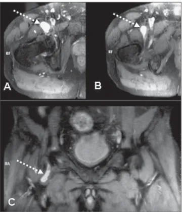
Sectional imaging methods should be indicated if a diagnosis is not achieved by radiography and ultrasonography. Mag-netic resonance imaging can eventually demonstrate inflammatory processes in the snapping tendon region or identifying a bursa probably associated with the ping (Figures 2 and 3). Intra-articular snap-ping syndrome can be investigated by means of magnetic resonance imaging (Figure 4) or one of the sectional imaging methods preceded by arthrography: magnetic resonance imaging and arthro-computed tomography(15).
Treatment is usually clinical and conser-vative, most of times with good outcomes.
Surgical management or arthroscopy is considered in cases where a good clinical response is not achieved, particularly in the presence of intra-articular causes(5,6,11,12,15).
SNAPPING SCAPULA
Palpable and, sometimes audible crepi-tation of the scapulo-thoracic joint should not always be considered as pathological. This is a relatively frequent and, most of times, painless alteration.
Scapulocostal syndrome can be identi-fied in overhead-throwing athletes(3).Some
authors, however, have not found any di-rect relation between symptoms and type of activity, sports of postural alterations, and diagnosis is reported even in sedentary patients or in non-dominant limbs(18).
The patients generally complain of pain with increase in activities involving the shoulder. One of the most consistent find-ings is crepitation with motion of the scapula. Inspection at clinical examination can demonstrate scapular asymmetry, and it is important to evaluate the minor pecto-ris, trapezium and elevator muscles in or-der to detect the presence of tension in these muscles of the affected side. Several bursae lying around represent potential causes of crepitation(3,8).
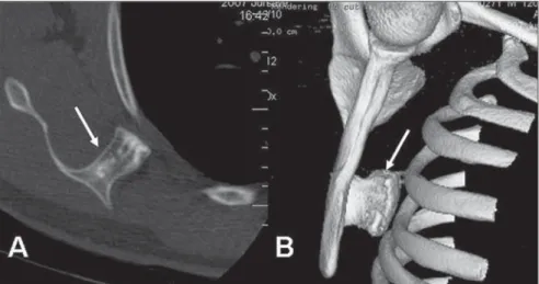
Although elastofibromas (Figure 5), os-teochondromas (Figure 6) and other tumors are reported as causes of this syndrome in the scapulothoracic region, associated
le-Figure 1. Sonographic images of a hip (trochanteric region). Cross sectional views obtained with internal (A) and external hip rotation (B). The dashed arrows indicate the fascia lata tensor displaced in relation to the trochanter and to the deepest gluteal tendon (long arrows).
Figure 4. Intra-articular snapping hip. Magnetic resonance imaging demon-strates loose intra-articular body (arrows) on coronal (A) and axial (B) fluid-sensitive sequences with fat-saturation.
Figure 3. Magnetic resonance imaging of right hip – axial, T2-weighted sequence with fat-saturation (A,B). Coronal view of the left hip on T2-weighted sequence with fat-saturation (C). The arrows indicate iliopsoas bursa disten-tion that may be eventually associated with medial snapping hip syndrome.
Figure 5.A: Histologically confirmed elastofibroma in the right infrascapular region. Axial computed tomography demonstrates heterogeneous lesion with soft parts and fat density. B: Magnetic resonance image of the same patient – axial, T2-weighted sequence demonstrating a heterogeneous mass adjacent to the costal grid. C: sagittal, T2-weighted sequence demonstrating that the heterogeneous mass presents hyposignal intensity in relation to the muscles. The arrows indicate the lesion.
sions or tumors are not easily identified, and the cause may be dynamic and related to repetitive microtraumas, bursitis and anatomical variations(3,8,9,19,20).
The initial step for radiological investi-gation of scapula snapping syndrome is
plain radiography to rule out obvious lesion of the scapula and ribs such as post-trau-matic exostosis, osteochondromas or other expansile bone lesions. Fluoroscopy can be utilized to visualize the snapping during motion. If any bone abnormality that could
resolution as its primary advantage to iden-tify soft parts abnormalities, for example, inflammatory processes or hemorrhage in the involved bursa(9,18).
Three-dimensional computed tomogra-phy can be utilized to evaluate the bone in-congruity between the anterior aspect of the scapula and the chest wall(21).
Elastofibroma is a non-neoplastic, reac-tive and rare type of lesion that is most fre-quently unilateral, although it can be uni-lateral or biuni-laterally found. Most frequently, this type of lesion is found deeply in rela-tion to the rhomboid and great dorsal muscles, between the subscapular area and the chest wall. Because of this typical lo-calization, this lesion is called dorsal elastofibroma. Typically, this lesion devel-ops in mid-aged or elderly patients. Al-though it can be identified by ultrasonog-raphy and computed tomogultrasonog-raphy, magnetic resonance imaging, if available, should be the method of choice.
Magnetic resonance imaging may sug-gest a presumptive diagnosis, particularly in cases where the lesion is found in the most typical site, i.e., the infrascapular re-gion, and especially when demonstrates a fibrotic and fatty content. The definite di-agnosis is achieved by means of histo-pathologic study.
The treatment of snapping scapula is generally clinical and conservative, the lat-ter including the utilization of anti-inflam-matory drugs, local analgesia, judicious uti-lization of corticosteroid infiltration and, mainly, muscular rehabilitation. In cases
where no clinical improvement is observed in three to six months, surgical manage-ment may be considered(3,8,9,18). Surgical
resection may be required in cases where an elastofibroma or osteochondroma is identified.
SNAPPING ELBOW
Snapping elbow syndrome is a less fre-quent clinical entity, and as such, is poorly recognized. The snapping elbow may progress with no other manifestation such as pain and neurological symptoms(1,2,6,7).
Several causes are reported, the most fre-quent one being the elbow relationship with medial anatomical structures. Other causes include inflammatory processes and anatomical alterations, particularly cubitus varus deformity, that may result in displace-ment of the ulnar nerve in combination or not with dislocation of the medial head of the triceps over the medial epicondyle with elbow flexion(1,2,4,22).
Most frequently, ultrasonography and magnetic resonance imaging are indicated for snapping elbow investigation(2,4,22).
Ul-trasonography (Figure 7), may be preferred because of its wide availability and dy-namic evaluation capability, and should be performed with a 7.0 MHZ (at minimum) Figure 6. Patient with snapping in the dorsal region. A: Right scapular osteochondroma seen on axial
computed tomography (arrow). B: Osteochondroma identified on 3D image reconstruction (arrow).
presentations. The dynamic evaluation of fibular tendons by ultrasonography should be performed with cross-sectional views on the retromalleolar region and the tendons are observed during passive or active foot flexion and eversion.
A recently published casuistic reports 14 cases of intrasheath subluxation of pero-neal tendons, and in this case the tendons remain in the retromalleolar region, but transitorily switch their relative positions with a reproducible painful click(30).
Most rarely, snapping syndrome can also be caused by a third fibular tendon(31)
or being related to the posterior tibial ten-don(32), in this latter case, with medial
snap-ping. In a systematic literature review(32)
published in 2008, the authors observed that 58.5% of lesions with luxation of the posterior tibial tendon were caused by sports trauma, and 53.1% of all the cases with luxation of the posterior tibial tendon have initially remained without a correct diagnosis. The authors have also observed that 35.6% of the patients experienced re-current snapping of the tendon. Surgical management with different techniques was the most frequent approach adopted (83.1%) and outcomes were considered as excellent and asymptomatic in 80% of cases and good in other 12%.
Like in other joints, the investigation of snapping of superficial tendons can be ef-fectively performed by ultrasonography. Computed tomography and magnetic reso-linear transducer. Before ultrasonography,
the olecranon and the medial epicondyle should be palpated with the limb in exten-sion with subtle supination. The transducer must be placed on the axis between these structures, for identification and measure-ment of the sectional area of the ulnar nerve inside its groove. Subsequently, the patient should be asked to flex the elbow with the transducer in the same position, and the diagnosis of the dislocation is made as the ulnar nerve passes the medial epicondyle apex, in association or not with displace-ment of the medial head of the triceps(2).
The flexion against resistance facilitates the diagnosis and should be utilized. In some cases, passive or active dynamic studies without counter-resistance may not repro-duce the snapping. With magnetic reso-nance imaging, the sequences should be acquired with the limb at maximum exten-sion and flexion for evaluation of the ini-tial and end positions of the ulnar nerve and medial head of the triceps(4,22).
It is very important for the radiologist to differentiate an isolate displacement of the ulnar nerve from a combination with dislocation of the medial head of the tri-ceps(2). A considerable number of patients
with snapping ulnar nerve are asymptom-atic or at least do not require surgical man-agement. However, the symptoms may persist after surgery, if a surgical transpo-sition of the ulnar nerve is indicated, and association with dislocation of the medial head of the triceps has not been identified. Lateral snapping of the elbow is much less frequently found and may be associ-ated with poster lateral rotator instability, a menisci interposition with the radio-humeral joint, snapping annular ligament over the radial head or even a lateral dis-placement of the distal triceps(1,23–25).
SNAPPING KNEE
In the knee, the snapping syndrome is most frequently associated with traumatic or non-traumatic meniscal lesions. Lateral discoid meniscus, which is the most fre-quently found abnormality of children, may be the cause, particularly the lateral discoid meniscus type III or Wrisberg’s ligament. This rare alteration occurs when the poste-rior fixation between the meniscus and the
Figure 8. Sonographic image on cross-section view oblique to the pes anserine. The flexor tendons of the pes anserine — semitendinosus (st), gracilis (g) e sartorius (sa) — can be identified on the most superficial plane. The semimembranosus tendon (sm) is localized in a slightly deeper plane. In this patient, dynamic images have demonstrated a sudden snapping of the gracilis in relation to the semi-membranosus tendon.
tibia is absent with a significantly higher meniscal mobility(7,26). Other less frequent
causes, such as tendon dislocation and in-tra-articular tumors have also been de-scribed(10,26–28).
Investigation with imaging methods should be initiated with conventional radi-ology. Evaluation of snapping tendons may be performed with ultrasonography (Fig-ure 8). On the other hand, the utilization of magnetic resonance imaging should be en-couraged if an intra-articular lesion is sus-pected. Arthroscopy, despite its invasive-ness, can also be utilized(7,26). The treatment
must be individualized, and can be clinical, by arthroscopy or open surgery, as the case requires(7,10,26–28).
SNAPPING ANKLE
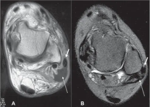
Snapping ankle is rare and may be as-sociated with subluxation or luxation of the tendon. Most rarely, it is reported in asso-ciation with intra-articular bodies. Among reports in the literature, the most frequently found situation is luxation or subluxation of fibular tendons, with or without concur-rent tendineal rupture(29). In most of
nance imaging (Figure 9) can statically demonstrate luxation or subluxation and also allow the evaluation of the fíbula or other lesions that may be associated.
SNAPPING WRIST
Cases of snapping wrist syndrome have been rarely reported. Tumors, loose articu-lar bodies, and dislocation of tendons can cause snapping(30,33–35). Also, another study
describes a case of snapping of superficial flexor tendon of the fifth finger over the hamate hook after surgical carpal tunnel decompression (36).
Instability of the extensor carpi ulnaris tendon may cause snapping during sublux-ation and luxsublux-ation and has been reported as the most frequent cause of snapping of the wrist. The evaluation of this condition can be performed by means of ultrasonography (Figure 10). This condition may eventually be clinically confused or being concurrent with subluxation of the distal radioulnar joint. In rheumatoid arthritis, the tendon may dislocate towards the volar region, while ulnar subluxation may occur poste-riorly to the distal radioulnar joint.
In normal wrists there is a certain ac-ceptable degree of subluxation of the ex-tensor carpi ulnaris tendon as related to its osseous groove(37). The literature reports
normality values for this dislocation in terms of percentage of the width of the tendon’s osseous groove. The tendon may displace by up to 40% of the osseous groove width towards the volar region with wrist flexion, and towards the dorsal region up to 33% with wrist extension.
Generally, the traumatic mechanism that causes subluxation of the extensor carpi ulnaris tendon corresponds to forced supi-nation, palmar flexion and ulnar disloca-tion. With the pronation, the tendon gen-erally returns to its usual position. Mag-netic resonance imaging can characterize the inflammatory process in the synovial sheath and, eventually, the inappropriate positioning of the tendon. Acute sublux-ation has been rarely reported in the litera-ture, and can be managed with immobili-zation in pronation and a slight radial de-viation of the wrist. Surgical reconstruction may be required in cases of symptomatic and chronic subluxation.
Figure 10.A: The arrows indicate the extensor carpi ulnaris tendon on an axial sonographic image with the forearm in pronation. B: Cross-sectional sonographic view with forced supination demonstrating the dislocation of the extensor carpi ulnaris tendon (arrows).
Figure 9.A: Axial magnetic resonance imaging of ankle – contrast-enhanced T1-weighted sequence of a patient with acute traumatic luxation of the fibular tendon. The white arrow indicates the fibular tendon anterior and laterally dislocated in relation to the lateral malleolus. The dashed arrow indicates post-traumatic collection. B: Another patient with chronic luxation of the fibular tendon on axial, T2-weighted sequence with fat-suppression. The white arrow indicates the tendon and the dashed arrow indicates the thickened retinaculum.
CONCLUSION
Snapping syndrome is a challenging clinical condition with several causes and relatively poorly commented. In most of cases, it is characterized by mild clinical manifestations, but eventually the syn-drome may cause a significant physical limitation. It may be difficult to define a diagnosis because of the absence of knowl-edge on the nature of this syndrome. The knowledge on this syndrome is relevant for
guiding in the selection of the appropriate imaging method. The present review dis-cusses the main causes of this syndrome and indicates the most appropriate diagnos-tic methods according to the joint involved and the clinical suspicion.
REFERENCES
1. Spinner RJ, An KN, Kim KJ, et al. Medial or lat-eral dislocation (snapping) of a portion of the distal triceps: a biomechanical, anatomic expla-nation. J Shoulder Elbow Surg. 2001;10:561–7.
dislocation and snapping triceps syndrome: diag-nosis with dynamic sonography – report of three cases. Radiology. 2001;220:601–5.
3. Manske RC, Reiman MP, Stovak ML. Nonoper-ative and operNonoper-ative management of snapping scapula. Am J Sports Med. 2004;32:1554–65. 4. Spinner RJ, Goldner RD. Snapping of the medial
head of the triceps and recurrent dislocation of the ulnar nerve. Anatomical and dynamic factors. J Bone Joint Surg Am. 1998;80:239–47.
5. White RA, Hughes MS, Burd T, et al. A new op-erative approach in the correction of external coxa saltans: the snapping hip. Am J Sports Med. 2004; 32:1504–8.
6. Provencher MT, Hofmeister EP, Muldoon MP. The surgical treatment of external coxa saltans (the snapping hip) by Z-plasty of the iliotibial band. Am J Sports Med. 2004;32:470–6.
7. Kelly BT, Green DW. Discoid lateral meniscus in children. Curr Opin Pediatr. 2002;14:54–61. 8. Nicholson GP, Duckworth MA. Scapulothoracic
bursectomy for snapping scapula syndrome. J Shoulder Elbow Surg. 2002;11:80–5.
9. Majó J, Gracia I, Doncel A, et al. Elastofibroma dorsi as a cause of shoulder pain or snapping scapula. Clin Orthop Relat Res. 2001;(388):200– 4.
10. Cooper DE. Snapping popliteus tendon syn-drome. A cause of mechanical knee popping in athletes. Am J Sports Med. 1999;27:671–4. 11. Pelsser V, Cardinal E, Hobden R, et al.
Extraartic-ular snapping hip: sonographic findings. AJR Am J Roentgenol. 2001;176:67–73.
12. Choi YS, Lee SM, Song BY. Dynamic sonography of external snapping hip syndrome. J Ultrasound Med. 2002;21:753–8.
13. Janzen DL, Partridge E, Logan PM, et al. The snapping hip: clinical and imaging findings in transient subluxation of the iliopsoas tendon. Can Assoc Radiol J. 1996;47:202–8.
14. Costa FP, Canto RST. Quadril em ressalto. Rev Bras Ortop. 1990;25:369–72.
15. Wunderbaldinger P, Bremer C, Matuszewski L. Efficient radiological assessment of the internal snapping hip syndrome. Eur Radiol. 2001;11: 1743–7.
16. Staple TW, Jung D, Mork A. Snapping tendon syndrome: hip tenography with fluoroscopic monitoring. Radiology. 1988;166:873–4.
17. Cardinal E, Buckwalter KA, Capello WN, et al. US of the snapping iliopsoas tendon. Radiology. 1996;198:521–2.
18. Carrera EF, Matsumoto MH, Archetti Netto N, et al. Elastofibroma dorsi: relato de casos e revisão da literatura. Rev Bras Ortop. 2004;39:468–75.
19. Edelson JG. Variations in the anatomy of the scapula with reference to the snapping scapula. Clin Orthop Relat Res. 1996;(322):111–5. 20. Hayes AJ, Alexander N, Clark MA, et al.
Elasto-fibroma: a rare soft tissue tumour with a pathog-nomonic anatomical location and clinical symp-tom. Eur J Surg Oncol. 2004;30:450–3.
21. Mozes G, Bickels J, Ovadia D, et al. The use of three-dimensional computed tomography in evaluating snapping scapula syndrome. Orthope-dics. 1999;22:1029–33.
22. Yiannakopoulos CK. Imaging diagnosis of the snapping triceps syndrome. Radiology. 2002;225: 607–8.
23. Huang GS, Lee CH, Lee HS, et al. A meniscus causing painful snapping of the elbow joint: MR imaging with arthroscopic and histologic corre-lation. Eur Radiol. 2005;15:2411–4.
24. Mehta JA, Bain GI. Posterolateral rotatory insta-bility of the elbow. J Am Acad Orthop Surg. 2004; 12:405–15.
25. Aoki M, Okamura K, Yamashita T. Snapping an-nular ligament of the elbow joint in the throwing arms of young brothers. Arthroscopy. 2003;19: E4–7.
26. Mine T, Ihara K, Taguchi T, et al. Snapping knee caused by intra-articular tumors. Arthroscopy. 2003;19:E21.
27. Lokiec F, Velkes S, Schindler A, et al. The snap-ping biceps femoris syndrome. Clin Orthop Relat Res. 1992;(283):205–6.
28. Bae DK, Kwon OS. Snapping knee caused by the gracilis and semitendinosus tendon. A case report. Bull Hosp Jt Dis. 1997;56:177–9.
29. Arrowsmith SR, Fleming LL, Allman FL. Trau-matic dislocations of the peroneal tendons. Am J Sports Med. 1983;11:142–6.
30. Raikin SM, Elias I, Nazarian LN. Intrasheath sub-luxation of the peroneal tendons. J Bone Joint Surg Am. 2008;90:992–9.
31. Sammarco GJ, Henning C. Peroneus tertius muscle as a cause of snapping and ankle pain: a case report. Am J Sports Med. 2007;35:1377–9. 32. Lohrer H, Nauck T. Posterior tibial tendon dislo-cation. A systematic review of the literature and presentation of a case. Br J Sports Med. 2008 Jan 16 Epub ahead of print.
33. Zachee B, DeSmet L, Fabry G. A snapping wrist due to a loose body. Arthroscopic diagnosis and treatment. Arthroscopy. 1993;9:117–8. 34. Rupnik E, Szloboda J. “Snapping wrist” caused
by tumor of the flexor tendon. Magy Traumatol Orthop Helyreallito Seb. 1991;34:57–8.
35. Renard M, Simonet J, Bencteux P, et al. Intermit-tent dislocation of the flexor hallucis longus ten-don. Skeletal Radiol. 2003;32:78–81. 36. Itsubo T, Uchiyama S, Takahara K, et al.
Snap-ping wrist after surgery for carpal tunnel syn-drome and trigger digit: a case report. J Hand Surg [Am]. 2004;29:384–6.